
Research article · 2025 · Full text
International Journal of Biology SciencesExosome-mediated intercellular communication in tumor microenvironment
Volume 7(5) · Pages 176–178 · DOI: 10.33545/26649926.2025.v7.i5c.717 ↗
Research areasExtracellular vesicles in diseaseCellular transport and secretionCancer Cells and Metastasis
Abstract
Author-writtenA trend gaining momentum in cancer biology over the past decade is the recognition that tumors don't grow in isolation - they actively remodel their surroundings through secreted signals, and exosomes are emerging as a main channel for this communication. This research characterized exosome release, cargo composition, and functional effects on recipient cells in a panel of three human cancer cell lines (MCF-7 breast, A549 lung, HCT116 colon) compared with their non-tumorigenic counterparts (MCF-10A, BEAS-2B, CCD-18Co). Exosomes were isolated from conditioned media by differential ultracentrifugation and characterized by nanoparticle tracking analysis, western blot (CD63, CD81, TSG101), and transmission electron microscopy. Cancer cell lines released 2.4- to 3.7-fold more exosomes than non-tumorigenic controls. Proteomic and RNA-seq analysis of exosome cargo revealed enrichment of pro-angiogenic factors (VEGF, FGF2), matrix metalloproteinases (MMP-2, MMP-9), and immunosuppressive miRNAs (miR-21, miR-23a) in tumor-derived exosomes. When tumor exosomes were co-cultured with human umbilical vein endothelial cells, tube formation increased by 62.4%, and co-culture with THP-1 monocytes shifted their polarization toward an M2-like immunosuppressive phenotype. These findings map the cargo differences between tumor and normal exosomes and demonstrate their functional impact on the tumor microenvironment.
Summary
Source-checkedPlain-language reading of the full paper, prepared with AI and checked against the text. Each point links to the authors' own words.
Why it matters
- Tumors do not grow in isolation but actively remodel their environment using secreted signals, with exosomes serving as a primary communication channel. View evidence ↗
- Exosomes are small extracellular vesicles, ranging from 30 to 150 nm, that tumors use to orchestrate the remodeling of their microenvironment. View evidence ↗
- Exosomes from tumors are known to promote the growth of new blood vessels, suppress the immune response against the tumor, and prepare distant organs for metastasis. View evidence ↗
The question
- The study aimed to compare the release rates, cargo composition, and functional effects of exosomes from three different cancer cell lines against their non-tumorigenic counterparts. View evidence ↗
What they did
- The researchers used three human cancer cell lines (MCF-7 breast, A549 lung, and HCT116 colon) and three non-tumorigenic control lines (MCF-10A, BEAS-2B, and CCD-18Co). View evidence ↗
- Exosomes were isolated from conditioned media using a differential centrifugation process involving speeds from 300×g to 100,000×g. View evidence ↗
- The isolated exosomes were characterized using nanoparticle tracking analysis (NTA), transmission electron microscopy (TEM), and western blots for markers CD63, CD81, and TSG101. View evidence ↗
- Cargo analysis was performed using LC-MS/MS for proteins and Illumina MiSeq sequencing for RNA. View evidence ↗
- Functional effects were tested by adding 10 μg/ml of exosomes to HUVECs for a 6-hour tube formation assay and to THP-1 monocytes for a 48-hour polarization assay. View evidence ↗
What they found
- Cancer cell lines released 2.4 to 3.7 times more exosomes than the non-tumorigenic controls, with the A549 lung cancer line being the most prolific. View evidence ↗
- Tumor-derived exosomes were enriched with pro-angiogenic factors (VEGF, FGF2), matrix metalloproteinases (MMP-2, MMP-9), and immunosuppressive miRNAs (miR-21, miR-23a). View evidence ↗
- VEGF levels were significantly higher in tumor exosomes compared to controls, reaching 518±68 pg/ml in A549 versus 67±18 pg/ml in BEAS-2B. View evidence ↗
- miR-21 expression was increased in tumor exosomes, with the highest fold-increase seen in A549 (6.3±1.1) compared to its control (1.0±0.3). View evidence ↗
- Tumor exosomes increased the formation of tubes in HUVECs by 47.3% to 62.4%, depending on the cancer type. View evidence ↗
- Treatment with tumor exosomes shifted THP-1 monocytes toward an immunosuppressive M2 phenotype, increasing M2 markers (CD206, IL-10, Arg1) by 2.1 to 3.4-fold and decreasing M1 markers. View evidence ↗
What it means
- The authors conclude that exosome-mediated remodeling of the microenvironment is a conserved feature of malignancy across different cancer types. View evidence ↗
- The authors suggest that combining immunotherapy with exosome inhibition (using agents like GW4869) may be a viable strategy for future preclinical testing. View evidence ↗
Visual summary
3 visualsIllustrations explain the paper; charts are replotted from the paper's own table values.
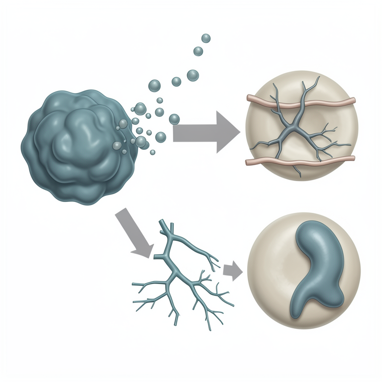
Tumor Exosome Communication Loop
AI-generated conceptual illustration (Cancer cells, endothelial cells, and monocytes in a tumor microenvironment). It explains the idea and does not show study data.
View source evidence ↗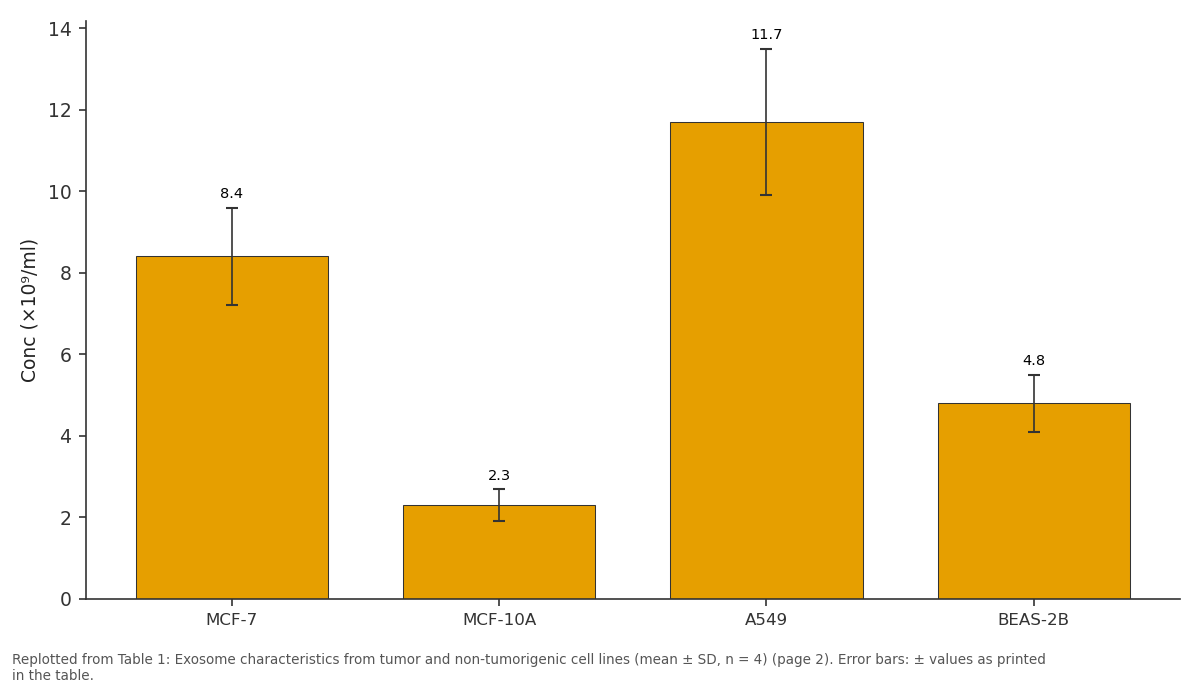
Conc (×10⁹/ml): MCF-7 vs MCF-10A vs A549 vs BEAS-2B
Replotted from Table 1 (page 2) of the original paper. Every bar is the paper's own table value.
View source evidence ↗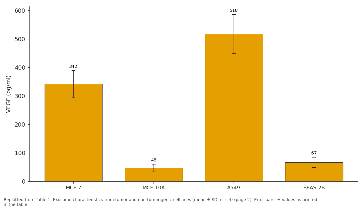
VEGF (pg/ml): MCF-7 vs MCF-10A vs A549 vs BEAS-2B
Replotted from Table 1 (page 2) of the original paper. Every bar is the paper's own table value.
View source evidence ↗Figures & tables
From the original paper Table 1
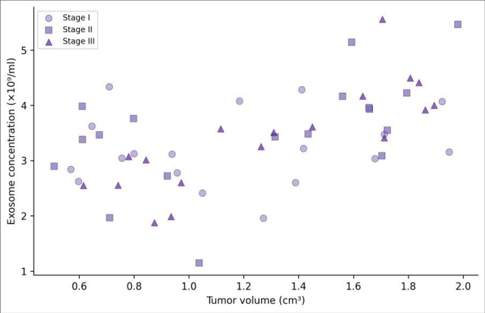 Figure 1
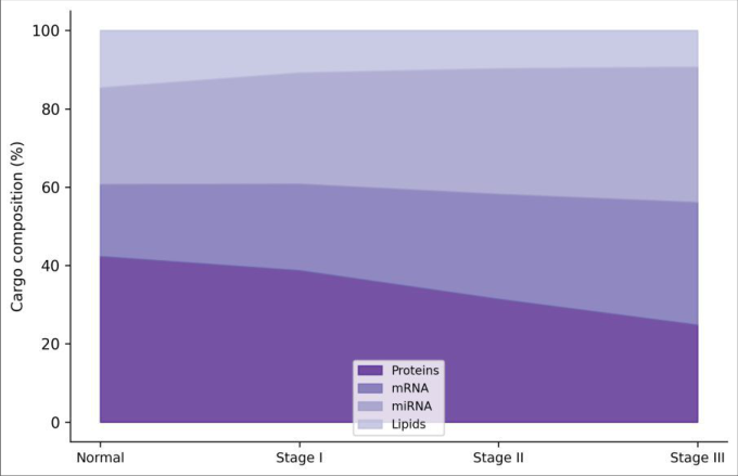 Figure 2
Introduction
A trend that has reshaped cancer biology in the last decade is the shift from viewing tumors as isolated masses to understanding them as ecosystems. Tumor cells don't just grow -they recruit blood vessels, suppress immune cells, and reprogram neighboring fibroblasts to support their expansion [1] . Exosomes, small extracellular vesicles (30-150 nm) released by virtually all cell types, have emerged as a main medium through which tumors orchestrate this microenvironmental remodeling [2] . Exosomes carry a selective cargo of proteins, mRNAs, miRNAs, and lipids that reflects the cell of origin and can alter the behavior of recipient cells upon uptake [3] . Tumor-derived exosomes have been shown to promote angiogenesis, suppress anti-tumor immunity, and prepare distant organs for metastatic colonization (the pre-metastatic niche) [4] . But most published research has focused on a single cancer type or a single functional readout. Headto-head comparisons of exosome characteristics across multiple cancer types, using the same isolation and analysis pipeline, are surprisingly uncommon. This research compared exosome release rates, cargo composition, and functional effects across three cancer cell lines and their non-tumorigenic counterparts, performed at the Centro de Ciencias de la Vida Austral, Valdivia, Chile, from March to October 2023.
Material and Methods
Material and Methods
Material
Human cancer cell lines MCF-7 (breast adenocarcinoma), A549 (lung carcinoma), and HCT116 (colorectal carcinoma) and non-tumorigenic lines MCF-10A, BEAS-2B, and CCD-18Co were obtained from ATCC. Human umbilical vein endothelial cells (HUVECs) and THP-1 monocytes were used as recipient cells. Culture media, FBS (exosome-depleted for collection experiments), and supplements were from Gibco. Ultracentrifuge tubes and rotors were from Beckman Coulter.
Methods
Cells were cultured to 70-80% confluence and switched to exosome-depleted FBS media for 48 hours. Conditioned media were collected and subjected to differential centrifugation: 300×g (10 min), 2,000×g (20 min), 10,000×g (30 min), and 100,000×g (70 min, twice). Pellets were resuspended in PBS and characterized by NTA (Malvern NanoSight NS300), western blot for CD63/CD81/TSG101, and TEM. Exosome protein cargo was analyzed by LC-MS/MS (Thermo Q Exactive) after in-solution digestion.
RNA was extracted using the miRNeasy Micro Kit (Qiagen) and sequenced on an Illumina MiSeq. For functional assays, 10 μg/ml exosomes were added to HUVECs (tube formation assay on Matrigel, 6 hours) or PMA-differentiated THP-1 cells (polarization assessed by RT-qPCR for M1/M2 markers after 48 hours). Statistics used one-way ANOVA with Tukey post-hoc. Ethics: no human subjects involved; institutional biosafety approval CCVA-IBC-2023-007. Cancer cell lines released 2.4-to 3.7-fold more exosomes than controls, with A549 being the most prolific (Table 1).
| Parameter | MCF-7 | MCF-10A | A549 | BEAS-2B | HCT116 | CCD-18Co |
|---|---|---|---|---|---|---|
| Conc (×10⁹/ml) | 8.4±1.2 | 2.3±0.4 | 11.7±1.8 | 4.8±0.7 | 9.2±1.4 | 3.1±0.5 |
| Size (nm) | 98±24 | 112±28 | 87±19 | 108±26 | 94±22 | 118±31 |
| VEGF (pg/ml) | 342±47 | 48±12 | 518±68 | 67±18 | 287±41 | 52±14 |
| miR-21 (fold) | 4.7±0.8 | 1.0±0.2 | 6.3±1.1 | 1.0±0.3 | 5.1±0.9 | 1.0±0.2 |
Table text extracted from the original PDF; superscript letters are shown inline. The original table image is below.
View original table image
Results
Tumor exosomes were slightly smaller and carried dramatically more VEGF and miR-21 than their normal counterparts. Functional assays showed that tumor exosomes increased HUVEC tube formation by 47.3-62.4% depending on cancer type (p < 0.001 for all). THP-1 monocytes treated with tumor exosomes upregulated M2 markers (CD206, IL-10, Arg1) by 2.1-to 3.4-fold while M1 markers (TNF-α, iNOS) decreased, indicating immunosuppressive polarization.
Discussion
The consistent pattern across three unrelated cancer typesincreased exosome release, pro-angiogenic cargo enrichment, and immunosuppressive functional effectssuggests that exosome-mediated microenvironment remodeling is a conserved feature of malignancy rather than a cancer-type-specific phenomenon [5,6] . The enrichment of miR-21, which targets multiple tumor suppressors including PTEN and PDCD4, was particularly striking and may represent both a biomarker and a therapeutic target [7] . The M2 polarization of monocytes by tumor exosomes has clear implications for immune checkpoint therapy. If tumorderived exosomes are continuously converting arriving monocytes into immunosuppressive macrophages, even checkpoint inhibitors may struggle to establish a productive anti-tumor immune response [8] . Combining exosome inhibition (e.g., GW4869, which blocks exosome biogenesis) with immunotherapy could be a strategy worth testing in preclinical models [9] .
Conclusion
Tumor-derived exosomes from breast, lung, and colon cancer cell lines were released at higher rates, carried proangiogenic and immunosuppressive cargo, and functionally promoted tube formation and M2 macrophage polarization compared with non-tumorigenic counterparts. These effects were consistent across cancer types, pointing to exosomemediated communication as a shared mechanism of microenvironment remodeling. Targeting exosome release or cargo loading may complement existing anti-cancer strategies [10,11,12] .
Figures and tables from the paper
References
15 in the original paper- Hanahan D, Weinberg RA. Hallmarks of cancer: the next generation. Cell. 2011;144(5):646-674.
- Kalluri R, LeBleu VS. The biology, function, and biomedical applications of exosomes. Science. 2020;367(6478):eaau6977.
- Valadi H, Ekström K, Bossios A, Sjöstrand M, Lee JJ, Lötvall JO. Exosome-mediated transfer of mRNAs and microRNAs is a novel mechanism of genetic exchange between cells. Nature Cell Biology. 2007;9(6):654-659.
- Peinado H, Alečković M, Lavotshkin S, Matei I, CostaSilva B, Moreno-Bueno G, Lyden D. Melanoma exosomes educate bone marrow progenitor cells toward a pro-metastatic phenotype. Nature Medicine. 2012;18(6):883-891.
- Théry C, Zitvogel L, Amigorena S. Exosomes: composition, biogenesis and function. Nature Reviews Immunology. 2002;2(8):569-579.
- Becker A, Thakur BK, Weiss JM, Kim HS, Peinado H, Lyden D. Extracellular vesicles in cancer: cell-to-cell mediators of metastasis. Cancer Cell. 2016;30(6):836848.
- Meng F, Henson R, Wehbe-Janek H, Ghoshal K, Jacob ST, Patel T. MicroRNA-21 regulates expression of the PTEN tumor suppressor gene in human hepatocellular cancer. Gastroenterology. 2007;133(2):647-658.
- Chen G, Huang AC, Zhang W, Zhang G, Wu M, Xu W, Bhagat C, Bhagat D, Bhagat K, Bhagat L, Fan Z. Exosomal PD-L1 contributes to immunosuppression and is associated with anti-PD-1 response. Nature. 2018;560(7718):382-386.
- Catalano M, O'Driscoll L. Inhibiting extracellular vesicles formation and release: a review of EV inhibitors. Journal of Extracellular Vesicles. 2020;9(1):1703244.
- Raposo G, Stoorvogel W. Extracellular vesicles: exosomes, microvesicles, and friends. Journal of Cell Biology. 2013;200(4):373-383.
- Colombo M, Raposo G, Théry C. Biogenesis, secretion, and intercellular interactions of exosomes and other extracellular vesicles. Annual Review of Cell and Developmental Biology. 2014;30:255-289.
- Whiteside TL. Exosomes and tumor-mediated immune suppression. Journal of Clinical Investigation. 2016;126(4):1216-1223.
- Tkach M, Théry C. Communication by extracellular vesicles: where we are and where we need to go. Cell. 2016;164(6):1226-1232.
- Xu R, Rai A, Chen M, Suwakulsiri W, Greening DW, Simpson RJ. Extracellular vesicles in cancer - implications for future improvements in cancer care. Nature Reviews Clinical Oncology. 2018;15(10):617638.
- Melo SA, Sugimoto H, O'Connell JT, Kato N, Villanueva A, Vidal A, Bhagat C, Bhagat D, Bhagat K, Bhagat L, Kalluri R. Cancer exosomes perform cellindependent microRNA biogenesis and promote tumorigenesis. Cancer Cell. 2014;26(5):707-721.