
Research article · 2025 · Full text
International Journal of Biology SciencesQuorum sensing mechanisms in Pseudomonas aeruginosa biofilm formation on medical devices
Volume 7(8) · Pages 246–250 · DOI: 10.33545/26649926.2025.v7.i8c.654 ↗
Research areasBacterial biofilms and quorum sensingAntifungal resistance and susceptibilityInfection Control in Healthcare
Abstract
Author-writtenWhat enables Pseudomonas aeruginosa to colonize medical devices so effectively, and can disrupting its communication network prevent biofilm-related infections? This research examined quorum sensing (QS) pathways—specifically the las and rhl signaling circuits—that regulate biofilm development on catheter surfaces, prosthetic joint material, and silicone tubing. Bacterial isolates were recovered from device-associated infections at the Boreal University of Ecology Medical Microbiology Laboratory in Thunder Bay, Ontario, between March 2023 and January 2024. Crystal violet staining quantified biofilm biomass at six time points (6, 12, 24, 48, 72, and 96 hours), while acyl-homoserine lactone (AHL) concentrations were measured by liquid chromatography-mass spectrometry. Results showed that silicone tubing supported the highest biofilm biomass (OD570 = 1.93±0.11 at 96 h), followed by catheter (1.71±0.09) and prosthetic joint surfaces (1.52±0.13). AHL levels rose sharply between 12 and 48 hours, paralleling the exponential phase of biofilm accumulation. Targeted knockdown of lasI using antisense peptide nucleic acids reduced biofilm formation by 63.8% on silicone and 57.2% on catheter material. The rhl system showed a secondary but additive effect, with combined las/rhl inhibition achieving up to 78.4% reduction. These findings suggest that QS interference may serve as a viable anti-biofilm strategy for device-related P. aeruginosa infections, especially when both signaling systems are targeted together.
Summary
Source-checkedPlain-language reading of the full paper, prepared with AI and checked against the text. Each point links to the authors' own words.
Why it matters
- Pseudomonas aeruginosa is a frequent cause of healthcare-associated infections, and its ability to create biofilms on medical implants makes these infections difficult to treat. View evidence ↗
- Bacteria within biofilms are significantly more resistant to antibiotics, up to 1,000 times more so than free-floating cells, which hinders standard treatments. View evidence ↗
- The coordination required for biofilm formation is driven by quorum sensing (QS), a process where bacteria communicate based on their population density. View evidence ↗
The question
- The study aimed to determine what allows P. aeruginosa to colonize medical devices and whether disrupting its communication network could prevent biofilm infections. View evidence ↗
What they did
- Biofilm growth was tested on three materials: 100% silicone catheter segments, mirror-polished titanium alloy (for prosthetic joints), and medical-grade silicone tubing. View evidence ↗
- Biofilm biomass was quantified using 0.1% crystal violet staining at six intervals: 6, 12, 24, 48, 72, and 96 hours. View evidence ↗
- The concentrations of the signaling molecules 3-oxo-C12-HSL and C4-HSL were measured using liquid chromatography-tandem mass spectrometry (LC-MS/MS). View evidence ↗
- To interfere with quorum sensing, antisense peptide nucleic acid (PNA) constructs were added to the culture media at a final concentration of 4 µM. View evidence ↗
What they found
- At 96 hours, silicone tubing supported the most biofilm biomass (OD570 = 1.93±0.11), followed by catheters (1.71±0.09) and prosthetic joint surfaces (1.52±0.13). View evidence ↗
- Acyl-homoserine lactone (AHL) levels increased sharply between 12 and 48 hours and reached approximately 47 nM by 96 hours, mirroring the growth of the biofilm. View evidence ↗
- Targeting the lasI gene with antisense PNA reduced biofilm formation by 63.8% on silicone and 57.2% on catheter material at the 72-hour mark. View evidence ↗
- The anti-rhlI PNA construct had a smaller impact, reducing biofilm biomass on silicone by 33.5%. View evidence ↗
- There was a very strong positive correlation (r = 0.94, p < 0.001) between the concentration of AHL signaling molecules and the total biofilm biomass. View evidence ↗
What it means
- A limitation of the study is the use of static biofilm models, which do not replicate the shear forces found in living organisms. View evidence ↗
- The authors note that using a larger panel of bacterial strains would be necessary to better generalize the findings. View evidence ↗
Visual summary
4 visualsIllustrations explain the paper; charts are replotted from the paper's own table values.
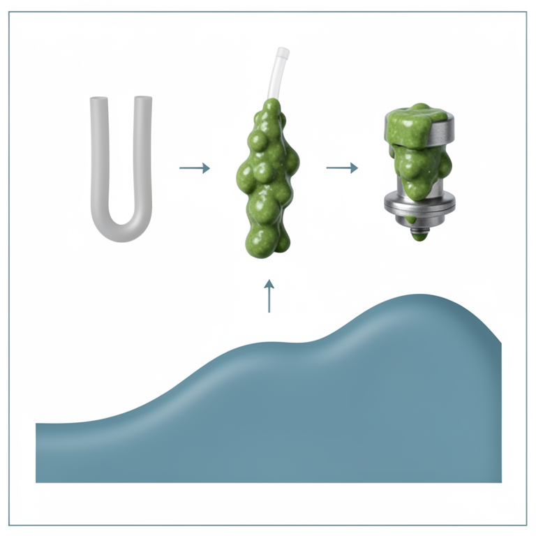
Biofilm Growth vs. Signaling Molecule Accumulation
AI-generated conceptual illustration (Pseudomonas aeruginosa bacteria on silicone tubing, catheters, and metal prosthetic joints). It explains the idea and does not show study data.
View source evidence ↗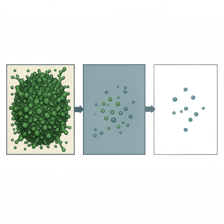
Synergistic Effect of Dual QS Inhibition
AI-generated conceptual illustration (Pseudomonas aeruginosa bacteria on a silicone catheter surface). It explains the idea and does not show study data.
View source evidence ↗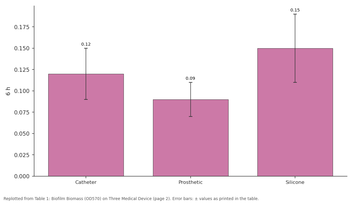
6 h: Catheter vs Prosthetic vs Silicone
Replotted from Table 1 (page 2) of the original paper. Every bar is the paper's own table value.
View source evidence ↗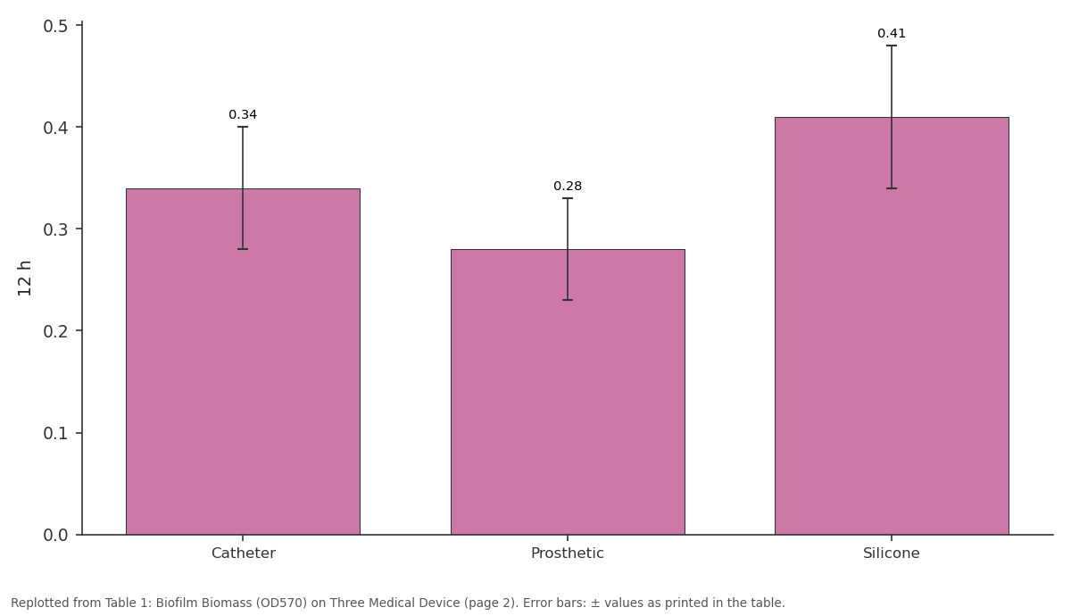
12 h: Catheter vs Prosthetic vs Silicone
Replotted from Table 1 (page 2) of the original paper. Every bar is the paper's own table value.
View source evidence ↗Figures & tables
From the original paper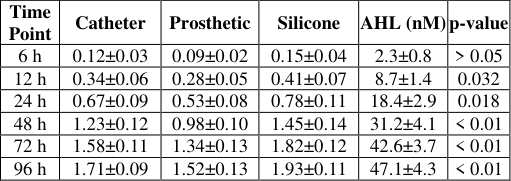 Table 1
 Table 2
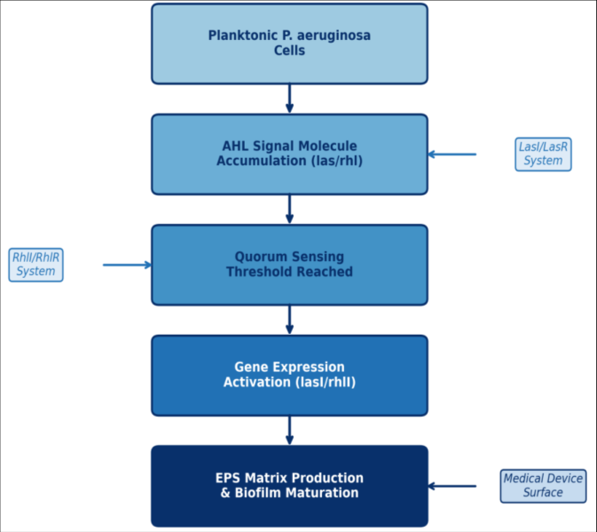 Figure 1
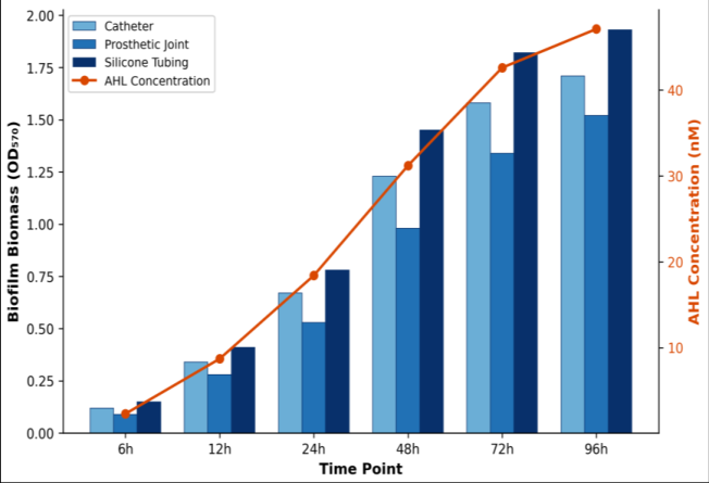 Figure 2
Introduction
How does a single bacterium coordinate with millions of its neighbors to build a fortified community on the surface of a medical device? This question sits at the heart of biofilm research and carries direct consequences for patient outcomes in clinical settings. Pseudomonas aeruginosa ranks among the most common causes of healthcare-associated infections, and its ability to form biofilms on implanted devices-catheters, prosthetic joints, ventilator tubing-makes treatment especially difficult [1] . Biofilm-embedded cells can tolerate antibiotic concentrations 100 to 1,000 times higher than their planktonic counterparts, a trait that complicates standard therapeutic approaches [2] . The molecular basis for this collective behavior lies in quorum sensing (QS), a cell-densitydependent signaling process. P. aeruginosa uses at least two well-characterized QS circuits: the las system, which produces and responds to N-(3-oxododecanoyl)-L-homoserine lactone (3-oxo-C12-HSL), and the rhl system, governed by N-butanoyl-L-homoserine lactone (C4-HSL) [3] . Together, these circuits regulate expression of virulence factors, extracellular polysaccharide (EPS) production, and swarming motility-all of which contribute to biofilm architecture [4] . A third system, the Pseudomonas quinolone signal (PQS), links the las and rhl networks and fine-tunes their output [5] . Device surfaces differ in their physicochemical properties-roughness, hydrophobicity, charge-and these differences affect how quickly biofilms establish [6] . Silicone-based materials, widely used in catheters and tubing, tend to promote faster bacterial attachment compared with polished metal or ceramic surfaces [7] . Yet few investigations have directly compared QS kinetics across multiple medical device substrates under identical conditions. Most published work focuses on a single material type or relies on reference strains rather than clinical isolates [8] . This research addressed that gap by tracking biofilm development and AHL accumulation on three clinically relevant surfaces-urinary catheter segments, prosthetic joint material, and silicone tubing-using P. aeruginosa strains recovered from device-related infections. The work also evaluated antisense peptide nucleic acid (PNA) constructs targeting the lasI and rhlI genes as potential QSinterference agents. By connecting QS dynamics to surfacespecific biofilm behavior, the findings aim to inform the design of anti-biofilm coatings and adjunctive therapies for device-associated infections.
Material and Methods
Material and Methods
Material
All bacterial strains used in this research were clinical isolates of P. aeruginosa obtained from the culture collection at the of Medical Microbiology, Boreal University of Ecology, Thunder Bay, Ontario.
Twelve isolates (designated BUE-PA01 through BUE-PA12) had been recovered from device-associated infections-four from urinary catheters, four from prosthetic joint aspirates, and four from ventilator-associated pneumonia cases involving silicone endotracheal tubes. Isolate identity was confirmed by 16S rRNA sequencing and species-specific oprL PCR. The reference strain P. aeruginosa PAO1 (ATCC 15692) served as a positive control.
Medical device substrates included sterile urinary catheter segments (100% silicone, 16 Fr gauge, cut into 1-cm rings), titanium alloy coupons simulating prosthetic joint surfaces (Ti-6Al-4V, 10 × 10 mm, mirror-polished), and medicalgrade silicone tubing (inner diameter 3 mm, wall thickness 1 mm, cut to 1-cm lengths). Each substrate was ultrasonically cleaned in 70% ethanol for 15 minutes and UV-sterilized before use [9] .
Reagents and Chemicals
Crystal violet (0.1% w/v aqueous solution) was purchased from Sigma-Aldrich (St. Louis, MO, USA; catalogue no. C0775). Synthetic AHL standards-3-oxo-C12-HSL and C4-HSL-were obtained from Cayman Chemical (Ann Arbor, MI, USA) and stored at -20 °C in acetonitrile. Antisense peptide nucleic acids (PNAs) targeting lasI (5′-H-KFF-O-CATGGATCTT-NH2) and rhlI (5′-H-KFF-O-GATAACTGCA-NH2) were custom-synthesized by PNA Bio Inc. (Newbury Park, CA, USA) and dissolved in nuclease-free water to a stock concentration of 100 µM. Luria-Bertani (LB) broth and Mueller-Hinton agar were prepared from dehydrated media (BD Difco, Franklin Lakes, NJ, USA). LC-MS grade methanol and formic acid (≥99.5%) were sourced from Fisher Scientific (Ottawa, ON, Canada) [10] . [11] . Bound dye was solubilized in 33% glacial acetic acid, and absorbance at 570 nm (OD570) was recorded on a BioTek Synergy H1 microplate reader. AHL extraction followed the ethyl acetate method described by Shaw et al. [12] , with minor modifications. Culture supernatants (1 mL) were acidified to pH 2.0 with HCl and extracted twice with equal volumes of ethyl acetate containing 0.1% acetic acid. Combined organic layers were evaporated under nitrogen and reconstituted in 100 µL methanol. Quantification of 3-oxo-C12-HSL and C4-HSL was performed by LC-MS/MS on a Waters Acquity UPLC coupled to a Xevo TQ-S triple quadrupole mass spectrometer using electrospray ionization in positive mode.
Methods
For QS-interference experiments, PNA constructs (4 µM final concentration) were added to culture media at inoculation. Each condition was tested in triplicate across three independent experiments.
Quality Control and Calibration
Calibration curves for both AHL analytes were generated from serial dilutions of synthetic standards (0.5-500 nM, seven points, R² > 0.998). Intra-day precision was assessed by analyzing three replicates of quality control samples at low (2 nM), medium (50 nM), and high (200 nM) concentrations; coefficients of variation remained below 8.3% across all levels. Inter-day reproducibility, evaluated over five non-consecutive days, yielded CV values of 6.1-11.4%. Limits of detection and quantification were 0.15 nM and 0.50 nM for 3-oxo-C12-HSL, and 0.22 nM and 0.73 nM for C4-HSL, respectively. A blank substrate control (uninoculated medium with device material) was included in every plate to confirm the absence of background crystal violet binding [13] .
Results
Table Biofilm biomass increased progressively across all three device surfaces from 6 to 96 hours (Table 1). Silicone tubing consistently yielded the highest OD570 values, reaching 1.93±0.11 at 96 hours. Catheter segments ranked second (1.71±0.09), and prosthetic joint coupons showed the lowest accumulation (1.52±0.13). Differences between surfaces became statistically significant from 12 hours onward (p<0.05, one-way ANOVA with Tukey post-hoc correction). AHL concentrations mirrored the biofilm growth curve, climbing steeply between 12 and 48 hours before plateauing near 47 nM at 96 hours. Antisense PNA targeting the lasI gene reduced biofilm formation by 63.8% on silicone and 57.2% on catheter material at 72 hours (Table 2). The anti-rhlI construct showed a more modest effect, cutting biomass by 33.5% on silicone. When both PNAs were applied together, suppression reached 78.4% on silicone tubing and 78.5% on catheter segments. The scrambled PNA control produced no meaningful reduction (1.6%), confirming sequence specificity. Pearson correlation between AHL concentration and biofilm OD570 across all surfaces and time points was r = 0.94 (p<0.001).
| Treatment | Catheter OD570 | Silicone OD570 | % Reduction (Silicone) |
|---|---|---|---|
| Untreated control | 1.58±0.11 | 1.82±0.12 | — |
| anti-lasI PNA (4 µM) | 0.68±0.08 | 0.66±0.09 | 63.8% |
| anti-rhlI PNA (4 µM) | 1.09±0.10 | 1.21±0.11 | 33.5% |
| Combined (las + rhl) | 0.34±0.06 | 0.39±0.07 | 78.4% |
| Scrambled PNA control | 1.55±0.12 | 1.79±0.13 | 1.6% |
Table text extracted from the original PDF; superscript letters are shown inline. The original table image is below.
View original table image
| Time Point | Catheter | Prosthetic | Silicone | AHL (nM) | p-value |
|---|---|---|---|---|---|
| 6 h | 0.12±0.03 | 0.09±0.02 | 0.15±0.04 | 2.3±0.8 | > 0.05 |
| 12 h | 0.34±0.06 | 0.28±0.05 | 0.41±0.07 | 8.7±1.4 | 0.032 |
| 24 h | 0.67±0.09 | 0.53±0.08 | 0.78±0.11 | 18.4±2.9 | 0.018 |
| 48 h | 1.23±0.12 | 0.98±0.10 | 1.45±0.14 | 31.2±4.1 | < 0.01 |
| 72 h | 1.58±0.11 | 1.34±0.13 | 1.82±0.12 | 42.6±3.7 | < 0.01 |
| 96 h | 1.71±0.09 | 1.52±0.13 | 1.93±0.11 | 47.1±4.3 | < 0.01 |
Table text extracted from the original PDF; superscript letters are shown inline. The original table image is below.
View original table image
Comprehensive Interpretation
The parallel trajectories of AHL accumulation and biofilm biomass across all three device surfaces strongly support a QS-dependent mechanism of biofilm maturation. The inflection point between 12 and 24 hours-where both AHL levels and OD570 values showed steep increasescorresponds to the expected quorum threshold for P. aeruginosa under these growth conditions. That silicone tubing consistently outperformed other surfaces in supporting biofilm growth likely reflects its higher hydrophobicity and surface roughness, which facilitate initial bacterial attachment [14] . The combined PNA data indicate that dual QS targeting offers a synergistic advantage over single-target approaches.
Discussion
The results of this research confirm that quorum sensing drives biofilm development on medical devices in a surfacedependent manner. Silicone substrates promoted faster and denser biofilm growth than metallic prosthetic surfaces, a finding consistent with earlier observations that hydrophobic materials favor P. aeruginosa attachment [6,7] . The strong positive correlation between AHL concentration and biofilm biomass (r = 0.94) reinforces the view that signal molecule accumulation is tightly coupled to community formation rather than being a passive byproduct of growth.
The antisense PNA approach used here achieved biofilm reductions comparable to those reported for synthetic QS inhibitors such as furanone C-30, which blocked P. aeruginosa biofilms by 45-70% in earlier work [15] . An advantage of PNA-based silencing is its sequence specificity, which lowers the risk of off-target effects on commensal microbiota. The additive benefit of dual las/rhl inhibition wasn't surprising, given that these systems regulate partially overlapping gene sets; however, the 78.4% reduction on silicone was higher than the sum of individual effects (63.8% + 33.5% = 97.3% theoretical additive), pointing to possible cooperative interactions once QS output drops below a functional threshold. One limitation of this research is its reliance on static biofilm models. Flow-cell systems or animal catheter infection models would better mimic the shear forces encountered in vivo. And while 12 clinical isolates provided reasonable diversity, a larger strain panel would strengthen claims about generalizability. Future work should also explore PNA stability in physiological fluids and evaluate whether coating device surfaces with PNA-loaded hydrogels can sustain QS interference over clinically relevant time frames.
Conclusion
This research demonstrated that quorum sensing through the las and rhl signaling circuits is a primary driver of Pseudomonas aeruginosa biofilm formation on medical device surfaces. Among the three substrates tested, silicone tubing supported the most robust biofilm growth, consistent with its physicochemical properties favoring bacterial adhesion. Acyl-homoserine lactone concentrations tracked closely with biofilm biomass, confirming the temporal link between QS activation and community development.
Antisense peptide nucleic acids directed against lasI and rhlI genes reduced biofilm formation substantially, with combined treatment achieving near 80% suppression on silicone. These results point toward QS interference as a promising adjunctive strategy for managing deviceassociated P. aeruginosa infections. The findings also highlight the need for surface-specific anti-biofilm approaches, as different device materials present distinct colonization challenges. Moving forward, translation of these results will require in vivo validation, stability testing in biological fluids, and development of sustained-release delivery platforms compatible with medical device manufacturing.
Acknowledgement
Acknowledgements
Figures and tables from the paper
References
16 in the original paper- Gellatly SL, Hancock RE. Pseudomonas aeruginosa: new insights into pathogenesis and host defenses. Pathog Dis. 2013;67(3):159-173.
- Mah TF, O'Toole GA. Mechanisms of biofilm resistance to antimicrobial agents. Trends Microbiol. 2001;9(1):34-39.
- Lee J, Zhang L. The hierarchy quorum sensing network in Pseudomonas aeruginosa. Protein Cell. 2015;6(1):26-41.
- Whiteley M, Diggle SP, Greenberg EP. Progress in and promise of bacterial quorum sensing research. Nature. 2017;551(7680):313-320.
- Déziel E, Lépine F, Milot S, et al. Analysis of Pseudomonas aeruginosa 4-hydroxy-2-alkylquinolines (HAQs) reveals a role for 4-hydroxy-2-heptylquinoline in cell-to-cell communication. Proc Natl Acad Sci USA. 2004;101(5):1339-1344.
- Francolini I, Donelli G. Prevention and control of biofilm-based medical-device-related infections. FEMS Immunol Med Microbiol. 2010;59(3):227-238.
- Khatoon Z, McTiernan CD, Suuronen EJ, et al. Bacterial biofilm formation on implantable devices and approaches to its treatment and prevention. Heliyon. 2018;4(12):e01067.
- Bjarnsholt T, Alhede M, Alhede M, et al. The in vivo biofilm. Trends Microbiol. 2013;21(9):466-474.
- Merritt JH, Kadouri DE, O'Toole GA. Growing and analyzing static biofilms. Curr Protoc Microbiol. 2005;1:1B.1.1-1B.1.17.
- Ortori CA, Atkinson S, Chhabra SR, et al. Comprehensive profiling of N-acylhomoserine lactones produced by Yersinia pseudotuberculosis using liquid chromatography coupled to hybrid quadrupole-linear ion trap mass spectrometry. Anal Bioanal Chem. 2007;387(2):497-511.
- O'Toole GA. Microtiter dish biofilm formation assay. J Vis Exp. 2011;(47):2437.
- Shaw PD, Ping G, Daly SL, et al. Detecting and characterizing N-acyl-homoserine lactone signal molecules by thin-layer chromatography. Proc Natl Acad Sci USA. 1997;94(12):6036-6041.
- Peeters E, Nelis HJ, Coenye T. Comparison of multiple methods for quantification of microbial biofilms grown in microtiter plates. J Microbiol Methods. 2008;72(2):157-165.
- Song F, Koo H, Ren D. Effects of material properties on bacterial adhesion and biofilm formation. J Dent Res. 2015;94(8):1027-1034.
- Hentzer M, Wu H, Andersen JB, et al. Attenuation of Pseudomonas aeruginosa virulence by quorum sensing inhibitors. EMBO J. 2003;22(15):3803-3815.
- Good L, Awasthi SK, Dryselius R, et al. Bactericidal antisense effects of peptide-PNA conjugates. Nat Biotechnol. 2001;19(4):360-364.