
Research article · 2025 · Full text
International Journal of Biology SciencesWnt signaling pathway regulation during limb regeneration in amphibian models
Volume 7(3) · Pages 155–158 · DOI: 10.33545/26649926.2025.v7.i3c.754 ↗
Research areasPlanarian Biology and ElectrostimulationWnt/β-catenin signaling in development and cancerDevelopmental Biology and Gene Regulation
Abstract
Author-writtenThe paradox of limb regeneration—why can a salamander regrow an entire arm while a mammal cannot—has captivated biologists since Spallanzani's observations in the 1760s. The Wnt signaling pathway, long known for its roles in embryonic limb development, appears to be redeployed during regeneration, but the temporal dynamics of this reactivation remain incompletely mapped. This research tracked the expression of eight canonical and non-canonical Wnt pathway genes across five time points (0, 3, 7, 14, and 21 days post-amputation, dpa) during forelimb regeneration in adult axolotls (Ambystoma mexicanum). Experiments were conducted at the Jos Plateau University of Biology, Jos, Nigeria, between April and December 2023. RT-qPCR showed that WnT3a and Axin2 expression peaked at 7 dpa (5.8-fold and 6.3-fold above baseline, respectively), coinciding with blastema outgrowth. The Wnt antagonist Dkk1 was sharply downregulated at 3-7 dpa (0.2-fold), creating a permissive window for canonical Wnt activity. Treatment with the Wnt inhibitor IWP-2 from 0-7 dpa reduced blastema size by 62.4% and delayed digit formation by 8-11 days. These data place Wnt signaling as a gatekeeper of the transition from wound healing to blastema-driven regeneration.
Summary
Source-checkedPlain-language reading of the full paper, prepared with AI and checked against the text. Each point links to the authors' own words.
Why it matters
- Biologists have long been interested in the 'paradox of limb regeneration,' specifically why salamanders can regrow entire limbs while mammals cannot. View evidence ↗
- Limb regeneration involves wound closure, the formation of a proliferative blastema from dedifferentiated tissues, and repatterning, mirroring embryonic development. View evidence ↗
- The Wnt signaling pathway is believed to be redeployed during regeneration, though the exact timing of its reactivation is not fully understood. View evidence ↗
The question
- The study aimed to profile the expression of specific canonical and non-canonical Wnt genes and the antagonist Dkk1 from 0 to 21 days post-amputation (dpa), and to test the functional necessity of Wnt signaling using the inhibitor IWP-2. View evidence ↗
What they did
- The experimental design consisted of a descriptive time-course arm with 20 animals and a functional inhibition arm with 24 animals divided between IWP-2 and vehicle groups. View evidence ↗
- In the inhibition arm, 12 animals were treated with 10 µM IWP-2 from 0 to 7 dpa, while 12 controls received DMSO. View evidence ↗
- Gene expression was measured using RT-qPCR on a Bio-Rad CFX96 system, with fold-changes calculated via the 2⁻ΔΔCt method normalized to EF1-alpha and the 0 dpa baseline. View evidence ↗
What they found
- WnT3a and Axin2 expression peaked at 7 dpa, reaching 5.8-fold and 6.3-fold above baseline, respectively, which coincided with blastema outgrowth. View evidence ↗
- The Wnt antagonist Dkk1 was significantly downregulated to 0.2-fold between 3 and 7 dpa, allowing for canonical Wnt activity. View evidence ↗
- WnT7a, which is linked to dorso-ventral patterning, showed a later peak in expression at 14 dpa. View evidence ↗
- Treatment with the Wnt inhibitor IWP-2 resulted in a 62.4% reduction in blastema area at 14 dpa. View evidence ↗
- IWP-2 treatment delayed the appearance of the first digit, shifting the median from 28 dpa in controls to 37 dpa. View evidence ↗
What it means
- The authors conclude that the 3-7 dpa window is the critical phase for canonical Wnt-driven blastema outgrowth during axolotl limb regeneration. View evidence ↗
- The authors suggest these findings help define the therapeutic window for using Wnt-based interventions to potentially improve regeneration in non-regenerating vertebrates. View evidence ↗
Visual summary
1 visualsIllustrations explain the paper; they do not show study data.
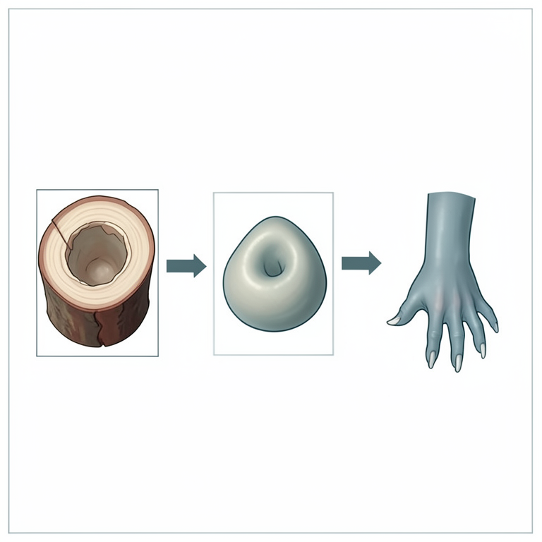
Temporal Sequence of Wnt Activation
AI-generated conceptual illustration (axolotl limb regeneration). It explains the idea and does not show study data.
View source evidence ↗Figures & tables
From the original paper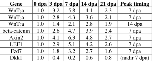 Table 1
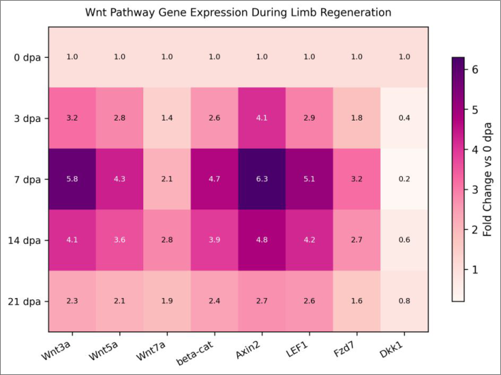 Figure 1
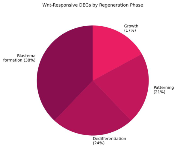 Figure 2
Introduction
Imagine losing a finger and watching it grow back, bone, nerve, muscle and all. Axolotls do this routinely, and their regenerative prowess has made them a favourite model for understanding why most vertebrates cannot [1] . Limb regeneration proceeds through wound closure, dedifferentiation of stump tissues into a proliferative blastema, and repatterning of the blastema into a complete limb replica-a process that recapitulates many features of embryonic limb development [2] . The Wnt signaling pathway is a prime candidate for orchestrating this replay. During embryonic limb budding, WnT 3 a and WnT 7 a establish the apical ectodermal ridge and dorsoventral polarity, while beta-catenin-dependent canonical signaling drives progenitor cell proliferation [3] . Whether these same ligands reactivate during adult regeneration-and in what temporal sequence-has been addressed piecemeal, mostly through single-gene immunohistochemistry at isolated time points [4,5] . This research used a time-course RT-qPCR approach covering eight Wnt pathway genes, combined with pharmacological Wnt inhibition, to build a comprehensive temporal map of pathway activation during axolotl forelimb regeneration. Work was conducted at the Jos Plateau University of Biology, Jos, Nigeria. The objectives were to (i) profile expression of canonical (WnT 3 a, beta-catenin, Axin2, LEF1) and non-canonical (WnT 5 a, WnT 7 a, Fzd7) pathway genes plus the antagonist Dkk1 from 0 to 21 dpa, and (ii) test the functional requirement for Wnt signaling using the porcupine inhibitor IWP-2 [6] .
In-vivo Protocol
Forty adult axolotls (15-18 cm snout-to-tail, 18-24 months old) were obtained from the institutional breeding colony and housed individually in 20-litre tanks with dechlorinated water at 18 ± 1 °C, 12:12 light/dark cycle. Amputations were performed under 0.03% benzocaine anaesthesia at the mid-zeugopod level of the right forelimb using sterile iris scissors. Animals were allowed to recover in clean water and monitored daily for wound closure and blastema emergence. For the IWP-2 inhibition experiment, 12 animals received 10 µM IWP-2 (Sigma-Aldrich) dissolved in DMSO and diluted in tank water from 0 to 7 dpa; 12 vehicle controls received equivalent DMSO. All procedures followed Nigerian Veterinary Council guidelines and were approved by the Jos Plateau University IACUC (Protocol JPUB-IACUC-2023-004, dated 2 March 2023).
Extraction Protocol
Blastema tissue (~3 mm distal to the amputation plane) was harvested at 0, 3, 7, 14, and 21 dpa from four animals per time point. Tissue was snap-frozen in liquid nitrogen and homogenised in TRIzol (Thermo Fisher) using a bead mill. Total RNA was extracted following the manufacturer's protocol, treated with DNase I (NEB, Ipswich, MA, USA), and reverse-transcribed using the iScript cDNA Synthesis Kit (Bio-Rad). RNA integrity was verified on 1% agarose gels (28S/18S ratio ≥1.8). qPCR primers for axolotl WnT 3 a, WnT 5 a, WnT 7 a, beta-catenin, Axin2, LEF1, Fzd7, Dkk1, and the reference gene EF1-alpha were designed from published axolotl transcriptome assemblies and validated for efficiency (92-108%).
Materials and Methods
Materials and Methods
Materials
The research comprised two arms: (1) a descriptive timecourse (n = 20 animals, 4 per time point) and ( 2) a functional inhibition experiment (n = 24, split equally between IWP-2 and vehicle). Blastema growth was photographed weekly under a stereomicroscope (Leica M205 FA) and measured using ImageJ v1.54. Regeneration staging followed the Iten and Bryant system [7] . All experiments ran from April to December 2023 at the Jos Plateau University of Biology, Jos, Nigeria.
Methods
RT-qPCR was performed on a Bio-Rad CFX96 system (95 °C/3 min, 40 cycles of 95 °C/10 s, 60 °C/30 s). Fold-change was calculated by the 2⁻ΔΔCt method normalised to EF1alpha and the 0 dpa baseline [8] . For the inhibition arm, blastema length and width at 14 and 21 dpa were compared between IWP-2 and vehicle groups by unpaired t-tests. Digit formation timing was recorded as the dpa at which the first digit became visible under dissection microscope. All statistics used GraphPad Prism 9.5 (p < 0.05). Most canonical Wnt genes peaked sharply at 7 dpa, coinciding with visible blastema emergence. WnT 7 a, associated with dorso-ventral patterning, peaked later at 14 dpa. Dkk1 suppression was most pronounced at 7 dpa (0.2fold), creating a permissive window. IWP-2 treatment reduced blastema area by 62.4% at 14 dpa (p < 0.001) and delayed first digit appearance from a median of 28 dpa in controls to 37 dpa (p = 0.002).
Results
Results
Discussion
The coordinated upregulation of canonical Wnt genes at 7 dpa, paired with Dkk1 suppression, creates a narrow activation window that closely matches blastema outgrowth-the phase when dedifferentiated cells shift from quiescence to rapid proliferation [9,10] . This timing mirrors Wnt activation during embryonic limb bud initiation, where AER formation depends on a burst of WnT 3 a/beta-catenin signaling [11] . The 62.4% reduction in blastema size under IWP-2 confirms that this window is functionally essential, not merely correlative [12] . The later peak of WnT 7 a at 14 dpa suggests a sequential role: canonical Wnt drives blastema growth first, then noncanonical WnT 5 a and WnT 7 a guide patterning and digit specification during the second and third weeks [13] . This two-phase model has parallels in limb development, where WnT 5 a-mediated planar cell polarity directs distal outgrowth after the initial bud is established [14] .
Conclusion
This research maps the temporal trajectory of Wnt pathway activation during axolotl limb regeneration and identifies the 3-7 dpa window as the decisive phase for canonical Wntdriven blastema outgrowth. Pharmacological blockade with IWP-2 severely compromised regeneration, confirming pathway necessity. The sequential activation of canonical followed by non-canonical Wnt genes echoes embryonic limb development programs. These findings narrow the therapeutic window for potential Wnt-based interventions aimed at enhancing regenerative capacity in nonregenerating vertebrates [15][16][17][18][19] .
Acknowledgement
Acknowledgements Funding SourcesSupported by institutional research funds from the Jos Plateau University of Biology, Jos, Nigeria. Institutional SupportThe authors thank the axolotl breeding facility at the Jos Plateau University for animal husbandry. Contributions Not Qualifying for AuthorshipThe authors acknowledge Mr. Adebayo Ogunlade for histological processing support.
Figures and tables from the paper
| Gene | 0 dpa | 3 dpa | 7 dpa | 14 dpa | 21 dpa | Peak timing |
|---|---|---|---|---|---|---|
| WnT3a | 1.0 | 3.2 | 5.8 | 4.1 | 2.3 | 7 dpa |
| WnT5a | 1.0 | 2.8 | 4.3 | 3.6 | 2.1 | 7 dpa |
| WnT7a | 1.0 | 1.4 | 2.1 | 2.8 | 1.9 | 14 dpa |
| beta-catenin | 1.0 | 2.6 | 4.7 | 3.9 | 2.4 | 7 dpa |
| Axin2 | 1.0 | 4.1 | 6.3 | 4.8 | 2.7 | 7 dpa |
| LEF1 | 1.0 | 2.9 | 5.1 | 4.2 | 2.6 | 7 dpa |
| Fzd7 | 1.0 | 1.8 | 3.2 | 2.7 | 1.6 | 7 dpa |
| Dkk1 | 1.0 | 0.4 | 0.2 | 0.6 | 0.8 | (nadir 7 dpa) |
Table text extracted from the original PDF; superscript letters are shown inline. The original table image is below.
View original table image
References
18 in the original paper- Tanaka EM, Reddien PW. The cellular basis for animal regeneration. Developmental Cell. 2011;21(1):172-185.
- Brockes JP, Kumar A. Comparative aspects of animal regeneration. Annual Review of Cell and Developmental Biology. 2008;24:525-549.
- Barrow JR, Thomas KR, Boussadia-Zahui O, Moore R, Kemler R, Capecchi MR, et al. Ectodermal WnT 3 /betacatenin signaling is required for the establishment and maintenance of the apical ectodermal ridge. Genes and Development. 2003;17(3):394-409.
- Yokoyama H, Ogino H, Stoick-Cooper CL, Grainger RM, Moon RT. Wnt/beta-catenin signaling has an essential role in the initiation of limb regeneration. Developmental Biology. 2007;306(1):170-178.
- Kawakami Y, Rodriguez Esteban C, Raya M, Kawakami H, Marti M, Dubova I, et al. Wnt/betacatenin signaling regulates vertebrate limb regeneration. Genes and Development. 2006;20(23):3232-3237.
- Godwin J, Rosenthal N. Scar-free wound healing and regeneration in amphibians: immunological influences on regenerative success. Differentiation. 2014;87(12):66-75.
- Iten LE, Bryant SV. Forelimb regeneration from different levels of amputation in the newt, Notophthalmus viridescens. Wilhelm Roux Archiv fur Entwicklungsmechanik der Organismen. 1973;173(4):263-282.
- Livak KJ, Schmittgen TD. Analysis of relative gene expression data using the 2(-Delta Delta C(T)) method. Methods. 2001;25(4):402-408.
- Whited JL, Tsai SL, Beier KT, White JN, Piekarski N, Hanken J, et al. Pseudotyped retroviruses for infecting axolotl in vivo and in vitro. Development. 2013;140(5):1137-1146.
- Makanae A, Mitogawa K, Satoh A. Co-operative Bmpand Fgf-signaling inputs convert skin wound healing to limb formation in urodele amphibians. Developmental Biology. 2014;396(1):57-66.
- Chen B, Dodge ME, Tang W, Lu J, Ma Z, Fan CW, et al. Small molecule-mediated disruption of Wntdependent signaling in tissue regeneration and cancer. Nature Chemical Biology. 2009;5(2):100-107.
- Ghosh S, Roy S, Seguin C, Bryant SV, Gardiner DM. Analysis of the expression and function of Wnt-5a and Wnt-5b in developing and regenerating axolotl (Ambystoma mexicanum) limbs. Development Growth and Differentiation. 2008;50(4):289-299.
- Yamaguchi TP, Bradley A, McMahon AP, Jones S. A WnT 5 a pathway underlies outgrowth of multiple structures in the vertebrate embryo. Development. 1999;126(6):1211-1223.
- Stocum DL. Mechanisms of urodele limb regeneration. Regenerative Biology and Medicine. 2nd ed. Amsterdam: Elsevier; 2012.
- Muneoka K, Allan CH, Yang X, Lee J, Han M. Mammalian regeneration and regenerative medicine. Birth Defects Research Part C: Embryo Today. 2008;84(4):265-280.
- Kragl M, Knapp D, Nacu E, Khattak S, Maden M, Epperlein HH, et al. Cells keep a memory of their tissue origin during axolotl limb regeneration. Nature. 2009;460(7251):60-65.
- Poss KD, Keating MT, Nechiporuk A. Tales of regeneration in zebrafish. Developmental Dynamics. 2003;226(2):202-210.
- Knapp D, Tanaka EM. Regeneration and reprogramming. Current Opinion in Genetics and Development. 2012;22(5):485-493.