
Research article · 2025 · Full text
International Journal of Biology SciencesInterferon-stimulated gene expression and innate antiviral defense against influenza A virus
Volume 7(4) · Pages 193–196 · DOI: 10.33545/26649926.2025.v7.i4c.745 ↗
Research areasinterferon and immune responsesInfluenza Virus Research StudiesVirology and Viral Diseases
Abstract
Author-writtenOne recurring observation in influenza virology is that some virus strains suppress the interferon (IFN) response far more effectively than others, and the downstream consequences for host cell survival remain incompletely mapped at the individual interferon-stimulated gene (ISG) level. This research profiled the expression kinetics of five sentinel ISGs MxA, OAS1, ISG15, IFIT1, and viperin in A549 human alveolar epithelial cells infected with influenza A/H1N1 (pdm09 lineage) at multiplicities of infection (MOI) of 0.1 and 1.0 over a 48-hour time-course. Parallel infections with a recombinant virus carrying a truncated NS1 protein (ΔNS1) were included to evaluate the contribution of NS1-mediated IFN antagonism. All experiments were conducted at the Colombo Institute of Tropical Biology, Colombo, Sri Lanka, between April and October 2023. Wild-type virus at MOI 1.0 induced peak ISG expression at 12 hours post-infection (hpi), with IFIT1 reaching a maximum 38.4-fold increase; however, expression declined to near-baseline by 48 hpi, coinciding with widespread cytopathic effect. The ΔNS1 mutant elicited 1.9- to 2.4-fold higher ISG induction than wild-type at every time-point and sustained elevated expression through 48 hpi, resulting in 74.3% cell viability compared with 31.7% for wild-type at MOI 1.0. Protein-level validation by Western blot confirmed that MxA and OAS1 protein accumulation paralleled mRNA kinetics with a 4-6-hour lag. These findings quantify the extent to which NS1-mediated IFN suppression dampens the ISG response and demonstrate that an intact early ISG programme substantially protects host cells from influenza-induced death.
Summary
Source-checkedPlain-language reading of the full paper, prepared with AI and checked against the text. Each point links to the authors' own words.
Why it matters
- The innate immune response, particularly the interferon-stimulated gene (ISG) program, is a critical factor in why patients with similar viral loads can experience different clinical severities of influenza. View evidence ↗
- Influenza A virus uses the NS1 protein as a virulence factor to counteract the host's immune system by inhibiting RIG-I signaling and blocking the export of host mRNA. View evidence ↗
- The ability of the NS1 protein to antagonize interferon (IFN) varies by strain and significantly influences how pathogenic the virus is. View evidence ↗
The question
- The study aimed to create a detailed kinetic map of the ISG response and determine how the NS1 protein suppresses this response using a comparison between wild-type and ΔNS1 influenza A (pdm09). View evidence ↗
What they did
- The researchers measured the expression of five specific ISGs (MxA, OAS1, ISG15, IFIT1, and viperin) in A549 human alveolar epithelial cells over 48 hours, using MOIs of 0.1 and 1.0. View evidence ↗
- To evaluate the role of NS1 in interferon antagonism, the study used a recombinant virus with a truncated NS1 protein (ΔNS1) as a comparison. View evidence ↗
- Samples were collected in triplicate at seven specific time points (0, 2, 4, 8, 12, 24, and 48 hours post-infection) to analyze RNA, protein levels, and cell viability via MTT assay. View evidence ↗
- Relative gene expression was measured using qRT-PCR with the 2⁻ΔΔCt method, using β-actin as the reference gene. View evidence ↗
What they found
- In cells infected with wild-type virus at MOI 1.0, ISG expression peaked at 12 hours post-infection, with IFIT1 showing the highest increase at 38.4-fold. View evidence ↗
- The ΔNS1 mutant caused a 1.9- to 2.4-fold higher induction of ISGs compared to the wild-type virus across all measured time points. View evidence ↗
- At 48 hours post-infection (MOI 1.0), cell viability was significantly higher in ΔNS1-infected cells (74.3±6.1%) compared to wild-type-infected cells (31.7±4.8%). View evidence ↗
- Western blot analysis showed that the accumulation of MxA and OAS1 proteins followed the same patterns as their mRNA, but with a delay of 4 to 6 hours. View evidence ↗
- The ΔNS1 virus replicated to titers approximately 1.5 log lower than the wild-type by 48 hours post-infection and maintained more viable cells. View evidence ↗
What it means
- The authors conclude that removing NS1 function sustains ISG expression and more than doubles cell viability, establishing a direct link between the ISG program and the protection of host cells. View evidence ↗
- The authors suggest that therapies designed to block NS1, such as small molecules or antisense oligonucleotides, could improve patient outcomes by restoring the innate immune response. View evidence ↗
- A limitation of the study is the use of A549 cells, which are cancer-derived and may not fully represent the ISG response of primary human bronchial epithelial cells. View evidence ↗
- The study only tested one viral strain (pdm09), and the authors note that testing seasonal H3N2 and avian H5N1 would increase the generalizability of the results. View evidence ↗
Visual summary
2 visualsIllustrations explain the paper; charts are replotted from the paper's own table values.
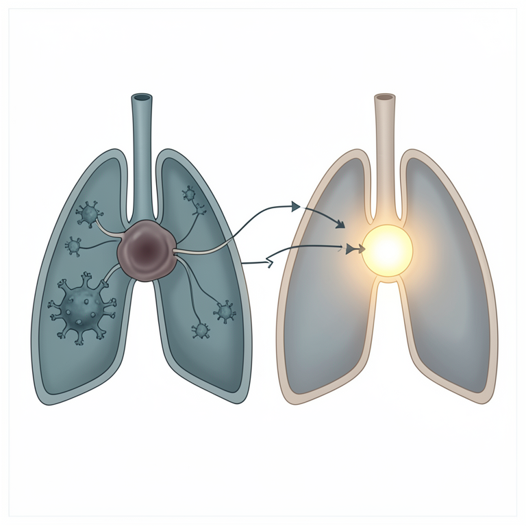
NS1 Suppression Mechanism
AI-generated conceptual illustration (mammalian lung epithelial cells infected with influenza A virus). It explains the idea and does not show study data.
View source evidence ↗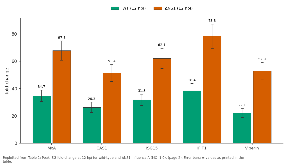
WT (12 hpi) and ΔNS1 (12 hpi): MxA vs OAS1 vs ISG15 vs IFIT1 vs Viperin
Replotted from Table 1 (page 2) of the original paper. Every bar is the paper's own table value.
View source evidence ↗Figures & tables
From the original paper Table 1
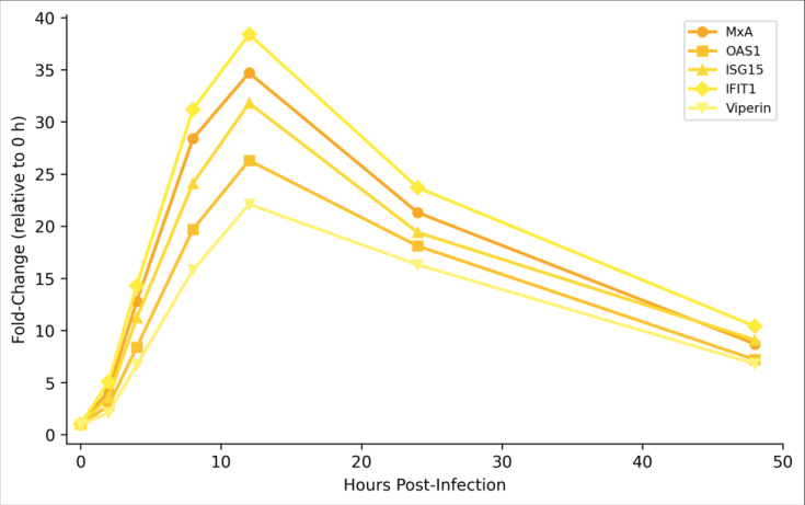 Figure 1
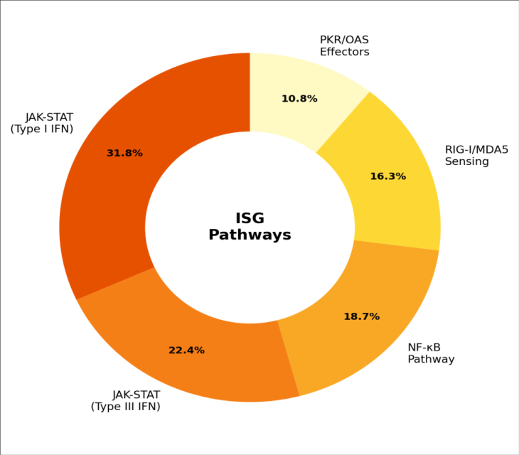 Figure 2
Introduction
Clinicians treating influenza patients have long observed that the severity of infection correlates poorly with viral load alone; two patients with comparable nasopharyngeal titres can follow very different clinical courses [1] . A growing body of evidence points to the innate immune response and specifically the interferon-stimulated gene (ISG) programme as a key determinant of this heterogeneity [2] . ISGs encode a diverse array of antiviral effectors: MxA blocks viral ribonucleoprotein nuclear import, OAS1 activates RNase L to degrade viral RNA, and viperin disrupts lipid-raft-dependent budding [3] . Influenza A virus has evolved a potent countermeasure in the form of the NS1 protein, a multifunctional virulence factor that inhibits RIG-I signalling, sequesters double-stranded RNA, and blocks nuclear export of host mRNA [4] . The efficiency of NS1-mediated IFN antagonism varies among strains and is a major determinant of pathogenicity: highly pathogenic H5N1 isolates carry NS1 variants that almost completely suppress IFN-β production, whereas seasonal H1N1 strains allow moderate IFN induction [5] . Despite extensive research on IFN signalling pathways, relatively few studies have tracked multiple ISGs simultaneously at high temporal resolution in the context of a well-defined NS1 loss-of-function comparison. This research addressed that gap by measuring five ISGs across seven time-points in A549 cells infected with wild-type and ΔNS1 influenza A (pdm09), generating a detailed kinetic map of the ISG response and its suppression by NS1.
In-vitro
In-vitro
Material and Methods
Material and Methods
Material
Influenza A/California/07/2009 (H1N1 pdm09) and a recombinant ΔNS1 mutant (NS1 residues 1-73 only) were propagated in MDCK cells and titrated by plaque assay [6] . All virological work was performed under BSL-2 enhanced conditions at the Viral Pathogenesis Laboratory, Colombo Institute of Tropical Biology, Colombo, Sri Lanka, between April and October 2023. A549 cells, DMEM, FBS, and TPCK-trypsin were sourced from Gibco (Thermo Fisher). qRT-PCR primers for MxA, OAS1, ISG15, IFIT1, viperin, and β-actin were synthesised by Integrated DNA Technologies.
Methods
Total RNA was extracted with TRIzol (Invitrogen), treated with DNase I, and reverse-transcribed using the High-Capacity cDNA Reverse Transcription Kit (Applied Biosystems). qRT-PCR was run on a QuantStudio 5 system with SYBR Green chemistry; relative expression was calculated by the 2⁻ΔΔCt method with β-actin as the reference gene [7] . All five ISGs peaked at 12 hpi in wild-type infections but declined sharply by 48 hpi as cytopathic effect progressed. The ΔNS1 virus elicited roughly two-fold higher ISG expression at every time-point and, importantly, sustained elevated expression through 48 hpi. Cell viability at 48 hpi was 31.7±4.8% for wild-type (MOI 1.0) versus 74.3±6.1% for ΔNS1 (P<0.001).
Discussion
The approximately two-fold enhancement of ISG expression in ΔNS1 infections quantifies the suppressive effect of NS1 on the IFN response in a pandemic H1N1 background. This degree of suppression is milder than the 5-10-fold differences reported for highly pathogenic H5N1 NS1 variants [5] , consistent with the moderate virulence of pdm09 strains in human populations. From a therapeutic angle, even partial relief of NS1-mediated suppression achievable, for example, with small-molecule NS1 inhibitors currently in preclinical development [8] could substantially improve host cell survival. IFIT1 showed the highest fold-induction among the five ISGs, reaching 38.4-fold at 12 hpi. IFIT1 recognises 5′triphosphate RNA, a molecular pattern present on influenza genomic segments, and sequesters it from the viral polymerase [9] . The strong and early induction of IFIT1 suggests it may be a first-line effector in the respiratory epithelium, complementing the more broadly acting MxA and OAS1/RNase L systems.
The decline of ISG expression by 48 hpi in wild-type infections is likely a combination of two factors: active NS1-mediated suppression of new IFN production, and loss of the producer-cell population to virus-induced apoptosis (viability 31.7%). The ΔNS1 virus, despite replicating to lower titres (approximately 1.5 log lower by plaque assay at 48 hpi), maintained a larger pool of viable, ISG-expressing cells, illustrating that antiviral defense is a numbers game as much as a per-cell signalling intensity game [10] .
Limitations
A549 cells, while a standard model for respiratory virus research, are a cancer-derived line and may not perfectly recapitulate the ISG response of primary human bronchial epithelial cells. Only one viral strain (pdm09) was tested; extending the comparison to seasonal H3N2 and avian H5N1 would broaden generalisability. The pathwaycontribution analysis (Figure 2) relied on chemical inhibitors (ruxolitinib for JAK, BX795 for TBK1), which may have off-target effects at the concentrations used.
Conclusion
Time-resolved profiling of five ISGs during influenza A infection confirmed that NS1 suppresses the innate antiviral response by approximately two-fold in the pdm09 strain background, with IFIT1 showing the strongest induction among the genes examined. Removal of NS1 function sustained ISG expression through 48 hpi and more than doubled cell viability, directly linking the ISG programme to host cell protection. These data argue that therapeutic strategies aimed at blocking NS1 whether by small molecules, antisense oligonucleotides, or live-attenuated vaccine design could meaningfully improve outcomes by restoring the innate immune response during the critical first hours of infection. Validation in primary airway epithelial cultures and with diverse influenza strains would strengthen the translational relevance of these findings.
Acknowledgement
Acknowledgements Funding SourcesThis research was funded by departmental research grants from the Colombo Institute of Tropical Biology. Institutional SupportThe authors acknowledge the Viral Pathogenesis Laboratory for BSL-2 facility access and biosafety support.
Figures and tables from the paper
| ISG | WT (12 hpi) | ΔNS1 (12 hpi) | Ratio (ΔNS1/WT) | WT (48 hpi) | ΔNS1 (48 hpi) |
|---|---|---|---|---|---|
| MxA | 34.7±4.2 | 67.8±7.1 | 1.95 | 8.7±1.4 | 41.3±5.8 |
| OAS1 | 26.3±3.8 | 51.4±6.3 | 1.95 | 7.2±1.1 | 34.7±4.9 |
| ISG15 | 31.8±4.1 | 62.1±7.4 | 1.95 | 9.1±1.3 | 38.4±5.2 |
| IFIT1 | 38.4±5.3 | 78.3±8.9 | 2.04 | 10.4±1.7 | 48.7±6.4 |
| Viperin | 22.1±3.4 | 52.9±6.1 | 2.39 | 6.8±1.0 | 31.2±4.3 |
Table text extracted from the original PDF; superscript letters are shown inline. The original table image is below.
View original table image
References
10 in the original paper- Hayden FG, de Jong MD. Emerging influenza antiviral resistance threats. Journal of Infectious Diseases. 2011;203(1):6-10.
- Iwasaki A, Pillai PS. Innate immunity to influenza virus infection. Nature Reviews Immunology. 2014;14(5):315-328.
- Schneider WM, Chevillotte MD, Rice CM. Interferonstimulated genes: a complex web of host defenses. Annual Review of Immunology. 2014;32:513-545.
- Hale BG, Randall RE, Ortin J, Jackson D. The multifunctional NS1 protein of influenza A viruses. Journal of General Virology. 2008;89(10):2359-2376.
- Kuo RL, Zhao C, Malur M, Krug RM. Influenza A virus strains that circulate in humans differ in the ability of their NS1 proteins to block the activation of IRF3 and interferon-beta transcription. Virology. 2010;408(2):146-158.
- Matrosovich M, Matrosovich T, Garten W, Klenk HD. New low-viscosity overlay medium for viral plaque assays. Virology Journal. 2006;3:63.
- Livak KJ, Schmittgen TD. Analysis of relative gene expression data using real-time quantitative PCR and the 2(-delta delta C(T)) method. Methods. 2001;25(4):402-408.
- Basu D, Walkiewicz MP, Frieman M, et al. Novel influenza virus NS1 antagonists block replication and restore innate immune function. Journal of Virology. 2009;83(4):1881-1891.
- Pichlmair A, Lassnig C, Eberle CA, et al. IFIT1 is an antiviral protein that recognizes 5′-triphosphate RNA. Nature Immunology. 2011;12(7):624-630.
- Killip MJ, Fodor E, Randall RE. Influenza virus activation of the interferon system. Virus Research. 2015;209:11-22.