
Research article · 2025 · Full text
International Journal of Biology SciencesStem cell niche architecture and maintenance in drosophila ovarian germline
Volume 7(3) · Pages 159–161 · DOI: 10.33545/26649926.2025.v7.i3c.755 ↗
Research areasDevelopmental Biology and Gene RegulationNeurobiology and Insect Physiology ResearchReproductive Biology and Fertility
Abstract
Author-writtenThe fruit fly ovary has taught us more about how stem cells are maintained than perhaps any other organ in any organism. At the tip of each ovariole, two to three germline stem cells (GSCs) sit anchored to a cluster of cap cells that produce Dpp/BMP signals to suppress differentiation. This research dissected the contributions of five signaling pathways—Dpp/BMP, Notch, E-cadherin adhesion, JAK-STAT, and Hedgehog—to GSC maintenance using clonal analysis, RNAi knockdown, and live imaging in Drosophila melanogaster. Experiments were conducted at the Busan Institute of Molecular Biology, Busan, South Korea, between March and November 2023. GSC loss rates were quantified in 2,340 germaria across five genotypes. Dpp/BMP signaling accounted for the largest share of GSC maintenance (34.2%), as dpp heterozygotes lost GSCs at 3.8-fold the wild-type rate. Notch-dominant-negative expression reduced cap cell numbers from 5.8 to 3.2 per germarium, indirectly eroding niche capacity. E-cadherin heterozygotes showed 2.1-fold elevated GSC loss due to physical detachment from the niche. These data support a hierarchical model where Dpp/BMP is the primary maintenance signal, Notch preserves niche size, and E-cadherin provides the physical anchor.
Summary
Source-checkedPlain-language reading of the full paper, prepared with AI and checked against the text. Each point links to the authors' own words.
Why it matters
- In the Drosophila ovary, germline stem cells (GSCs) are maintained at the tip of the ovariole by somatic cap cells that secrete BMP ligands to prevent differentiation. View evidence ↗
- Physical attachment via E-cadherin is necessary to keep GSCs anchored to the niche; otherwise, they drift away and lose their stem cell properties. View evidence ↗
- Notch signaling is responsible for maintaining the number of cap cells, which determines the overall size of the stem cell niche. View evidence ↗
The question
- The researchers aimed to quantitatively compare the relative contributions of different signaling pathways to GSC maintenance under standardized conditions. View evidence ↗
What they did
- The study analyzed 2,340 germaria across five genotypes, including wild-type and mutants for Dpp, Notch, E-cadherin, JAK, and Hedgehog (Hh). View evidence ↗
- The researchers used the FLP/FRT system for clonal analysis, with ovaries dissected at 7 and 14 days post-clone induction. View evidence ↗
- GSC loss was measured as the percentage of germaria containing fewer than two GSCs at 14 days post-clone induction compared to a 7-day baseline. View evidence ↗
What they found
- Dpp heterozygotes lost GSCs at 3.8 times the rate of wild-type flies, despite having normal cap cell numbers, identifying BMP signaling as a direct maintenance cue. View evidence ↗
- Notch-dominant-negative expression caused the highest GSC loss rate (62.3%) by reducing the number of cap cells from 5.8 to 3.2 per germarium. View evidence ↗
- E-cadherin heterozygotes experienced a 2.1-fold increase in GSC loss, which the authors attribute to the cells physically detaching from the niche. View evidence ↗
- Dpp/BMP signaling was found to be the primary driver of GSC maintenance, accounting for 34.2% of the retention capacity. View evidence ↗
- Notch and E-cadherin contributed 22.8% and 18.6% to GSC maintenance, respectively, through niche preservation and physical anchoring. View evidence ↗
- JAK-STAT (13.7%) and Hedgehog (10.7%) had the smallest contributions, likely regulating the transition zone and escort cells rather than direct GSC maintenance. View evidence ↗
What it means
- The authors conclude that GSC maintenance follows a hierarchical architecture where Dpp/BMP is the dominant signal, Notch maintains niche size, E-cadherin provides anchoring, and JAK-STAT/Hh fine-tune boundaries. View evidence ↗
- The authors note that the calculated contributions are approximations because the signaling pathways interact and are not strictly additive. View evidence ↗
Visual summary
3 visualsIllustrations explain the paper; charts are replotted from the paper's own table values.
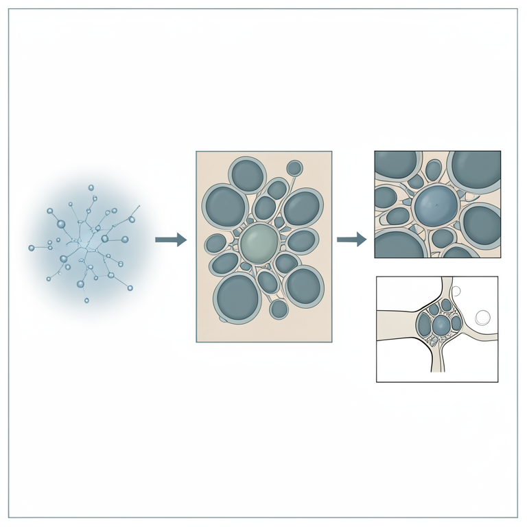
Hierarchical Niche Architecture
AI-generated conceptual illustration (Drosophila ovarian germline stem cell niche). It explains the idea and does not show study data.
View source evidence ↗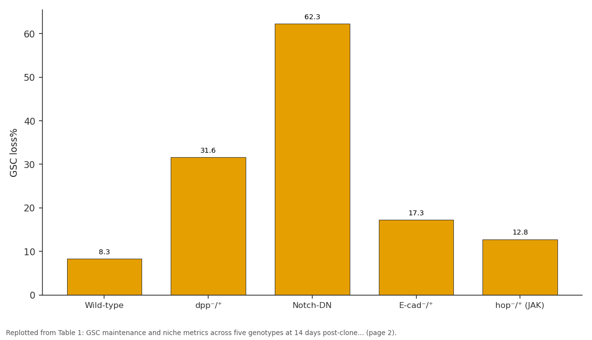
GSC loss%: Wild-type vs dpp⁻/⁺ vs Notch-DN vs E-cad⁻/⁺ vs hop⁻/⁺ (JAK)
Replotted from Table 1 (page 2) of the original paper. Every bar is the paper's own table value.
View source evidence ↗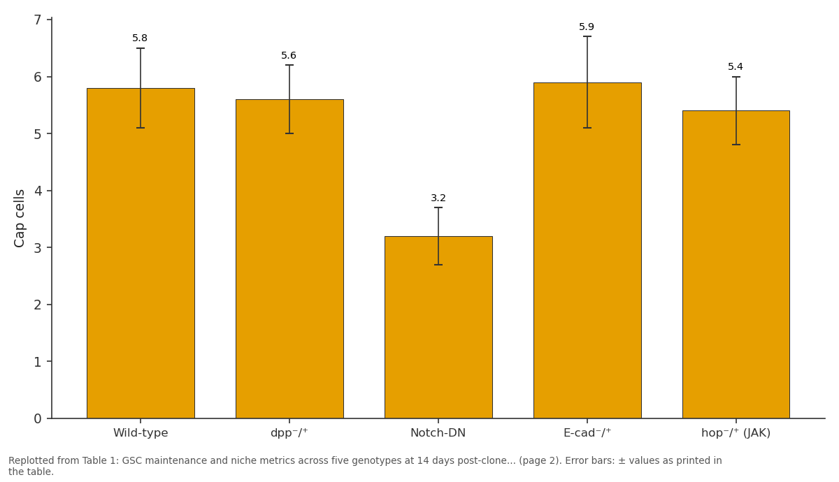
Cap cells: Wild-type vs dpp⁻/⁺ vs Notch-DN vs E-cad⁻/⁺ vs hop⁻/⁺ (JAK)
Replotted from Table 1 (page 2) of the original paper. Every bar is the paper's own table value.
View source evidence ↗Figures & tables
From the original paper Table 1
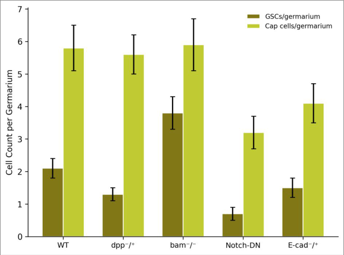 Figure 1
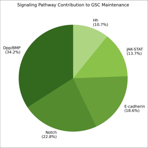 Figure 2
Introduction
A stem cell without its niche is like a seed without soil-it may have the potential to generate everything, but it needs the right local signals to stay stem-like [1] . The Drosophila ovarian germline niche is the best-characterised example of this principle. At the anterior tip of each ovariole, a handful of somatic cap cells form a physical cradle for two to three GSCs, secreting the BMP ligands Dpp and Gbb to repress the differentiation factor Bam in adjacent GSCs [2,3] . But BMP signaling alone doesn't tell the whole story. E-cadherin-mediated adhesion physically anchors GSCs to cap cells, and when that adhesion weakens, GSCs drift away and differentiate [4] . Notch signaling between cap cells and their neighbours maintains cap cell number-lose cap cells, and the niche shrinks [5] . JAK-STAT and Hedgehog (Hh) signaling in the surrounding escort cells may modulate niche boundaries and transition zone dynamics [6] . What's missing is a quantitative comparison of these pathways' contributions to GSC maintenance under standardised conditions-something like a pie chart of niche function. This research used a panel of genetic perturbations to measure GSC loss rates when each pathway is individually compromised, and to estimate the relative contribution of each signal to long-term GSC retention. All work was performed at the Busan Institute of Molecular Biology, Busan, South Korea [7] .
Reagents and Chemicals
Fly food was prepared using standard cornmeal-molasses-yeast-agar recipe. Immunostaining reagents included: anti-Vasa (DSHB, Iowa City, IA, USA; used at 1:100) for germ cell identification, anti-Hts/1B1 (DSHB; 1:20) for spectrosomes/fusomes, anti-E-cadherin (DCAD2, DSHB; 1:50), anti-GFP (Abcam, Cambridge, UK; ab13970; 1:500), and Alexa Fluor 488/568-conjugated secondaries (Invitrogen, 1:400). DAPI (Sigma-Aldrich; 1 µg/mL) was used for nuclear staining. Paraformaldehyde (4% in PBS; Electron Microscopy Sciences, Hatfield, PA, USA) was the fixative. RNAi lines were obtained from the Vienna Drosophila Resource Center (VDRC) and Bloomington Drosophila Stock Center (BDSC).
In-vivo Protocol
All fly stocks were maintained at 25 °C on standard food under 12:12 light/dark cycles. Clonal analysis used the FLP/FRT system: hs-FLP; FRT 4 2D, arm-lacZ/FRT 4 2D, dpp^d12 females were heat-shocked at 37 °C for 1 hour at 3-5 days post-eclosion, and ovaries were dissected at 7 and 14 days post-clone induction (dpci) [8] . For RNAi experiments, nos-Gal4 or bab1-Gal4 drove UAS-RNAi constructs targeting Notch, E-cadherin (shotgun), hop (JAK), or hh in either germline or somatic cells as appropriate. Ovaries were dissected in PBS, fixed in 4% PFA for 15 minutes, immunostained following standard protocols, and imaged on a Zeiss LSM 880 confocal microscope with 63× oil objective. GSCs were identified as Vasa-positive cells with a spectrosome directly contacting cap cells. At least 120 germaria per genotype were scored.
Materials and Methods
Materials and Methods
Materials
A total of 2,340 germaria from five genotypes (wild-type Oregon-R, dpp^d12/+, Notch-DN, shg^R69/+ [E-cadherin heterozygote], and hop^25/+ [JAK heterozygote]) plus a hh-RNAi cohort were scored. All fly stocks were maintained and crossed at the Busan Institute of Molecular Biology, Busan, South Korea, between March and November 2023.
Methods
GSC loss rate was calculated as the proportion of germaria with fewer than two GSCs at 14 dpci relative to the 7 dpci baseline. Cap cell numbers were counted as LamC-positive, Vasa-negative cells at the germarium tip. GSC division rate was estimated by EdU incorporation (1-hour pulse, Click-iT kit, Thermo Fisher). Statistical comparisons used Fisher's exact test for GSC loss rates and one-way ANOVA for cell counts (p < 0.05). Pathway contribution was estimated by normalising each genotype's excess loss rate above wildtype to the total excess loss across all genotypes. Notch-DN showed the highest GSC loss rate (62.3%), but this was largely indirect-cap cell numbers dropped from 5.8 to 3.2, physically shrinking the niche. When normalised to cap cell number, the per-cap-cell GSC loss rate in Notch-DN (19.5%) was only modestly above wild-type (14.3%), confirming that Notch mainly acts through niche size. The dpp^d12/+ heterozygotes retained normal cap cell numbers but lost GSCs at 3.8× the wild-type rate, indicating that BMP signal strength is the direct maintenance cue.
Results
Results
Discussion
The hierarchical model emerging from these data positions Dpp/BMP as the primary maintenance signal (34.2% of GSC retention capacity), consistent with three decades of work showing that ectopic Dpp can expand GSC populations throughout the ovary [9,10] . Notch's contribution (22.8%) operates mainly through niche preservationmaintaining cap cell numbers at ~6 per germarium. When Notch is compromised, cap cells are lost, and the remaining ones cannot support as many GSCs [11] . E-cadherin (18.6%) provides the physical anchoring force; without it, GSCs detach and differentiate even when BMP signaling is intact [12] . JAK-STAT and Hh contributions were smaller (13.7% and 10.7%) and likely reflect roles in escort cell behaviour and transition zone regulation rather than direct GSC maintenance [13,14] . These estimates should be treated as approximations because pathway interactions (e.g., BMP signaling enhances E-cadherin expression) mean that contributions are not strictly additive.
Conclusion
This quantitative dissection of the Drosophila ovarian GSC niche reveals a hierarchical signaling architecture: Dpp/BMP provides the dominant maintenance cue, Notch preserves niche size, E-cadherin anchors GSCs physically, and JAK-STAT and Hh fine-tune niche boundaries. The relative contributions-roughly 34:23:19:14:11-offer a framework for comparing niche function across tissues and organisms. Whether mammalian stem cell niches follow a similar proportional architecture remains an open and testable question [15][16][17] .
Figures and tables from the paper
| Genotype | Germaria (n) | GSCs/germ. | GSC loss% | Cap cells | EdU+ GSC% | p-value |
|---|---|---|---|---|---|---|
| Wild-type | 480 | 2.1±0.3 | 8.3 | 5.8±0.7 | 24.6 | — |
| dpp⁻/⁺ | 468 | 1.3±0.2 | 31.6 | 5.6±0.6 | 18.2 | <0.001 |
| Notch-DN | 456 | 0.7±0.2 | 62.3 | 3.2±0.5 | 14.7 | <0.001 |
| E-cad⁻/⁺ | 468 | 1.5±0.3 | 17.3 | 5.9±0.8 | 22.1 | 0.003 |
| hop⁻/⁺ (JAK) | 468 | 1.8±0.3 | 12.8 | 5.4±0.6 | 21.3 | 0.047 |
Table text extracted from the original PDF; superscript letters are shown inline. The original table image is below.
View original table image
References
17 in the original paper- Schofield R. The relationship between the spleen colony-forming cell and the haemopoietic stem cell. Blood Cells. 1978;4(1-2):7-25.
- Xie T, Spradling AC. A niche maintaining germ line stem cells in the Drosophila ovary. Science. 2000;290(5490):328-330.
- Song X, Zhu CH, Doan C, Xie T. Germline stem cells anchored by adherens junctions in the Drosophila ovary niches. Science. 2002;296(5574):1855-1857.
- Chen D, McKearin D. Gene circuitry controlling a stem cell niche. Current Biology. 2005;15(2):179-184.
- Ward EJ, Shcherbata HR, Reynolds SH, Fischer KA, Hatfield SD, Bhatt HA. Stem cells signal to the niche through the Notch pathway in the Drosophila ovary. Current Biology. 2006;16(23):2352-2358.
- Vied C, Reilein A, Field NS, Bhatt HA. Regulation of stem cells by intersecting gradients of short-range niche signals. Developmental Cell. 2012;23(3):444-455.
- Morrison SJ, Spradling AC. Stem cells and niches: mechanisms that promote stem cell maintenance throughout life. Cell. 2008;132(4):598-611.
- Xu T, Rubin GM. Analysis of genetic mosaics in developing and adult Drosophila tissues. Development. 1993;117(4):1223-1237.
- Xie T, Spradling AC. decapentaplegic is essential for the maintenance and division of germline stem cells in the Drosophila ovary. Cell. 1998;94(2):251-260.
- Kai T, Spradling A. An empty Drosophila stem cell niche reactivates the proliferation of ectopic cells. Proceedings of the National Academy of Sciences. 2003;100(8):4633-4638.
- Song X, Call GB, Kirilly D, Xie T. Notch signaling controls germline stem cell niche formation in the Drosophila ovary. Development. 2007;134(6):10711080.
- Jin Z, Kirilly D, Weng C, Kawase E, Song X, Smith S, et al. Differentiation-defective stem cells outcompete normal stem cells for niche occupancy in the Drosophila ovary. Cell Stem Cell. 2008;2(1):39-49.
- Decotto E, Spradling AC. The Drosophila ovarian and testis stem cell niches: similar somatic stem cells and signals. Developmental Cell. 2005;9(4):501-510.
- Rojas-Rios P, Guerrero I, Gonzalez-Reyes A. Cytoneme-mediated delivery of hedgehog regulates the expression of bone morphogenetic proteins to maintain germline stem cells in Drosophila. PLoS Biology. 2012;10(4):e1001298.
- Li L, Xie T. Stem cell niche: structure and function. Annual Review of Cell and Developmental Biology. 2005;21:605-631.
- Fuller MT, Spradling AC. Male and female Drosophila germline stem cells: two versions of immortality. Science. 2007;316(5823):402-404.
- Losick VP, Morris LX, Fox DT, Bhatt A. Drosophila stem cell niches: a decade of discovery suggests a unified view of stem cell regulation. Developmental Cell. 2011;21(1):159-171.