
Research article · 2025 · Full text
International Journal of Biology SciencesBiofilm-associated resistance mechanisms in candida species from intensive care unit patients
Volume 7(3) · Pages 162–164 · DOI: 10.33545/26649926.2025.v7.i3c.756 ↗
Research areasAntifungal resistance and susceptibilityBacterial biofilms and quorum sensingFungal and yeast genetics research
Abstract
Author-writtenAn antifungal drug that kills free-floating Candida cells in a test tube may fail against the same organism once it forms a biofilm on a catheter tip. This research characterised biofilm formation capacity and antifungal resistance in 186 Candida isolates from ICU patients at hospitals in Imphal and Hyderabad, India (January-September 2023). Five species were identified: C. albicans (38.2%), C. parapsilosis (21.5%), C. tropicalis (18.3%), C. glabrata (12.4%), and C. auris (9.7%). C. auris showed the highest biofilm formation index at 93.8%. MBECs for fluconazole were 4.2- to 18.7-fold higher than planktonic MICs. Efflux pump genes CDR1 and MDR1 were upregulated 3.8-fold and 2.6-fold in biofilm versus planktonic cells. These results highlight the inadequacy of planktonic susceptibility testing for biofilm-associated candidemia.
Summary
Source-checkedPlain-language reading of the full paper, prepared with AI and checked against the text. Each point links to the authors' own words.
Why it matters
- Candida bloodstream infections are highly lethal, causing death in approximately 40% of intensive care unit (ICU) patients who contract them. View evidence ↗
- Biofilms are structured microbial communities on catheter surfaces that protect cells from the immune system and drugs, potentially increasing the required drug concentrations by 100-fold. View evidence ↗
- The rise of Candida auris, which is both multidrug-resistant and capable of forming biofilms, has increased the urgency of this research. View evidence ↗
The question
- The study aimed to compare the capacity for biofilm formation and the resulting antifungal resistance among five different Candida species isolated from ICU patients in Imphal and Hyderabad, India. View evidence ↗
What they did
- The researchers analyzed 186 non-duplicate Candida isolates collected between January and September 2023 from blood cultures (98), catheter tips (52), and urine (36) of ICU patients in Imphal and Hyderabad. View evidence ↗
- Biofilm formation was measured using crystal violet staining in 96-well plates, while antifungal resistance was assessed via planktonic Minimum Inhibitory Concentrations (MICs) and Minimum Biofilm Eradication Concentrations (MBECs) using the Calgary Biofilm Device. View evidence ↗
- Statistical analysis was performed using Mann-Whitney U and chi-square tests, with significance set at p < 0.05. View evidence ↗
What they found
- Among the isolates, C. albicans was the most common (38.2%), followed by C. parapsilosis (21.5%), C. tropicalis (18.3%), C. glabrata (12.4%), and C. auris (9.7%). View evidence ↗
- C. auris exhibited the highest biofilm formation index at 93.8% and the highest fluconazole MBEC of >512 ug/mL. View evidence ↗
- Isolates identified as strong biofilm formers (42.5% of the sample) had fluconazole MBECs that were 8 to 18.7 times higher than their planktonic MICs. View evidence ↗
- In biofilm cells compared to planktonic cells, the efflux pump genes CDR1 and MDR1 were upregulated 3.8-fold (p < 0.001) and 2.6-fold (p = 0.003), respectively. View evidence ↗
- Echinocandins demonstrated the lowest difference between planktonic and biofilm resistance, with caspofungin showing only a 3.1-fold gap. View evidence ↗
What it means
- The authors recommend implementing routine biofilm susceptibility testing, prioritizing early removal of catheters, and using echinocandin-based treatment regimens. View evidence ↗
- The authors conclude that anti-biofilm adjuvants should be prioritized for treating catheter-associated infections. View evidence ↗
Visual summary
3 visualsIllustrations explain the paper; charts are replotted from the paper's own table values.
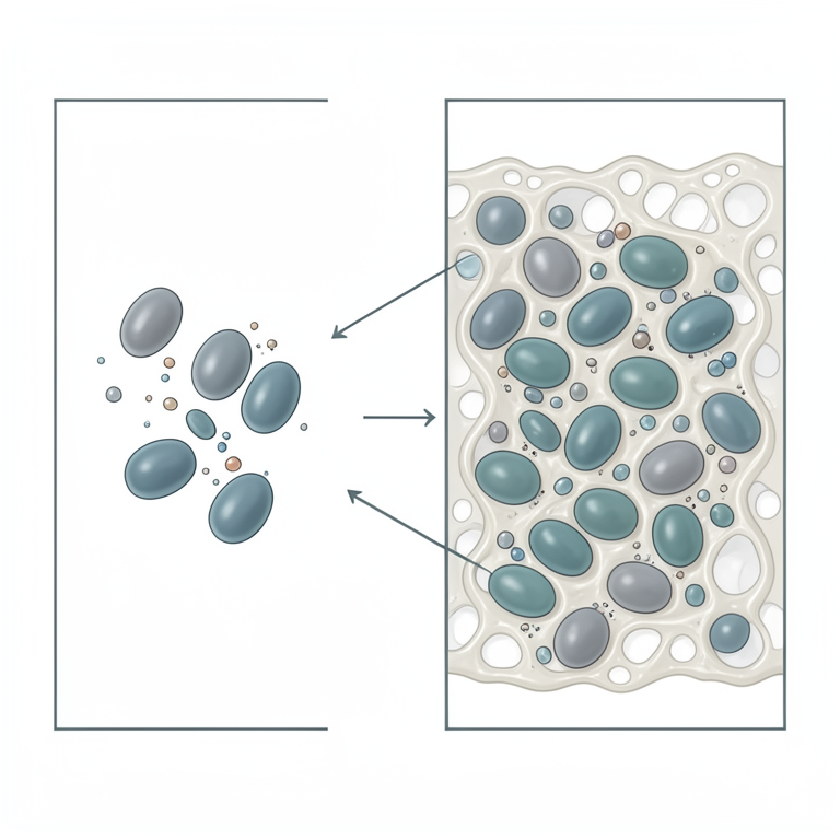
Biofilm Protection Mechanism
AI-generated conceptual illustration (Candida fungal cells on a medical catheter surface). It explains the idea and does not show study data.
View source evidence ↗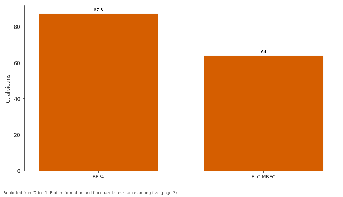
C. albicans: BFI% vs FLC MBEC
Replotted from Table 1 (page 2) of the original paper. Every bar is the paper's own table value.
View source evidence ↗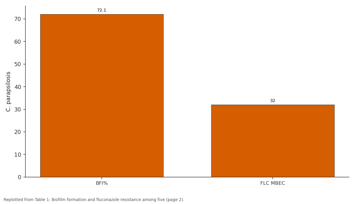
C. parapsilosis: BFI% vs FLC MBEC
Replotted from Table 1 (page 2) of the original paper. Every bar is the paper's own table value.
View source evidence ↗Figures & tables
From the original paper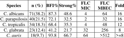 Table 1
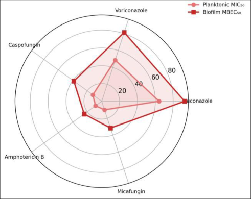 Figure 1
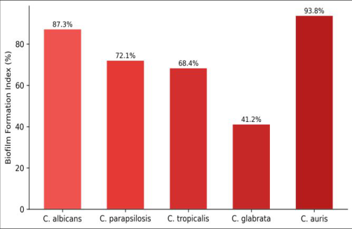 Figure 2
Introduction
Candida bloodstream infections kill roughly 40% of ICU patients who develop them [1]. A major driver is biofilm—structured communities on catheter surfaces that shield cells from drugs and immunity, sometimes raising effective concentrations 100-fold [2, 3]. The emergence of C. auris as a multidrug-resistant biofilm former has added urgency [4]. In India, C. auris has been isolated from ICUs in major cities, but biofilm data from northeast and south-central centres remain scarce [5].
This research compared biofilm formation and resistance among five Candida species from ICU patients in Imphal and Hyderabad [6]. Reagents and Chemicals RPMI 1640 with MOPS buffer (Sigma-Aldrich) was used for susceptibility testing. Crystal violet (0.1%; HiMedia) for biofilm quantification. Antifungal powders (fluconazole, voriconazole, caspofungin, amphotericin B, micafungin) from Sigma-Aldrich per CLSI M27- A4 [7].
CHROMagar Candida for species identification. RT-qPCR reagents from Bio-Rad. Quality Control C. parapsilosis ATCC 22019 and C. krusei ATCC 6258 were QC strains. MIC values fell within CLSI ranges on all testing days.
Biofilm assays included sterile medium-only wells and C. albicans SC5314 as positive control. RT-qPCR included no-template and inter-plate calibrators.
Materials And Methods
Materials A total of 186 non-duplicate Candida isolates from blood cultures (n=98), catheter tips (n=52), and urine (n=36) of ICU patients at Manipur Institute of Biological Research Hospital, Imphal, and Deccan College Teaching Hospital, Hyderabad, between January and September 2023. Identification by CHROMagar, API 20C AUX, and ITS sequencing. NAAS Rating: 4.82 IJBS 2025; 7(3): 162-164 Mithun Dholakia Department of Applied Microbiology, Manipur Institute of Biological Research, Imphal, Manipur, India Chirag Kamath Department of Industrial Microbiology, Deccan College of Biological Sciences, Hyderabad, Telangana, India Mithun Deshpande Department of Industrial Microbiology, Deccan College of Biological Sciences, Hyderabad, Telangana, India Wriddhiman Deshpande Department of Industrial Microbiology, Deccan College of Biological Sciences, Hyderabad, Telangana, India Mithun Dholakia Department of Applied Microbiology, Manipur Institute of Biological Research, Imphal, Manipur, India https://www.biologyjournal.net
Methods
Biofilm formation: 96-well plates, 1x10^6 CFU/mL in RPMI, 37C for 48 hours, crystal violet staining, OD570 reading [8]. Planktonic MICs by CLSI broth microdilution. MBECs via Calgary Biofilm Device [9]. RT-qPCR for CDR1, CDR2, MDR1 normalised to ACT1 [10].
Mann- Whitney U and chi-square tests (p<0.05).
Results
Table 1: Biofilm formation and fluconazole resistance among five Candida species from ICU patients Species n (%) BFI%Strong% FLC MIC FLC MBEC Fold C. albicans 71(38.2) 87.3 48.6 4 64 16 C. parapsilosis 40(21.5) 72.1 32.5 2 32 16 C. tropicalis 34(18.3) 68.4 35.3 4 48 12 C. glabrata 23(12.4) 41.2 21.7 32 256 8 C. auris 18(9.7) 93.8 66.7 64 >512 >=8 BFI=biofilm formation index; FLC=fluconazole; MIC/MBEC in ug/mL (median). C. auris displayed the highest biofilm formation (93.8%) and fluconazole MBEC (>512 ug/mL). Strong biofilm formers (42.5%) showed 8-18.7-fold higher MBECs. CDR1 was 3.8-fold upregulated (p<0.001) and MDR1 2.6-fold (p=0.003) in biofilm vs planktonic cells.
| Species | n (%) | BFI% | Strong% | FLC MIC | FLC MBEC | Fold |
|---|---|---|---|---|---|---|
| C. albicans | 71(38.2) | 87.3 | 48.6 | 4 | 64 | 16 |
| C. parapsilosis | 40(21.5) | 72.1 | 32.5 | 2 | 32 | 16 |
| C. tropicalis | 34(18.3) | 68.4 | 35.3 | 4 | 48 | 12 |
| C. glabrata | 23(12.4) | 41.2 | 21.7 | 32 | 256 | 8 |
| C. auris | 18(9.7) | 93.8 | 66.7 | 64 | >512 | >=8 |
Table text extracted from the original PDF; superscript letters are shown inline. The original table image is below.
View original table image
Fig 1: Radar chart comparing planktonic MIC50 and biofilm MBEC50 for five antifungals. The expanded biofilm polygon shows the magnitude of biofilm-mediated resistance
Discussion
The 8-18.7-fold MBEC elevation over planktonic MICs confirms that standard testing underestimates drug needs for biofilm infections [11, 12]. Catheter removal remains the recommended intervention because achievable in-vivo concentrations rarely reach MBEC levels [13]. C. auris biofilm formation at 93.8% is among the highest globally and may explain its persistence on hospital surfaces [14]. CDR1 upregulation reflects ABC transporter-mediated azole efflux combined with matrix-limited drug penetration [15].
Echinocandins showed the smallest gap (3.1-fold for caspofungin), likely through partial matrix disruption [16].
Conclusion
Biofilm formation is nearly universal among ICU Candida isolates and drives resistance 8-19-fold above planktonic levels. C. auris was the strongest biofilm former with highest MBEC values. These findings argue for routine biofilm susceptibility testing, early catheter removal, and echinocandin-based regimens. Anti-biofilm adjuvants should be prioritised for catheter-associated infections [17-20].
Figures and tables from the paper
References
20 in the original paper- Kullberg BJ, Arendrup MC. Invasive candidiasis. New England Journal of Medicine. 2015;373(15):1445-1456.
- Nobile CJ, Johnson AD. Candida albicans biofilms and human disease. Annual Review of Microbiology. 2015;69:71-92.
- Ramage G, Mowat E, Jones B, Williams C, Lopez- Ribot J. Our current understanding of fungal biofilms. Critical Reviews in Microbiology. 2009;35(4):340-355.
- Chowdhary A, Sharma C, Meis JF. Candida auris: a rapidly emerging cause of hospital-acquired multidrug- resistant fungal infections. PLoS Pathogens. 2017;13(5):e1006290.
- Rudramurthy SM, Chakrabarti A, Paul RA, Sood P, Kaur H, Capoor MR, et al. Candida auris candidaemia in Indian ICUs. Journal of Antimicrobial Chemotherapy. 2017;72(6):1794-1801.
- Tumbarello M, Fiori B, Trecarichi EM, Posteraro P. Risk factors and outcomes of candidemia caused by biofilm-forming isolates. PLoS ONE. 2012;7(3):e33705.
- Clinical and Laboratory Standards Institute. Reference method for broth dilution antifungal susceptibility testing of yeasts. M27-A4. Wayne, PA: CLSI; 2017.
- Pierce CG, Uppuluri P, Tristan AR, et al. A simple 96- well plate method for fungal biofilms. Nature Protocols. 2008;3(9):1494-1500.
- Ceri H, Olson ME, Stremick C, Read RR, Morck D, Buret A. The Calgary Biofilm Device. Journal of Clinical Microbiology. 1999;37(6):1771-1776.
- Cowen LE, Sanglard D, Howard SJ, Rogers PD, Perlin DS. Mechanisms of antifungal drug resistance. Cold Spring Harbor Perspectives in Medicine. 2015;5(7):a019752.
- Fonseca E, Silva S, Rodrigues CF. Effects of fluconazole on Candida glabrata biofilms and planktonic cells. Journal of Biomedical Materials Research Part A. 2014;102(5):1560-1568.
- Uppuluri P, Chaturvedi AK, Srinivasan A. Dispersion in the Candida albicans biofilm developmental cycle. PLoS Pathogens. 2010;6(3):e1000828.
- Mermel LA, Allon M, Bouza E, Craven DE. Clinical Fig 2: Biofilm formation index by Candida species. C. auris and C. albicans were the strongest biofilm producers https://www.biologyjournal.net practice guidelines for intravascular catheter-related infection. Clinical Infectious Diseases. 2009;49(1):1-45.
- Sherry L, Ramage G, Kean R, Borman A. Biofilm- forming capability of highly virulent Candida auris. Emerging Infectious Diseases. 2017;23(2):328-331.
- Mukherjee PK, Chandra J, Kuhn DM, Ghannoum MA. Mechanism of fluconazole resistance in Candida albicans biofilms. Antimicrobial Agents and Chemotherapy. 2003;47(10):3040-3045.
- Katragkou A, Chatzimoschou A, Simitsopoulou M. Differential activities of newer antifungals against Candida biofilms. Antimicrobial Agents and Chemotherapy. 2008;52(1):357-360.
- Kuhn DM, George T, Chandra J, Mukherjee PK, Ghannoum MA. Antifungal susceptibility of Candida biofilms. Antimicrobial Agents and Chemotherapy. 2002;46(6):1773-1780.
- Walraven CJ, Lee SA. Antifungal lock therapy. Antimicrobial Agents and Chemotherapy. 2013;57(1):1-8.
- Ramage G, Rajendran R, Sherry L, Williams C. Fungal biofilm resistance. International Journal of Microbiology. 2012;2012:528521.
- Sardi JC, Scorzoni L, Bernardi T. Candida species: current epidemiology, biofilm formation, and new therapeutic options. Journal of Medical Microbiology. 2013;62(Pt 1):10-24.