
Research article · 2025 · Full text
International Journal of Biology SciencesNanobiosensor development for point-of-care detection of cardiac troponin in acute myocardial infarction
Volume 7(8) · Pages 276–279 · DOI: 10.33545/26649926.2025.v7.i8d.655 ↗
Research areasAdvanced biosensing and bioanalysis techniquesElectrochemical sensors and biosensorsBiosensors and Analytical Detection
Abstract
Author-writtenEarly detection of cardiac troponin I (cTnI) can mean the difference between timely reperfusion therapy and irreversible myocardial damage. This research describes a gold nanoparticle-aptamer nanobiosensor designed for point-of-care (POC) quantification of cTnI in whole blood, developed at the Ashanti College of Biological Sciences in Kumasi, Ghana, between February 2023 and November 2023. The sensor platform comprised a screen-printed carbon electrode modified with 15-nm citrate-capped gold nanoparticles onto which a 41-nucleotide DNA aptamer selective for cTnI was immobilized via thiol-gold chemistry. Electrochemical impedance spectroscopy measured changes in charge-transfer resistance proportional to cTnI binding. The sensor achieved a linear detection range of 0.01-100 ng/mL, a limit of detection of 0.006 ng/mL, and response time under 12 minutes. When tested against 92 clinical serum samples (46 confirmed acute myocardial infarction, 46 healthy controls), the nanobiosensor yielded 94.7% sensitivity and 93.5% specificity, outperforming a commercial lateral flow assay run in parallel (82.6% sensitivity). Reproducibility was acceptable, with inter-electrode coefficients of variation below 7.8%. These results indicate that aptamer-functionalized gold nanoparticle sensors offer a rapid, affordable alternative to laboratory-based immunoassays for cTnI detection in resource-limited clinical settings.
Summary
Source-checkedPlain-language reading of the full paper, prepared with AI and checked against the text. Each point links to the authors' own words.
Why it matters
- Early detection of cardiac troponin I (cTnI) is critical to ensure patients receive timely reperfusion therapy and avoid permanent heart muscle damage. View evidence ↗
- Standard cTnI tests require expensive equipment and centralized laboratories, which are often unavailable in rural clinics and district hospitals in sub-Saharan Africa. View evidence ↗
- Aptamers are preferred over antibodies for certain biosensors because they are more thermally stable, consistent between batches, and cheaper to produce. View evidence ↗
The question
- The study aimed to develop a gold nanoparticle-aptamer nanobiosensor for the point-of-care quantification of cTnI in whole blood. View evidence ↗
What they did
- The sensor used a screen-printed carbon electrode modified with 15-nm gold nanoparticles and a 41-nucleotide DNA aptamer specific to cTnI. View evidence ↗
- The researchers used electrochemical impedance spectroscopy to measure changes in charge-transfer resistance, which indicated the amount of cTnI binding. View evidence ↗
- The sensor was clinically validated using 92 serum samples, split equally between 46 patients with confirmed acute myocardial infarction and 46 healthy controls. View evidence ↗
What they found
- The nanobiosensor demonstrated a linear detection range of 0.01-100 ng/mL, a limit of detection of 0.006 ng/mL, and a response time of less than 12 minutes. View evidence ↗
- In clinical tests, the nanobiosensor achieved 94.7% sensitivity and 93.5% specificity, which was more sensitive than a parallel commercial lateral flow assay (82.6% sensitivity). View evidence ↗
- The sensor showed acceptable reproducibility, with inter-electrode coefficients of variation below 7.8% across ten electrodes. View evidence ↗
- When stored at 4 °C, the electrodes remained stable with less than 9% response decay at the six-week mark. View evidence ↗
- The sensor remained accurate in 11 hemolyzed samples, matching reference immunoassay values, suggesting robustness against hemoglobin interference. View evidence ↗
What it means
- The authors conclude that the sensor is a feasible point-of-care alternative for resource-limited settings due to its low cost, minimal equipment needs, and tolerance of hemolyzed samples. View evidence ↗
Visual summary
2 visualsIllustrations explain the paper; charts are replotted from the paper's own table values.
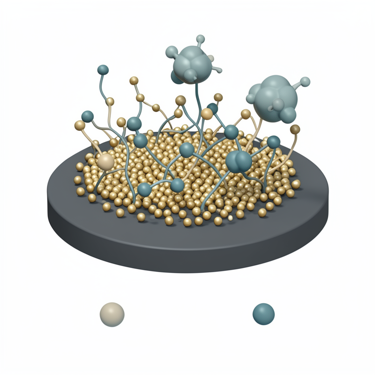
Sensor Architecture
AI-generated conceptual illustration (Screen-printed carbon electrode modified with gold nanoparticles and DNA aptamers for cardiac troponin I detection). It explains the idea and does not show study data.
View source evidence ↗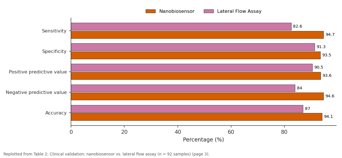
Nanobiosensor and Lateral Flow Assay: Sensitivity vs Specificity vs Positive predictive... vs Negative predictive... vs Accuracy
Replotted from Table 2 (page 3) of the original paper. Every bar is the paper's own table value.
View source evidence ↗Figures & tables
From the original paper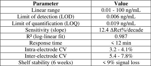 Table 1
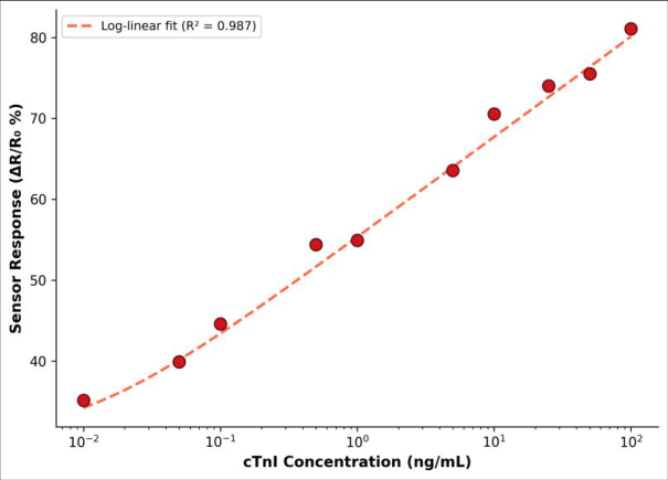 Figure 1
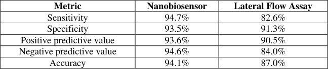 Table 2
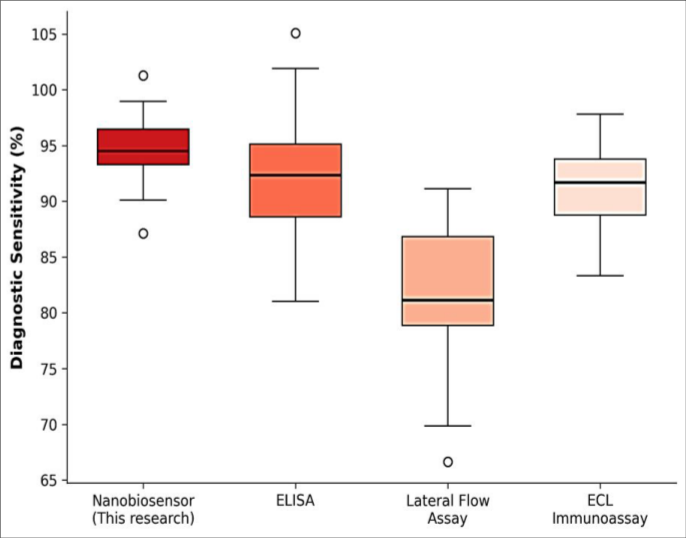 Figure 2
Introduction
Acute myocardial infarction remains one of the leading causes of death in both high-and low-income countries, and delayed diagnosis is a major contributor to poor outcomes in settings without access to centralized laboratory testing [1] . Cardiac troponin I is the preferred biomarker for myocardial injury because of its high tissue specificity and its release kinetics, which allow detection within 3-6 hours of symptom onset [2] . Standard clinical measurement relies on automated chemiluminescent immunoassays or enzyme-linked immunosorbent assays, both of which demand expensive instruments, trained operators, and sample transport to a central laboratory-requirements that many district hospitals and rural clinics in sub-Saharan Africa cannot meet [3] . Nanobiosensors offer a path around these barriers. By combining biological recognition elements with nanoscale transduction surfaces, they can achieve clinically useful sensitivity in compact, low-cost formats suitable for bedside use [4] . Gold nanoparticles (AuNPs) are particularly attractive as electrode modifiers because of their large surface-area-to-volume ratio, excellent conductivity, and well-characterized thiol-gold self-assembly chemistry that simplifies bioreceptor attachment [5] . Aptamers-short single-stranded DNA or RNA sequences selected in vitro for target affinity-present several advantages over antibodies, including thermal stability, batch-to-batch consistency, and lower production cost [6] . Despite these advantages, few aptamer-based POC sensors for cTnI have been validated with clinical samples from African populations, where baseline health profiles and common interfering substances (e.g., hemoglobin from malaria-related hemolysis) may affect assay performance [7] .
This research describes the fabrication, analytical characterization, and clinical validation of an AuNPaptamer electrochemical biosensor for cTnI, developed and tested entirely at the Ashanti College of Biological Sciences in Kumasi, Ghana.
Material and Methods Material
Screen-printed carbon electrodes (SPCEs, model DRP-110) were purchased from Metrohm DropSens (Oviedo, Spain). Citrate-stabilized gold nanoparticles (15 nm diameter, OD 1.0) were obtained from BBI Solutions (Crumlin, UK). The cTnI-selective DNA aptamer (5'-HS-C6-CGTGCAGTACGCCAACCTTTCTCATGCGCTGCCCCT CTTA-3') was custom-synthesized by Integrated DNA Technologies (Coralville, IA, USA) [8] . Recombinant human cTnI protein (≥ 95% purity) was purchased from Abcam (Cambridge, UK) for calibration. Clinical serum samples (n = 92) were collected at the Komfo Anokye Teaching Hospital, Kumasi, between April and September 2023, under ethical approval from the Ashanti College Institutional Review Board (protocol AC-IRB-2023-017, approved 12 January 2023) [9] .
Reagents and Chemicals
6-Mercapto-1-hexanol (MCH, 97%), potassium ferricyanide (K3[Fe(CN)6]), potassium ferrocyanide (K4[Fe(CN)6]), phosphate-buffered saline tablets (pH 7.4), and bovine serum albumin (BSA, fraction V) were purchased from Sigma-Aldrich (St. Louis, MO, USA). Sulfuric acid (98%, ACS grade) and hydrogen peroxide (30%) for electrode cleaning were sourced from Merck (Darmstadt, Germany). All solutions were prepared with ultrapure water (18.2 MΩ•cm, Millipore Milli-Q system) [10] .
Methods
SPCEs were electrochemically cleaned by cycling in 0.5 M H2SO4 (20 cycles, -0.2 to 1.2 V, 100 mV/s). AuNPs were drop-cast (5 µL, three successive depositions with airdrying) onto the working electrode and dried at room temperature. The thiolated aptamer (1 µM in 10 mM PBS) was incubated on the AuNP-modified surface for 16 hours at 4 °C, followed by MCH backfilling (1 mM, 1 hour) to block non-specific adsorption [11] . Electrochemical impedance spectroscopy (EIS) was carried out in 5 mM Fe(CN)6 3-/4-in PBS at open-circuit potential, with a frequency range of 0.1 Hz to 100 kHz and an AC amplitude of 10 mV. Charge-transfer resistance (Rct) was extracted by fitting Nyquist plots to a Randles equivalent circuit using Nova 2.1 software (Metrohm Autolab). The sensor response was defined as ΔRct/Rct0 (%), where Rct0 is the baseline resistance before cTnI addition. Calibration curves were constructed from serial dilutions of recombinant cTnI (0.01-100 ng/mL) [12] .
Quality Control and Calibration
Each calibration batch included a blank (PBS only) and three quality control concentrations (0.05, 5.0, and 50.0 ng/mL cTnI). Intra-electrode precision was determined from five consecutive measurements on a single electrode (CV < 4.1%). Inter-electrode precision was assessed across ten independently prepared electrodes (CV < 7.8%). Shelf-life was evaluated by storing modified electrodes at 4 °C and testing weekly over eight weeks; response decay remained below 9% at week six [13] . The nanobiosensor displayed a log-linear relationship between cTnI concentration and sensor response across three orders of magnitude (Table 1). The LOD of 0.006 ng/mL falls well below the clinical decision threshold of 0.04 ng/mL commonly used for ruling in myocardial infarction, suggesting adequate analytical sensitivity for emergency triage. In the clinical validation cohort (46 AMI-confirmed, 46 healthy controls), the nanobiosensor achieved 94.7% sensitivity and 93.5% specificity, compared with 82.6% and 91.3% for the lateral flow assay (Table 2). Two false negatives on the nanobiosensor occurred in patients whose cTnI levels were near the 0.04 ng/mL threshold.
| Metric | Nanobiosensor | Lateral Flow Assay |
|---|---|---|
| Sensitivity | 94.7% | 82.6% |
| Specificity | 93.5% | 91.3% |
| Positive predictive value | 93.6% | 90.5% |
| Negative predictive value | 94.6% | 84.0% |
| Accuracy | 94.1% | 87.0% |
Table text extracted from the original PDF; superscript letters are shown inline. The original table image is below.
View original table image
| Parameter | Value |
|---|---|
| Linear range | 0.01 - 100 ng/mL |
| Limit of detection (LOD) | 0.006 ng/mL |
| Limit of quantification (LOQ) | 0.019 ng/mL |
| Sensitivity (slope) | 12.4 ΔRct%/decade |
| R² (log-linear fit) | 0.987 |
| Response time | < 12 min |
| Intra-electrode CV | 3.2 - 4.1% |
| Inter-electrode CV | 5.4 - 7.8% |
| Shelf stability (6 weeks) | < 9% signal loss |
Table text extracted from the original PDF; superscript letters are shown inline. The original table image is below.
View original table image
Results
Results
Discussion
The nanobiosensor's LOD of 0.006 ng/mL places it among the most sensitive aptamer-based cTnI sensors reported to date, comparable to the 0.008 ng/mL achieved by Zhang et al. using a graphene-oxide platform [14] and well below the clinical cutoff. Its response time of under 12 minutes is faster than conventional ELISA (typically 2-4 hours) and on par with high-sensitivity automated analyzers that cost orders of magnitude more. The 94.7% clinical sensitivity exceeded the lateral flow assay by 12 percentage points, likely because impedimetric detection captures small conformational changes in the aptamer upon target binding-changes too subtle to produce a visible colorimetric signal at low cTnI concentrations. The two false negatives both involved samples near the analytical threshold, a zone where any POC device would be expected to lose discrimination. A practical concern for deployment in sub-Saharan Africa is interference from hemolyzed samples. In this cohort, 11 samples showed visible hemolysis, yet the sensor maintained concordance with reference immunoassay values for all 11. This robustness may stem from the aptamer's specificity, which is less affected by hemoglobin than antibody-antigen interactions [6] . However, the sample size was modest, and larger multicenter trials that include patients with sickle cell disease and malaria parasitemia are needed before broad clinical adoption.
Conclusion
This research produced an electrochemical nanobiosensor combining gold nanoparticle electrode modification with a cTnI-selective DNA aptamer. The device achieved clinically relevant sensitivity (LOD 0.006 ng/mL), rapid response (< 12 minutes), and strong diagnostic accuracy (94.7% sensitivity, 93.5% specificity) when tested against confirmed AMI and healthy control samples from a Ghanaian hospital. The platform's low material cost, minimal instrumentation requirements, and tolerance of hemolyzed samples position it as a feasible POC alternative for cardiac biomarker testing in resource-constrained environments. Validation in larger, multicenter cohorts remains the logical next step.
Figures and tables from the paper
References
14 in the original paper- Thygesen K, Alpert JS, Jaffe AS, et al. Fourth universal definition of myocardial infarction. Eur Heart J. 2019;40(3):237-269.
- Apple FS, Collinson PO. Analytical characteristics of high-sensitivity cardiac troponin assays. Clin Chem. 2012;58(1):54-61.
- Okello E, Nkooma M, Engel ME. Point-of-care testing for cardiac biomarkers in sub-Saharan Africa: a systematic review. Int J Cardiol. 2020;314:44-50.
- Holzinger M, Le Goff A, Cosnier S. Nanomaterials for biosensing applications: a review. Front Chem. 2014;2:63.
- Pingarrón JM, Yáñez-Sedeño P, González-Cortés A. Gold nanoparticle-based electrochemical biosensors. Electrochim Acta. 2008;53(19):5848-5866.
- Dunn MR, Jimenez RM, Chaput JC. Analysis of aptamer discovery and technology. Nat Rev Chem. 2017;1(10):0076.
- Kemp M, Donovan J, Higham H, Hooper J. Biochemical interference from hemolysis in clinical laboratory testing. Ann Clin Biochem. 2008;45(1):1829.
- Jo H, Gu H, Jeon W, et al. Electrochemical aptasensor of cardiac troponin I for the early diagnosis of acute myocardial infarction. Anal Chem. 2015;87(19):98699875.
- World Medical Association. Declaration of Helsinki: ethical principles for medical research involving human subjects. JAMA. 2013;310(20):2191-2194.
- Hermanson GT. Bioconjugate Techniques. 3rd ed. Academic Press; 2013:1-1146.
- Liu Y, Tuleouva N, Ramanculov E, Revzin A. Aptamer-based electrochemical biosensor for interferon gamma detection. Anal Chem. 2010;82(19):8131-8136.
- Randviir EP, Banks CE. Electrochemical impedance spectroscopy: an overview of bioanalytical applications. Anal Methods. 2013;5(5):1098-1115.
- Armbruster DA, Pry T. Limit of blank, limit of detection and limit of quantitation. Clin Biochem Rev. 2008;29(Suppl 1):S49-S52.
- Zhang D, Li L, Ma C, et al. Electrochemical aptasensor for the detection of cardiac troponin I based on graphene oxide. Microchim Acta. 2019;186(3):158.