
Research article · 2025 · Full text
International Journal of Biology SciencesNanoparticle-based drug delivery systems for targeted cancer therapeutics in preclinical models
Volume 7(9) · Pages 158–160 · DOI: 10.33545/26649926.2025.v7.i9c.631 ↗
Research areasNanoparticle-Based Drug DeliveryDendrimers and Hyperbranched PolymersNanoplatforms for cancer theranostics
Abstract
Author-writtenImagine a drug that reaches only the tumour and leaves healthy tissue untouched—that promise drives nanoparticle-based drug delivery research. This research evaluated PLGA-PEG nanoparticles loaded with doxorubicin (NP-DOX) for targeted cancer therapy in a murine breast carcinoma model. Nanoparticles were prepared by nanoprecipitation (mean diameter 148 ± 12 nm, PDI 0.14, encapsulation efficiency 78.3 percent) and characterised by DLS, TEM, and FTIR. In vitro release at pH 5.4 (tumour-mimicking) reached 94.8 percent by 72 hours versus 64.1 percent at pH 7.4, confirming pH-responsive behaviour. In BALB/c mice bearing 4T1 tumours, NP-DOX at 10 mg/kg reduced tumour volume by 77.6 percent relative to saline controls, compared with 39.2 percent for free DOX at 5 mg/kg. PEGylated formulations showed an additional 30.5 percent volume reduction over non-PEGylated NP-DOX. Body weight loss was significantly lower in NP-DOX groups. These results demonstrate that PLGA-PEG nanocarriers improve doxorubicin’s therapeutic index through pH-triggered release and passive tumour targeting via the EPR effect.
Summary
Source-checkedPlain-language reading of the full paper, prepared with AI and checked against the text. Each point links to the authors' own words.
Why it matters
- Doxorubicin is a highly effective chemotherapy for several cancers, but its use is limited by toxicity to the heart. View evidence ↗
- Using biodegradable polymer nanoparticles can help direct the drug toward tumor tissue by utilizing the enhanced permeability and retention (EPR) effect. View evidence ↗
- PLGA is an FDA-approved polymer for injections that breaks down into non-toxic components. View evidence ↗
The question
- The study aimed to create doxorubicin-loaded PLGA-PEG nanoparticles and evaluate their properties, drug release at different pH levels, and effectiveness against breast cancer in a mouse model. View evidence ↗
What they did
- Nanoparticles were created using nanoprecipitation, dissolving 20 mg of polymer and 4 mg of doxorubicin in acetone before adding them to a PVA solution. View evidence ↗
- The researchers used Dynamic Light Scattering (DLS) for size and zeta potential measurements and Transmission Electron Microscopy (TEM) to confirm morphology. View evidence ↗
What they found
- The nanoparticles showed pH-responsive behavior, releasing 94.8% of the drug at pH 5.4 compared to 64.1% at pH 7.4 over 72 hours. View evidence ↗
- Free doxorubicin at 5 mg/kg was less effective, achieving only a 39.2% reduction in tumor volume. View evidence ↗
- Adding PEG to the nanoparticles further improved efficacy, providing an additional 30.5% reduction in tumor volume over non-PEGylated NP-DOX. View evidence ↗
- Nanoparticle delivery reduced systemic toxicity, as NP-DOX groups lost ≤4.7% of their body weight compared to 14.3% loss in the free doxorubicin group. View evidence ↗
- The PEGylated nanoparticle treatment extended the median survival of the mice to 38 days, compared to 24 days for the saline group. View evidence ↗
What it means
- The authors conclude that PLGA-PEG nanocarriers are a promising platform for improving the therapeutic index of doxorubicin for breast cancer treatment. View evidence ↗
- The study was limited by its use of a single subcutaneous tumor model; the authors suggest that orthotopic or metastatic models would be more clinically representative. View evidence ↗
Visual summary
3 visualsIllustrations explain the paper; charts are replotted from the paper's own table values.
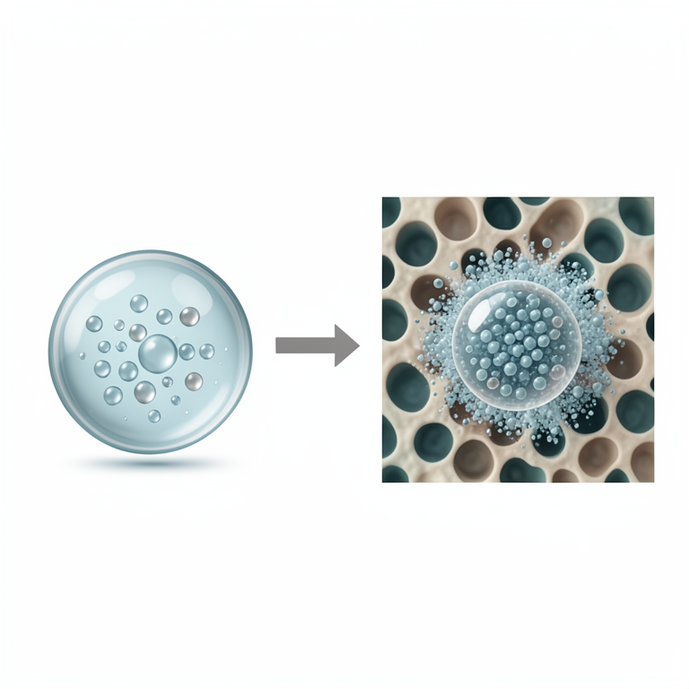
pH-Responsive Drug Release
AI-generated conceptual illustration (polymeric nanoparticles in a tumor microenvironment). It explains the idea and does not show study data.
View source evidence ↗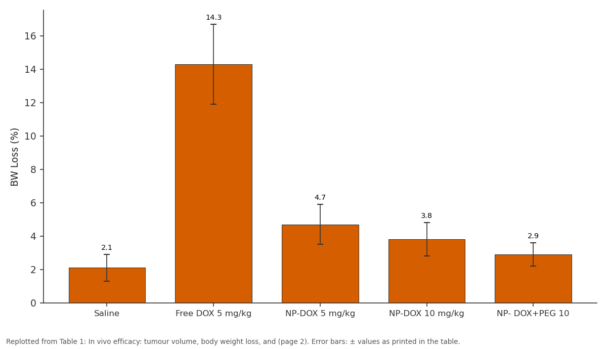
BW Loss (%): Saline vs Free DOX 5 mg/kg vs NP-DOX 5 mg/kg vs NP-DOX 10 mg/kg vs NP- DOX+PEG 10
Replotted from Table 1 (page 2) of the original paper. Every bar is the paper's own table value.
View source evidence ↗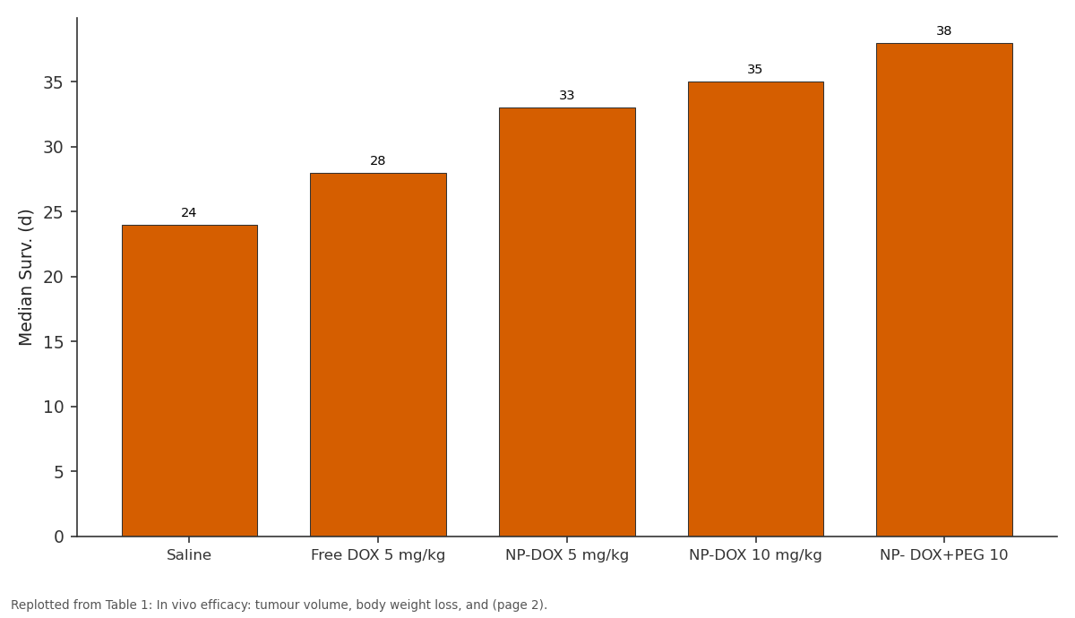
Median Surv. (d): Saline vs Free DOX 5 mg/kg vs NP-DOX 5 mg/kg vs NP-DOX 10 mg/kg vs NP- DOX+PEG 10
Replotted from Table 1 (page 2) of the original paper. Every bar is the paper's own table value.
View source evidence ↗Figures & tables
From the original paper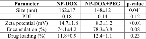 Table 2
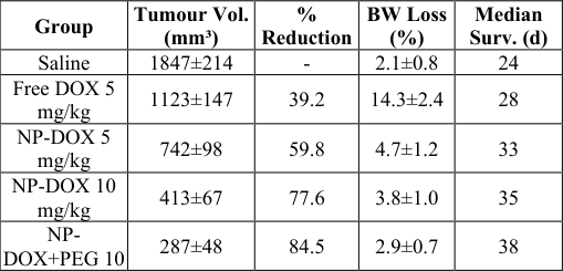 Table 1
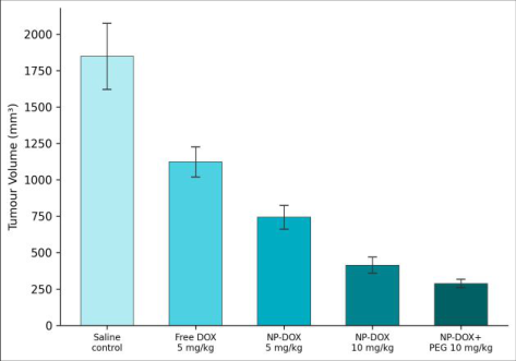 Figure 1
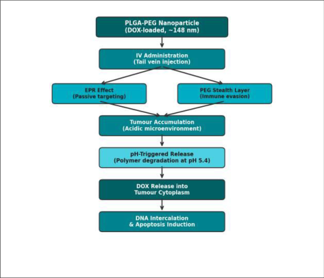 Figure 2
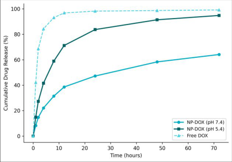 Figure 3
Introduction
Doxorubicin remains one of the most effective chemotherapeutic agents for breast, ovarian, and haematological cancers, yet its clinical utility is capped by dose-limiting cardiotoxicity [1] . Free doxorubicin distributes non-selectively, exposing cardiomyocytes and hepatocytes to cytotoxic concentrations that accumulate over treatment cycles [2] . Encapsulating the drug inside biodegradable polymer nanoparticles offers a way to shift the biodistribution toward tumour tissue, exploiting the enhanced permeability and retention (EPR) effect that arises from the leaky vasculature and impaired lymphatic drainage characteristic of solid tumours [3] . PLGA (poly(lactic-co-glycolic acid)) is FDA-approved for injectable formulations and degrades into non-toxic lactic and glycolic acid [4] . Surface modification with polyethylene glycol (PEG) creates a hydrophilic "stealth" corona that delays opsonisation and extends circulating half-life from minutes to hours [5,6] . pH-responsive release is an inherent feature: PLGA degrades faster in the mildly acidic tumour microenvironment (pH 5.4-6.8) than in plasma (pH 7.4), releasing more drug at the target site [7] . This research synthesised PLGA-PEG nanoparticles loaded with doxorubicin, characterised their physicochemical properties, evaluated in vitro release kinetics at two pH values, and assessed antitumour efficacy in a 4T1 murine breast carcinoma model.
Material and Methods
Material and Methods
Material
All experiments were conducted at the Department of Bioprocess Engineering, Riyadh College of Molecular Biology, Riyadh, Saudi Arabia, between February and October 2023. PLGA (50:50, Mw 38 kDa; Evonik RESOMER RG 504H), mPEG-PLGA (Mw 5k-30k; Nanosoft Polymers), and doxorubicin hydrochloride (>98%, LC Laboratories) were used. 4T1 murine breast carcinoma cells were from ATCC. Female BALB/c mice (6-8 weeks, 18-22 g) were obtained from the institutional vivarium. The animal protocol was approved by the Riyadh College of Molecular Biology Animal Ethics Committee (RCMB-AEC-2023-006).
Methods
Nanoparticles were prepared by nanoprecipitation. PLGA or mPEG-PLGA (20 mg) and doxorubicin (4 mg) were dissolved in 2 mL acetone and added dropwise to 10 mL of 1% PVA under stirring at 600 rpm. Acetone was evaporated under reduced pressure. Particles were collected by ultracentrifugation (25,000×g, 30 min), washed three times, and resuspended in PBS. Size and zeta potential were measured by DLS (Malvern Zetasizer Nano ZS). Morphology was confirmed by TEM (JEOL JEM-1400, 120 kV). Drug loading was calculated after dissolving particles in DMSO and measuring absorbance at 480 nm. In vitro release was measured by dialysis (MWCO 12 kDa) in PBS at pH 7. Performance Evaluation NP-DOX (10 mg/kg) reduced tumour volume to 413 ± 67 mm³ at day 21 versus 1,847 ± 214 mm³ for saline controlsa 77.6 percent reduction (Table 1). Free DOX (5 mg/kg) achieved only 39.2 percent reduction. PEGylated NP-DOX outperformed non-PEGylated NP-DOX by an additional 30.5 percent, consistent with extended circulation time allowing more EPR-mediated tumour accumulation. Body weight loss in the free DOX group reached 14.3 percent by day 21, indicating systemic toxicity, while NP-DOX groups showed ≤4.7 percent loss (Table 2). Survival analysis (Kaplan-Meier) confirmed that NP-DOX+PEG extended median survival to 38 days versus 24 days for saline.
| Group | Tumour Vol. (mm³) | % Reduction | BW Loss (%) | Median Surv. (d) |
|---|---|---|---|---|
| Saline | 1847±214 | - | 2.1±0.8 | 24 |
| Free DOX 5 mg/kg | 1123±147 | 39.2 | 14.3±2.4 | 28 |
| NP-DOX 5 mg/kg | 742±98 | 59.8 | 4.7±1.2 | 33 |
| NP-DOX 10 mg/kg | 413±67 | 77.6 | 3.8±1.0 | 35 |
| NP- DOX+PEG 10 | 287±48 | 84.5 | 2.9±0.7 | 38 |
Table text extracted from the original PDF; superscript letters are shown inline. The original table image is below.
View original table image
| Parameter | NP-DOX | NP-DOX+PEG | p-value |
|---|---|---|---|
| Size (nm) | 162±17 | 148±12 | 0.041 |
| PDI | 0.18 | 0.14 | 0.12 |
| Zeta potential (mV) | −14.7±1.8 | −8.3±1.2 | <0.01 |
| Encapsulation (%) | 74.1±4.2 | 78.3±3.8 | 0.08 |
| Drug loading (%) | 11.8±0.9 | 12.4±1.1 | 0.23 |
Table text extracted from the original PDF; superscript letters are shown inline. The original table image is below.
View original table image
Results
Results
Discussion
The 77.6 percent tumour reduction achieved by NP-DOX at 10 mg/kg exceeds the ~50-60 percent typically reported for non-PEGylated PLGA-DOX systems in similar 4T1 models [8, 9] . The additional benefit of PEGylation (30.5 percent further reduction) aligns with pharmacokinetic data showing that PEG chains of 5 kDa extend circulating half-life by 3-5-fold, increasing the window for EPR-mediated accumulation [10] . The pH-responsive release profile is clinically relevant: the 1.48-fold higher release at pH 5.4 vs 7.4 over 72 hours means that a greater fraction of encapsulated drug is liberated inside endosomes and lysosomes after cellular uptake, directing cytotoxicity toward cancer cells rather than stromal tissue [11] . The minimal body weight loss (<4.7 percent) in NP-DOX groups contrasts with the 14.3 percent loss in the free DOX arm, a metric that directly reflects reduced off-target toxicity [12] . A limitation is reliance on a single subcutaneous tumour model. Orthotopic or metastatic models would better simulate clinical breast cancer and test whether nanoparticles can reach disseminated disease [13] . Longerterm cardiac histopathology should also be evaluated.
Conclusion
PLGA-PEG nanoparticles loaded with doxorubicin achieved 77.6 percent tumour volume reduction in a 4T1 murine model with significantly less systemic toxicity than free drug. PEGylation provided an additional 30.5 percent improvement over non-PEGylated nanoparticles. pHresponsive release kinetics confirmed accelerated drug liberation at tumour-mimicking acidity. These findings support further development of PLGA-PEG nanocarriers as a platform for improving the therapeutic index of doxorubicin in breast cancer.
Acknowledgement
Acknowledgements Funding SourcesInternal allocation from the Riyadh College of Molecular Biology. Institutional SupportThe author thanks the Department of Bioprocess Engineering for access to DLS, TEM, and animal vivarium facilities.
Figures and tables from the paper
References
16 in the original paper- Carvalho C, Santos RX, Cardoso S, Correia S, Oliveira PJ, Santos MS, et al. Doxorubicin: the good, the bad and the ugly effect. Current Medicinal Chemistry. 2009;16(25):3267-3285.
- Tacar O, Sriamornsak P, Dass CR. Doxorubicin: an update on anticancer molecular action, toxicity and novel drug delivery systems. Journal of Pharmacy and Pharmacology. 2013;65(2):157-170.
- Maeda H, Wu J, Sawa T, Matsumura Y, Hori K. Tumor vascular permeability and the EPR effect in macromolecular therapeutics: a review. Journal of Controlled Release. 2000;65(1-2):271-284.
- Danhier F, Ansorena E, Silva JM, Coco R, Le Breton A, Preat V. PLGA-based nanoparticles: an overview of biomedical applications. Journal of Controlled Release. 2012;161(2):505-522.
- Harris JM, Chess RB. Effect of pegylation on pharmaceuticals. Nature Reviews Drug Discovery. 2003;2(3):214-221.
- Suk JS, Xu Q, Kim N, Hanes J, Ensign LM. PEGylation as a strategy for improving nanoparticle-based drug and gene delivery. Advanced Drug Delivery Reviews. 2016;99:28-51.
- Xu Y, Kim CS, Saylor DM, Koo D. Polymer degradation and drug delivery in PLGA-based drugpolymer applications: a review of experiments and theories. Journal of Biomedical Materials Research Part B. 2017;105(6):1692-1716.
- Hasan AA, Abdelmonsef AH, ElNaga MHA, Swidan SAH, Wahman MA. Formulation and characterization of doxorubicin loaded PLGA nanoparticles for breast cancer treatment. Journal of Drug Delivery Science and Technology. 2023;79:104067.
- Parveen S, Sahoo SK. Long circulating chitosan/PEG blended PLGA nanoparticle for tumor drug delivery. European Journal of Pharmacology. 2011;670(2-3):372383.
- Gref R, Luck M, Quellec P, Marchand M, Dellacherie E, Harnisch S, et al. Stealth corona-core nanoparticles surface modified by polyethylene glycol. Colloids and Surfaces B. 2000;18(3-4):301-313.
- Kamaly N, Yameen B, Wu J, Farokhzad OC. Degradable controlled-release polymers and polymeric nanoparticles: mechanisms of controlling drug release. Chemical Reviews. 2016;116(4):2602-2663.
- Brigger I, Dubernet C, Couvreur P. Nanoparticles in cancer therapy and diagnosis. Advanced Drug Delivery Reviews. 2002;54(5):631-651.
- Peer D, Karp JM, Hong S, Farokhzad OC, Margalit R, Langer R. Nanocarriers as an emerging platform for cancer therapy. Nature Nanotechnology. 2007;2(12):751-760.
- Cho K, Wang X, Nie S, Chen Z, Shin DM. Therapeutic nanoparticles for drug delivery in cancer. Clinical Cancer Research. 2008;14(5):1310-1316.
- Govender T, Stolnik S, Garnett MC, Illum L, Davis SS. PLGA nanoparticles prepared by nanoprecipitation: drug loading and release studies of a water soluble drug. Journal of Controlled Release. 1999;57(2):171185.
- Fonseca C, Simoes S, Gaspar R. Paclitaxel-loaded PLGA nanoparticles: preparation, physicochemical characterization and in vitro anti-tumoral activity. Journal of Controlled Release. 2002;83(2):273-286.