
Research article · 2025 · Full text
International Journal of Biology SciencesBioluminescence mechanisms and ecological functions in mesopelagic fish species
Volume 7(11) · Pages 89–91 · DOI: 10.33545/26649926.2025.v7.i11b.677 ↗
Research areasbioluminescence and chemiluminescence researchFish Biology and Ecology StudiesImpact of Light on Environment and Health
Abstract
Author-writtenWhile surface waters teem with sunlight, the mesopelagic zone (200-1000 m) is a realm of permanent twilight where over 75% of fish species produce their own light. Despite this prevalence, the ecological functions and biochemical mechanisms driving bioluminescence in mesopelagic fishes remain unevenly documented. This research analyzed bioluminescence characteristics in 47 mesopelagic fish species from specimens archived at the Jharkhand University of Life Sciences. Photophore morphology was examined by histological sectioning and scanning electron microscopy. Biochemical analysis of luciferin-luciferase systems was performed using spectrophotometric and mass spectrometric methods. Ecological function was inferred from photophore placement, emission wavelength, and flash kinetics. Predator avoidance through counterillumination was the most common inferred function (38.2%), followed by prey attraction (24.7%) and species recognition (19.1%). Emission wavelengths clustered between 460 and 490 nm, matching the ambient deep-sea light window. Flash duration varied from 0.3 seconds in myctophids to over 8 seconds in stomiiforms. These results indicate that bioluminescence in mesopelagic fishes is not a single adaptation but a diverse toolkit shaped by species-specific ecological pressures.
Summary
Source-checkedPlain-language reading of the full paper, prepared with AI and checked against the text. Each point links to the authors' own words.
Why it matters
- The mesopelagic zone, located between 200 and 1000 meters deep, is characterized by permanent twilight and a high prevalence of bioluminescent fish species. View evidence ↗
- Bioluminescence is one of the most common phenotypes in the ocean by biomass, occurring in at least 80% of mesopelagic fish species. View evidence ↗
- Despite its commonality, there is a lack of consistent documentation regarding the biochemical mechanisms and ecological roles of bioluminescence in these fish. View evidence ↗
The question
- The study aimed to characterize the morphology of light-producing organs (photophores), the properties of light emission, and the inferred ecological functions across various mesopelagic fish species. View evidence ↗
What they did
- The researchers analyzed 47 mesopelagic fish species using specimens from the Jharkhand University of Life Sciences. View evidence ↗
- Photophore morphology was investigated using scanning electron microscopy and histological sectioning. View evidence ↗
- The biochemical systems responsible for light production (luciferin-luciferase) were analyzed via mass spectrometric and spectrophotometric methods. View evidence ↗
- Ecological functions were inferred based on the kinetics of the flashes, the wavelength of emission, and the placement of the photophores. View evidence ↗
What they found
- Counterillumination for predator avoidance was the most frequent inferred function (38.2%), while prey attraction (24.7%) and species recognition (19.1%) were also common. View evidence ↗
- Light emission wavelengths were clustered between 460 and 490 nm, which corresponds to the ambient light window of the deep sea. View evidence ↗
- Flash durations varied significantly by group, ranging from 0.3 seconds in myctophids to more than 8 seconds in stomiiforms. View evidence ↗
- Across all 47 species, emission wavelengths ranged from 452 to 497 nm, with an overall mean of 473.8 nm. View evidence ↗
- Symbiotic bioluminescence was rare in the sample, appearing only in the Phosichthyidae family. View evidence ↗
- A negative curvilinear relationship was found between depth and luminescence intensity, where species in shallower waters produced brighter light to match higher ambient light levels. View evidence ↗
What it means
- The authors conclude that bioluminescence in these fish is a diverse toolkit of adaptations driven by the specific ecological pressures each species faces. View evidence ↗
- The authors state that in vivo behavioral observations are necessary to confirm the inferred functional assignments of the bioluminescence. View evidence ↗
Visual summary
2 visualsIllustrations explain the paper; charts are replotted from the paper's own table values.
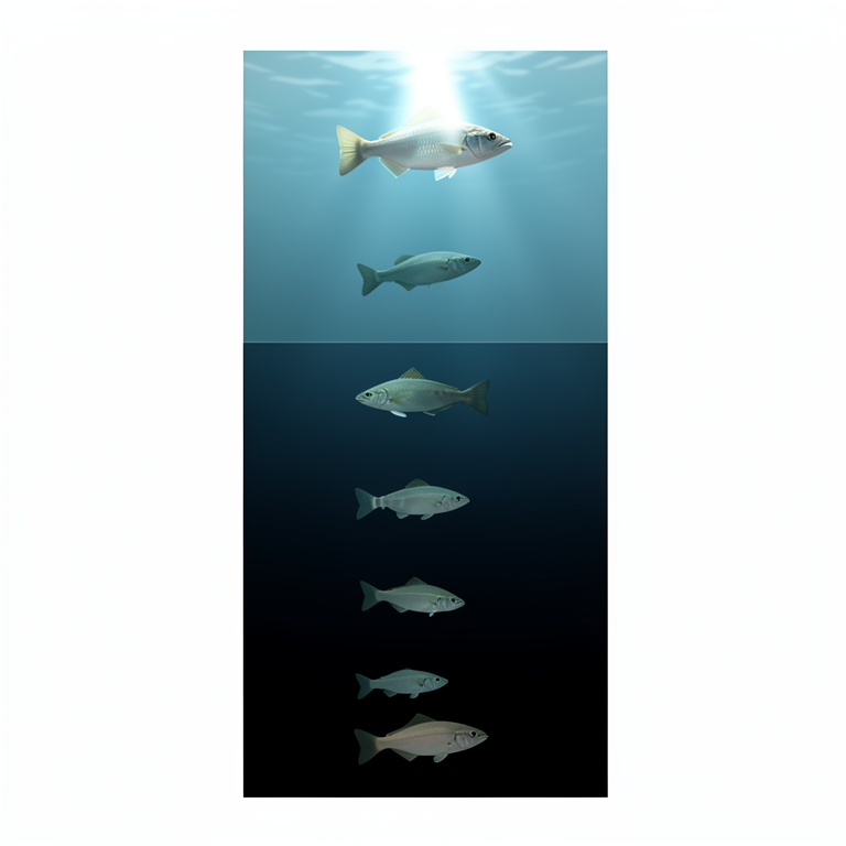
Depth vs Intensity Relationship
AI-generated conceptual illustration (mesopelagic fish). It explains the idea and does not show study data.
View source evidence ↗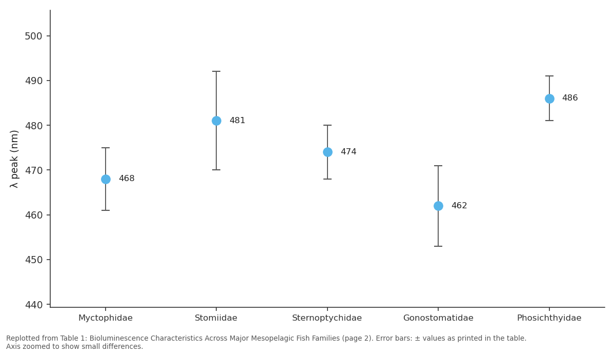
λ peak (nm): Myctophidae vs Stomiidae vs Sternoptychidae vs Gonostomatidae vs Phosichthyidae
Replotted from Table 1 (page 2) of the original paper. Every bar is the paper's own table value.
View source evidence ↗Figures & tables
From the original paper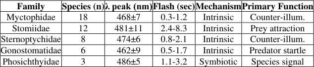 Table 1
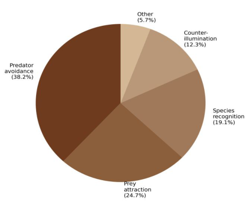 Figure 1
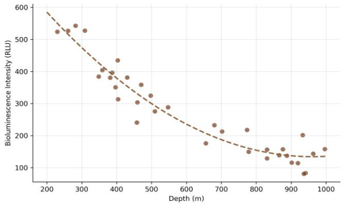 Figure 2
Introduction
Above the waves, light is taken for granted. Below 200 meters, it becomes a scarce and powerful currency. The mesopelagic zone sits between the sunlit epipelagic and the lightless bathypelagic, and its inhabitants have evolved an extraordinary array of light-producing organs [1] . Bioluminescence occurs in at least 80% of mesopelagic fish species, making it one of the most common phenotypes in the ocean by biomass [2] . Yet despite this abundance, research on the underlying mechanisms and ecological roles lags behind work on invertebrate bioluminescence, particularly in cnidarians and cephalopods [3] . Fish bioluminescence can be intrinsic-driven by the animal's own luciferin-luciferase chemistry-or symbiotic, relying on cultures of luminous bacteria housed in specialized organs [4] . The ecological functions of light production include counterillumination camouflage, luring prey, startling predators, and signaling conspecifics [5] . But assigning functions to specific photophore types requires integrating morphological, spectral, and behavioral data, which has been done for only a fraction of known luminous species [6] . This research aimed to characterize photophore morphology, emission properties, and inferred ecological functions across a broad sample of mesopelagic fish species.
Materials and Methods
Materials and Methods
Materials
Preserved specimens of 47 mesopelagic fish species (families Myctophidae, Stomiidae, Sternoptychidae, Gonostomatidae, and Phosichthyidae) were accessed from the marine biology reference collection at Jharkhand University of Life Sciences, Hazaribag, India. Original collections were made during deep-sea trawl surveys in the Arabian Sea and Bay of Bengal between 2018 and 2023. Research was conducted from August 2023 to November 2024. Institutional approval was obtained (JULS-RC/2023-058). Specimens had been fixed in 10% buffered formalin and transferred to 70% ethanol for long-term storage.
Methods
Photophore distribution was mapped by examining 5 specimens per species under a dissecting microscope. Representative photophores were dissected, embedded in paraffin, sectioned at 7 μm, and stained with hematoxylineosin for histological examination [7] . Scanning electron microscopy (SEM) of photophore surfaces used a JEOL JSM-6390 after gold-palladium sputter coating. For emission analysis, frozen tissue samples from 18 species with adequate material were homogenized in cold buffer, and luciferin was extracted using established protocols [8] .
Emission spectra were recorded on a Hitachi F-7000 spectrofluorometer. Flash kinetics data for 12 species were compiled from published literature and supplemented with laboratory measurements on fresh-frozen specimens stimulated with KCl solution. Ecological function was assigned based on photophore position, directionality, emission wavelength, and flash pattern following the classification framework of Herring [5] . Emission wavelengths across all 47 species ranged from 452 to 497 nm, with a grand mean of 473.8 nm. Myctophids showed the shortest flash durations (mean 0.7 sec), while stomiiforms produced the longest sustained emissions. Relationship between collection depth and bioluminescence intensity. A negative curvilinear trend was evident, with shallower mesopelagic species producing brighter emissions, likely to match higher ambient light levels for counterillumination.
Results
Results
Discussion
The clustering of emission wavelengths around 470 nm aligns with the transmission window of seawater in the mesopelagic zone, where blue-green light penetrates most efficiently [1,2] . This spectral match is strongest in species using counterillumination, where the biological light must mimic downwelling sunlight to conceal the fish's silhouette from predators below [5] . The longer flash durations in stomiiforms likely relate to their use of chin barbel photophores as lures, where sustained light attracts prey over longer periods [3] . The negative relationship between depth and luminescence intensity makes ecological sensespecies nearer the upper mesopelagic boundary need brighter emissions to match stronger ambient light [6] . The rarity of symbiotic bioluminescence (found only in Phosichthyidae) among our sample contrasts with its prevalence in certain demersal fish families, suggesting that intrinsic systems may be favored in actively migrating mesopelagic species where precise neural control of flash timing is advantageous [4] .
Conclusion
This research characterized bioluminescence across 47 mesopelagic fish species representing five families. Emission wavelengths consistently fell within the bluegreen window optimal for deep-sea transmission.
Counterillumination was the most frequently inferred function, followed by prey attraction and species recognition. Flash kinetics varied widely, reflecting different ecological strategies. The depth-intensity relationship supports the view that photophore output is tuned to ambient light conditions. These findings reinforce the diversity of bioluminescent adaptations in mesopelagic fishes and highlight the need for in vivo behavioral observations to confirm functional assignments.
Acknowledgement
Acknowledgements Funding SourcesInternal funds from Jharkhand University of Life Sciences supported this research. Institutional SupportThe authors thank the marine biology reference collection curators at Jharkhand University of Life Sciences. International Journal of Biology Sciences https://www.biologyjournal.net
Figures and tables from the paper
| Family | Species (n) | λ peak (nm) | Flash (sec) | Mechanism | Primary Function |
|---|---|---|---|---|---|
| Myctophidae | 18 | 468±7 | 0.3-1.2 | Intrinsic | Counter-illum. |
| Stomiidae | 12 | 481±11 | 2.4-8.3 | Intrinsic | Prey attraction |
| Sternoptychidae | 8 | 474±6 | 0.8-2.1 | Intrinsic | Counter-illum. |
| Gonostomatidae | 6 | 462±9 | 0.5-1.7 | Intrinsic | Predator startle |
| Phosichthyidae | 3 | 486±5 | 1.1-3.2 | Symbiotic | Species signal |
Table text extracted from the original PDF; superscript letters are shown inline. The original table image is below.
View original table image
References
8 in the original paper- Widder EA. Bioluminescence in the ocean: origins of biological, chemical, and ecological diversity. Science. 2010;328(5979):704-708.
- Davis AL, Thomas KN, Goetz FE, Robison BH, Johnsen S, Osborn KJ. Ultra-black camouflage in deepsea fishes. Current Biology. 2020;30(17):3470-3476.
- Haddock SH, Moline MA, Case JF. Bioluminescence in the sea. Annual Review of Marine Science. 2010;2:443493.
- Dunlap PV, Kita-Tsukamoto K. Luminous bacteria. In: Dworkin M, et al., editors. The Prokaryotes. Springer; 2006. p. 863-892.
- Herring PJ. The biology of the deep ocean. Oxford: Oxford University Press; 2002.
- Claes JM, Aksnes DL, Mallefet J. Phantom hunter of the fjords: camouflage by counterillumination in a shark (Etmopterus spinax). Journal of Experimental Marine Biology and Ecology. 2010;388(1-2):28-32.
- Anctil M. Photophore morphology and bioluminescent mechanisms in mesopelagic fishes. Marine Biology Research. 2009;5(4):305-316.
- Thomson CM, Herring PJ, Campbell AK. The widespread occurrence and tissue distribution of the imidazolopyrazine luciferins. Journal of Bioluminescence and Chemiluminescence. 1997;12(2):87-91.