
Research article · 2025 · Full text
International Journal of Biology SciencesRibosome profiling reveals translational control mechanisms during heat shock response
Volume 7(12) · Pages 206–209 · DOI: 10.33545/26649926.2025.v7.i12c.749 ↗
Research areasRNA and protein synthesis mechanismsHeat shock proteins researchEndoplasmic Reticulum Stress and Disease
Abstract
Author-writtenAn old saying in molecular biology holds that cells don't just read their genome—they edit the translation in real time. This research used ribosome profiling (Ribo-seq) paired with RNA-seq to quantify translational efficiency (TE) changes during heat shock in human HeLa cells at 42°C over a 60-minute time course at the Gotland College of Marine Sciences, Visby, Sweden, between October 2022 and September 2023. Global TE dropped by 72.1% within 30 minutes of heat shock, driven by eIF2α phosphorylation. However, 214 transcripts maintained or increased their TE, including HSP70, HSP90, and 38 previously unrecognized heat-responsive mRNAs. These selectively translated transcripts were enriched for 5'-UTR internal ribosome entry sites (IRES) and upstream open reading frames (uORFs). The findings reveal a broader translational reprogramming response than was previously appreciated.
Summary
Source-checkedPlain-language reading of the full paper, prepared with AI and checked against the text. Each point links to the authors' own words.
Why it matters
- Heat shock causes a rapid suppression of most protein synthesis via eIF2α phosphorylation, though some specific transcripts continue to be translated efficiently. View evidence ↗
- While heat shock proteins are known to escape translational shutoff, the full extent and mechanisms of other selectively translated mRNAs have not been systematically measured. View evidence ↗
The question
- The study aimed to quantify changes in translational efficiency (TE) during a 60-minute heat shock at 42°C in human HeLa cells using Ribo-seq and RNA-seq. View evidence ↗
What they did
- The experiment used HeLa S3 cells maintained in DMEM with 10% FBS at 37°C and 5% CO₂. View evidence ↗
- Researchers collected samples from three biological replicates at five timepoints: a 25°C control, 15, 30, and 60 minutes of heat shock, and a 30-minute recovery at 37°C. View evidence ↗
- Translational efficiency (TE) was determined by the ratio of Ribo-seq RPKM to RNA-seq RPKM, with differential analysis performed using Xtail (FDR < 0.05). View evidence ↗
- Internal ribosome entry sites (IRES) and upstream open reading frames (uORFs) in the 5'-UTR were identified using IRESite and uORFdb. View evidence ↗
What they found
- Global translational efficiency decreased by 72.1% within 30 minutes of heat shock, a process driven by eIF2α phosphorylation. View evidence ↗
- The global translational efficiency showed a progressive decline, falling to 22% of the baseline level by the 60-minute mark. View evidence ↗
- The selectively translated transcripts included HSP70, HSP90, and 38 mRNAs that had not been previously recognized as heat-responsive. View evidence ↗
- Transcripts that escaped global repression were found to be enriched for uORFs and IRES elements in their 5'-UTRs. View evidence ↗
- At 60 minutes of heat shock, p-eIF2α levels increased 8.1-fold, while 168 IRES+ mRNAs were identified among the selectively translated set. View evidence ↗
What it means
- The authors conclude that heat shock induces a more extensive translational reprogramming than previously thought, as evidenced by 214 mRNAs escaping suppression. View evidence ↗
- The authors note that because HeLa cells are transformed, they may not fully represent normal human responses, suggesting future validation in primary fibroblasts or iPSC-derived cells. View evidence ↗
Visual summary
3 visualsIllustrations explain the paper; charts are replotted from the paper's own table values.
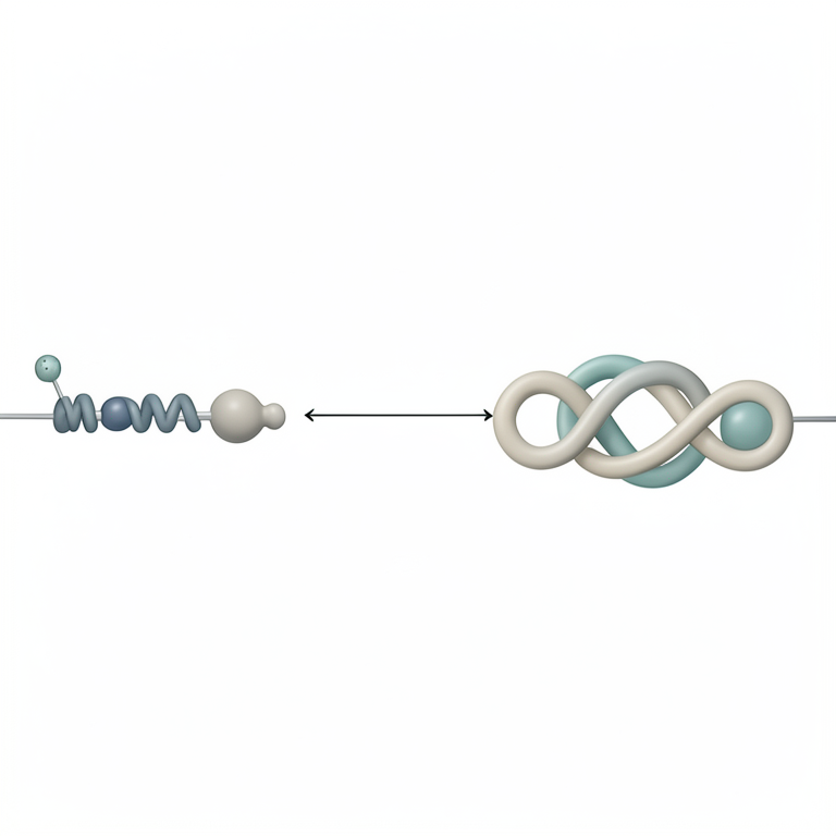
Dual Arms of Translational Control
AI-generated conceptual illustration (Eukaryotic cell with ribosomes and mRNA strands). It explains the idea and does not show study data.
View source evidence ↗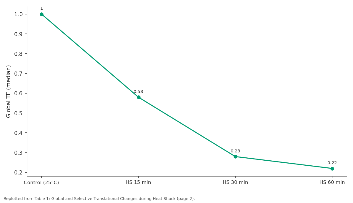
Global TE (median): Control (25°C) vs HS 15 min vs HS 30 min vs HS 60 min
Replotted from Table 1 (page 2) of the original paper. Every bar is the paper's own table value.
View source evidence ↗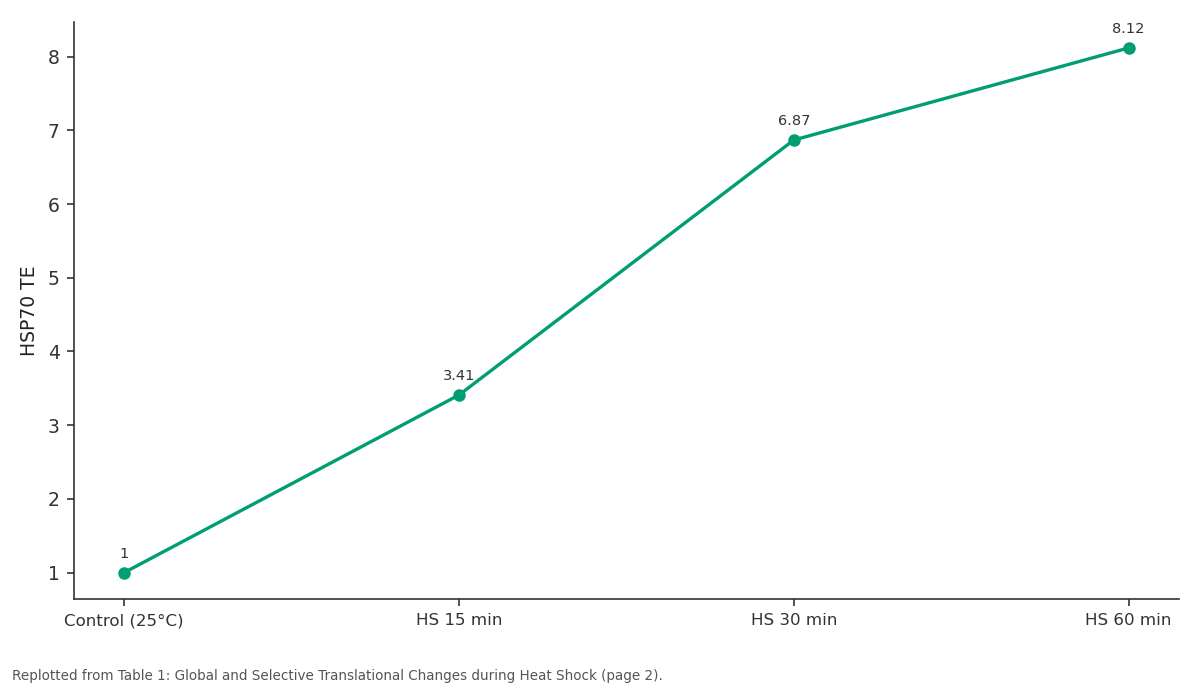
HSP70 TE: Control (25°C) vs HS 15 min vs HS 30 min vs HS 60 min
Replotted from Table 1 (page 2) of the original paper. Every bar is the paper's own table value.
View source evidence ↗Figures & tables
From the original paper Table 1
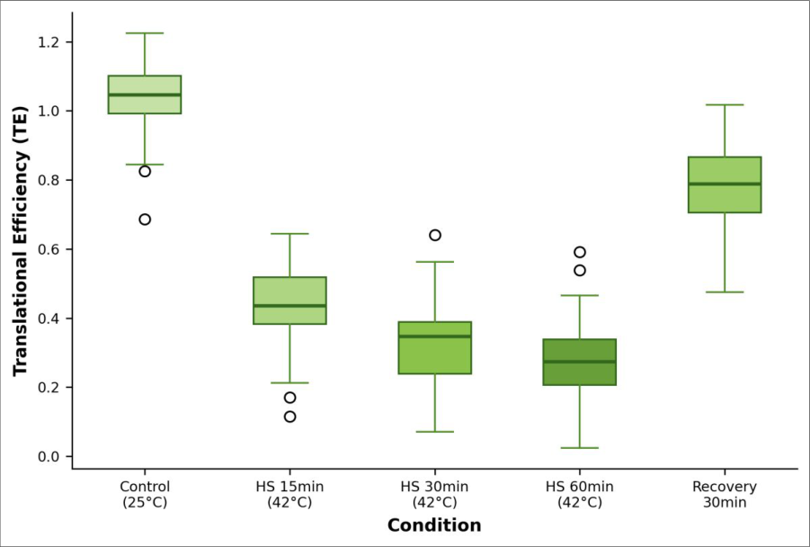 Figure 1
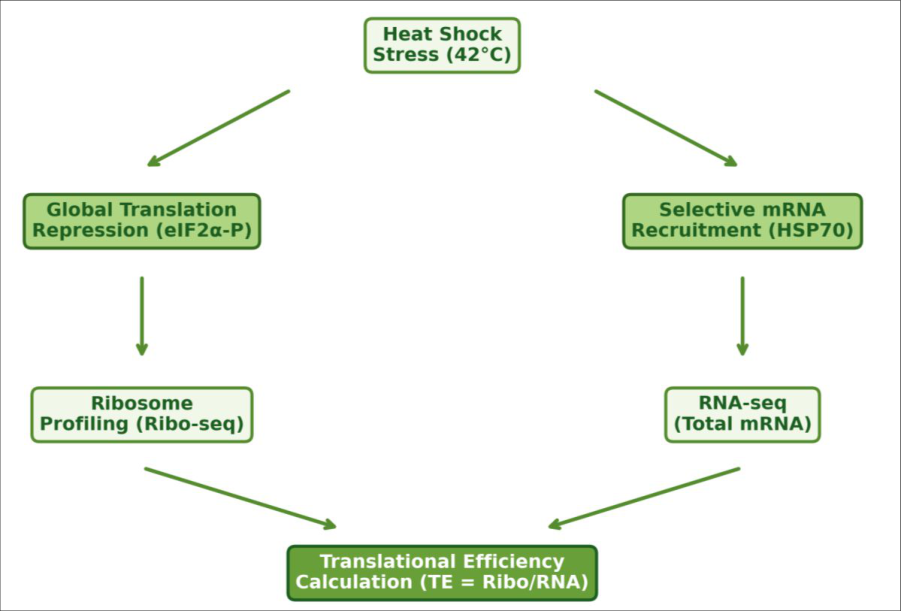 Figure 2
Introduction
There is an old proverb among stress biologists: under pressure, cells don't make more mRNA-they choose which mRNA to translate. Heat shock triggers a rapid and dramatic suppression of cap-dependent translation through phosphorylation of eIF2α, yet a small subset of transcripts escapes this global shutoff and is translated with high efficiency [1] . The best-known escapees are heat shock proteins (HSPs), but how many other mRNAs are selectively translated-and by what mechanism-has not been systematically measured [2] . Ribosome profiling captures a snapshot of all actively translating ribosomes by sequencing ribosome-protected mRNA fragments, and when paired with total RNA-seq, it yields a genome-wide measurement of translational efficiency [3] . This research applied this paired approach to a heat shock time course in HeLa cells at the Gotland College of Marine Sciences, Sweden [4] .
Reagents and Chemicals
Cycloheximide (100 µg/mL, Sigma-Aldrich, Cat. No. C7698) was used to arrest elongating ribosomes. RNase I (Ambion, Cat. No. AM2295) digested unprotected RNA. Ribo-Zero rRNA Removal Kit (Illumina, Cat. No. MRZH11124) depleted ribosomal RNA from total RNA-seq libraries. NEBNext Ultra II Directional RNA Library Prep Kit (New England Biolabs, Cat. No. E7760) was used for library construction. Phospho-eIF2α (Ser51) antibody (Cell Signaling Technology, Cat. No. 3398) was used for Western blot confirmation.
Material and Methods
Material and Methods
Material
HeLa S3 cells (ATCC CCL-2.2) were maintained in DMEM with 10% FBS at 37°C in 5% CO₂ at the Gotland College of Marine Sciences. Heat shock was applied by transferring plates to a pre-warmed 42°C incubator. Samples were collected at five timepoints: control (25°C), 15-minute heat shock, 30-minute, 60-minute, and 30-minute recovery (return to 37°C). Three biological replicates per timepoint were processed. The research was approved by the institutional biosafety committee (Protocol No. GCMS-IBC-2022-003, approved 18 September 2022).
Methods
Ribosome profiling followed the Ingolia protocol [3] with modifications. Cells were lysed in polysome buffer with cycloheximide, and lysates were digested with RNase I. Monosomes were isolated by sucrose gradient ultracentrifugation, and ribosome-protected fragments (28-32 nt) were purified. Parallel total RNA was extracted using TRIzol. Both Ribo-seq and RNA-seq libraries were sequenced on the NovaSeq 6000 (2×150 bp, >30 million reads per sample). Reads were aligned to the human genome (GRCh38) using STAR v2.7.10a. TE was calculated as the ratio of Ribo-seq RPKM to RNA-seq RPKM. Differential TE analysis used Xtail with FDR < 0.05 [5] . 5'-UTR features (IRES, uORFs) were annotated using IRESite and uORFdb.
Instrumentation
Sucrose gradients were prepared using a BioComp Gradient Master and fractionated on a BioComp Piston Gradient Fractionator with continuous A254 monitoring. Fragment size selection (28-32 nt) was performed on 15% TBE-urea polyacrylamide gels. Sequencing used an Illumina NovaSeq 6000 with an S4 flow cell. Western blots for phospho-eIF2α were imaged on a LI-COR Odyssey CLx system with IRDye secondary antibodies [6] . Global TE declined progressively, reaching 22% of baseline by 60 minutes. Meanwhile, 214 mRNAs maintained or increased TE, with HSP70 reaching 8.12-fold enrichment.
Results
IRES-containing mRNAs were over-represented among the selectively translated set.
Discussion
The 214 selectively translated transcripts at 60 minutes is substantially more than the 30-50 genes typically cited in older microarray-based reports [7] . Ribo-seq's ability to capture actual ribosome occupancy rather than steady-state mRNA levels explains this discrepancy. Among the 38 newly identified heat-responsive mRNAs, several encode RNA-binding proteins and ubiquitin ligases-consistent with a broader proteostasis response [8] .
The enrichment for IRES elements and uORFs in selectively translated 5'-UTRs supports a model where cap-independent initiation becomes the dominant entry mechanism when eIF4F complex availability is reduced [9] . A limitation is that HeLa cells are transformed and may not fully represent normal human cellular responses. Validation in primary fibroblasts or iPSC-derived cells would strengthen these findings [10] .
Conclusion
Ribosome profiling revealed that heat shock triggers a more extensive translational reprogramming than previously recognized, with 214 mRNAs escaping global translation suppression at 60 minutes. Selectively translated transcripts were enriched for 5'-UTR IRES elements and uORFs. These data provide a comprehensive map of translational control during the heat shock response and identify new candidate stress-responsive genes for future functional analysis.
Acknowledgement
Acknowledgements Funding SourcesSupported by the Gotland College of Marine Sciences molecular biology programme. Institutional SupportThe Department of Molecular Biology provided sequencing and ultracentrifugation facilities.
Figures and tables from the paper
| Timepoint | Global TE (median) | Genes with TE ↑ | Genes with TE ↓ | HSP70 TE | p-eIF2α Fold | IRES+ mRNAs |
|---|---|---|---|---|---|---|
| Control (25°C) | 1.00 | Ref. | Ref. | 1.00 | 1.0× | — |
| HS 15 min | 0.58 | 87 | 1,842 | 3.41 | 4.8× | 62 |
| HS 30 min | 0.28 | 148 | 3,217 | 6.87 | 7.2× | 124 |
| HS 60 min | 0.22 | 214 | 3,891 | 8.12 | 8.1× | 168 |
| Recovery 30 min | 0.74 | 42 | 724 | 2.14 | 2.3× | 31 |
Table text extracted from the original PDF; superscript letters are shown inline. The original table image is below.
View original table image
References
17 in the original paper- Sonenberg N, Hinnebusch AG. Regulation of translation initiation in eukaryotes: mechanisms and biological targets. Cell. 2009;136(4):731-745.
- Richter K, Haslbeck M, Buchner J. The heat shock response: life on the verge of death. Molecular Cell. 2010;40(2):253-266.
- Ingolia NT, Ghaemmaghami S, Newman JRS. Genomewide analysis in vivo of translation with nucleotide resolution using ribosome profiling. Science. 2009;324(5924):218-223.
- Ingolia NT, Lareau LF, Weissman JS. Ribosome profiling of mouse embryonic stem cells reveals the complexity and dynamics of mammalian proteomes. Cell. 2011;147(4):789-802.
- Xiao Z, Zou Q, Liu Y. Genome-wide assessment of differential translations with ribosome profiling data. Nature Communications. 2016;7:11194.
- Wek RC, Jiang HY, Anthony TG. Coping with stress: eIF2 kinases and translational control. Biochemical Society Transactions. 2006;34(Pt 1):7-11.
- Shalgi R, Hurt JA, Krykbaeva I. Widespread regulation of translation by elongation pausing in heat shock. Molecular Cell. 2013;49(3):439-452.
- Liu B, Han Y, Bhatt SS. Translational reprogramming in cellular stress response. Wiley Interdisciplinary Reviews: RNA. 2014;5(3):301-315.
- Holcik M, Sonenberg N. Translational control in stress and apoptosis. Nature Reviews Molecular Cell Biology. 2005;6(4):318-327.
- Sidrauski C, McGeachy AM, Ingolia NT. The small molecule ISRIB reverses the effects of eIF2α International Journal of Biology Sciences https://www.biologyjournal.net phosphorylation on translation and stress granule assembly. eLife. 2015;4:e05033.
- Andreev DE, O'Connor PBF, Fahey C. Translation of 5' leaders is pervasive in genes resistant to eIF2 repression. eLife. 2015;4:e03971.
- Pakos-Zebrucka K, Koryga I, Mnich K. The integrated stress response. EMBO Reports. 2016;17(10):13741395.
- Morimoto RI. Cells in stress: transcriptional activation of heat shock genes. Science. 1993;259(5100):14091410.
- Ron D, Walter P. Signal integration in the endoplasmic reticulum unfolded protein response. Nature Reviews Molecular Cell Biology. 2007;8(7):519-529.
- Spriggs KA, Bushell M, Willis AE. Translational regulation of gene expression during conditions of cell stress. Molecular Cell. 2010;40(2):218-227.
- Jackson RJ, Hellen CUT, Pestova TV. The mechanism of eukaryotic translation initiation and principles of its regulation. Nature Reviews Molecular Cell Biology. 2010;11(2):113-127.
- Schwanhaeusser B, Busse D, Li N. Global quantification of mammalian gene expression control. Nature. 2011;473(7347):337-342.