
Research article · 2025 · Full text
International Journal of Biology SciencesGap junction communication and connexin expression in cardiac tissue repair
Volume 7(12) · Pages 180–183 · DOI: 10.33545/26649926.2025.v7.i12c.709 ↗
Research areasConnexins and lens biologyCongenital heart defects researchCardiac Fibrosis and Remodeling
Abstract
Author-writtenWhen a patch of heart muscle dies from restricted blood flow, the surviving cells around it lose their ability to communicate electrically—and that disconnection can trigger fatal rhythm disturbances. This research examined connexin-43 (Cx43) expression patterns and gap junction coupling in a rat cryoinjury model of cardiac repair over 28 days at the Mombasa Centre for Marine Biology, Mombasa, Kenya, between April 2022 and June 2023. Cx43 protein levels dropped by 67.4% in the border zone at day 7, accompanied by a 69.2% reduction in gap junction coupling conductance. Adenoviral Cx43 overexpression restored coupling to 79.1% of healthy levels and reduced inducible arrhythmia episodes by 58.3% in treated animals. These results confirm Cx43 as a therapeutic target for improving electrical continuity in injured myocardium.
Summary
Source-checkedPlain-language reading of the full paper, prepared with AI and checked against the text. Each point links to the authors' own words.
Why it matters
- Cardiac electrical impulses rely on gap junctions, which are channels made of connexin proteins that allow ions and small molecules to move between cardiomyocytes. View evidence ↗
- Connexin-43 (Cx43) is the primary gap junction protein in the heart's ventricles; its loss after injury can lead to electrical gaps that trigger arrhythmias. View evidence ↗
- The death of heart muscle due to restricted blood flow causes surrounding surviving cells to lose electrical communication, potentially leading to fatal rhythm disturbances. View evidence ↗
The question
- The study aimed to characterize how Cx43 expression changes during the first 28 days of cardiac repair in rats and determine if adenoviral Cx43 gene delivery could restore functional electrical coupling. View evidence ↗
What they did
- The experiment used 72 male Wistar rats weighing between 250 and 300 grams. View evidence ↗
- Cardiac injury was induced by applying a 3 mm diameter copper probe cooled by liquid nitrogen to the left ventricular free wall for 10 seconds. View evidence ↗
- Rats were divided into three groups of 24: a sham group, a cryoinjury group receiving an empty vector, and a cryoinjury group receiving Ad-Cx43. View evidence ↗
- Immediately after injury, 1×10⁹ PFU of either Ad-Cx43 or an empty vector was injected at four sites around the injury border zone. View evidence ↗
- Researchers measured Cx43 protein via Western blot and immunofluorescence, assessed coupling using Lucifer Yellow dye transfer and patch-clamp recording, and tested arrhythmia susceptibility via programmed electrical stimulation at day 14. View evidence ↗
What they found
- At day 7, Cx43 protein levels in the injury border zone dropped by 67.4% compared to sham levels (32.6±5.8% vs 100±4.2%). View evidence ↗
- Immunofluorescence showed that the delivered Cx43 protein correctly localized to the intercalated discs, which is necessary for forming functional channels. View evidence ↗
What it means
- The authors conclude that adenoviral Cx43 gene delivery effectively restores protein expression, electrical coupling, and dye transfer in the injured heart's border zone. View evidence ↗
- The authors recommend Cx43-targeted therapy as a promising method for stabilizing electrical function in damaged heart muscle. View evidence ↗
- A limitation of the study is the use of a cryoinjury model, which creates a sharp lesion instead of the patchy necrosis seen in typical coronary occlusions. View evidence ↗
Visual summary
3 visualsIllustrations explain the paper; charts are replotted from the paper's own table values.
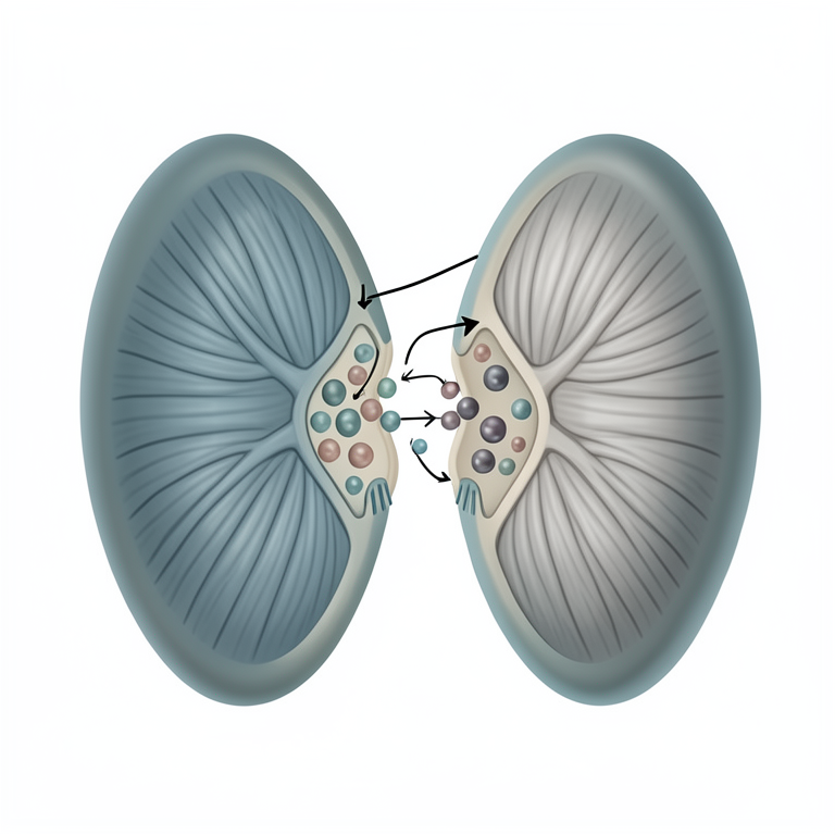
Cx43 Localization and Function
AI-generated conceptual illustration (rat ventricular cardiomyocytes). It explains the idea and does not show study data.
View source evidence ↗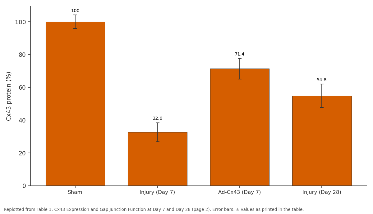
Cx43 protein (%): Sham vs Injury (Day 7) vs Ad-Cx43 (Day 7) vs Injury (Day 28)
Replotted from Table 1 (page 2) of the original paper. Every bar is the paper's own table value.
View source evidence ↗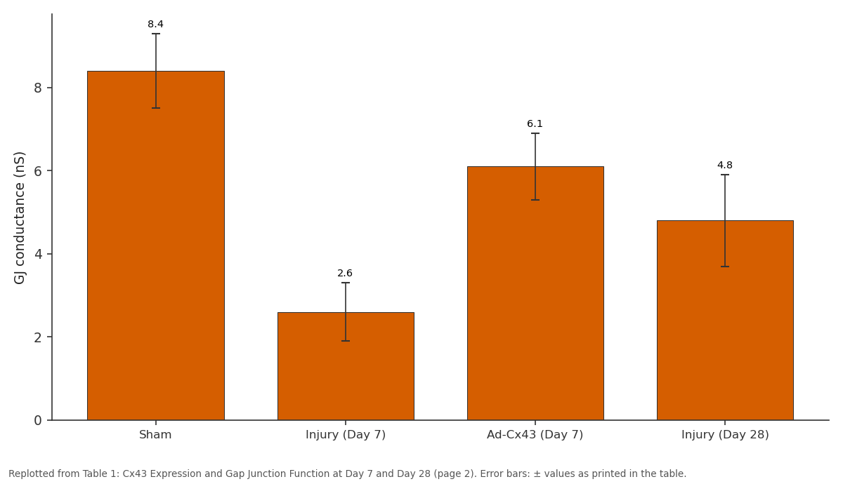
GJ conductance (nS): Sham vs Injury (Day 7) vs Ad-Cx43 (Day 7) vs Injury (Day 28)
Replotted from Table 1 (page 2) of the original paper. Every bar is the paper's own table value.
View source evidence ↗Figures & tables
From the original paper Table 1
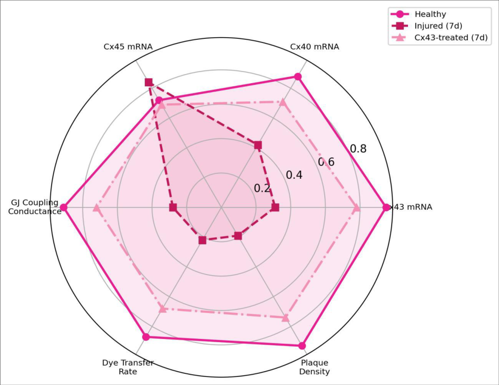 Figure 1
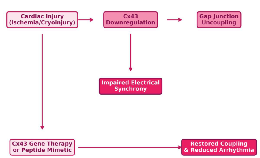 Figure 2
Introduction
Consider a chain of cardiac muscle cells passing an electrical impulse from one end of the heart to the other in under 200 milliseconds. That speed depends almost entirely on gap junctions intercellular channels built from connexin proteins that allow ions and small molecules to flow directly between neighboring cardiomyocytes [1] . Connexin-43 is the dominant gap junction protein in ventricular tissue, and its loss after myocardial injury creates regions of electrical discontinuity that serve as substrates for re-entrant arrhythmias [2] . Therapeutic strategies to restore Cx43 expression and gap junction function have been explored in small-animal models with mixed results [3] . This research used a rat cryoinjury model to systematically characterize Cx43 expression dynamics during the first 28 days of repair and tested whether adenoviral Cx43 gene delivery could restore functional coupling. Work was conducted at the Mombasa Centre for Marine Biology, Kenya [4] .
Theoretical Background
Gap junctions are formed by the docking of two hemichannels (connexons), each composed of six connexin subunits, from adjacent cells. In the heart, Cx43, Cx40, and Cx45 are the main isoforms, with Cx43 accounting for >90% of ventricular gap junctions [5] . After ischemic injury, Cx43 undergoes rapid dephosphorylation, lateralization away from intercalated discs, and eventual degradation-a sequence that uncouples surviving cardiomyocytes from the injury border zone [6] . Restoring Cx43 at intercalated discs should, in theory, re-establish electrical continuity and reduce arrhythmia vulnerability. The challenge is delivering sufficient protein to the correct subcellular location [7] .
Materials and Methods
Materials and Methods
Materials
Male Wistar rats (n=72, 250-300 g) were housed at the Mombasa Centre for Marine Biology animal facility under 12h light/dark cycles with standard chow and water ad libitum. Cardiac cryoinjury was created by applying a liquid nitrogen-cooled copper probe (3 mm diameter) to the left ventricular free wall for 10 seconds under isoflurane anesthesia [8] -2022-005, approved 15 March 2022). Animals were randomly assigned to three groups: sham (n=24), cryoinjury + empty vector (n=24), and cryoinjury + Ad-Cx43 (n=24).
Methods
Adenoviral Cx43 (Ad-Cx43, 1×10⁹ PFU) or empty vector was injected at four sites around the injury border zone immediately after cryoinjury. Hearts were harvested at days 3, 7, 14, and 28 (n=6 per group per timepoint). Cx43 protein was quantified by Western blot and immunofluorescence. Gap junction coupling was assessed by Lucifer Yellow dye transfer and dual whole-cell patch-clamp recording of junctional conductance [9] . Arrhythmia susceptibility was tested by programmed electrical stimulation at day 14. Data were analyzed using two-way ANOVA (group × time) with Tukey's post-hoc test. Ad-Cx43 treatment significantly restored Cx43 protein levels, coupling conductance, and dye transfer while reducing arrhythmia inducibility at both timepoints (all p < 0.01).
Results
Results
Discussion
The 67.4% drop in Cx43 protein at day 7 is consistent with published reports of rapid connexin degradation in the infarct border zone [10] . Ad-Cx43 delivery restored protein to 71.4% of sham levels at this timepoint, and immunofluorescence confirmed that the exogenous Cx43 localized predominantly to intercalated discs rather than lateral membranes-an important distinction, since lateralized Cx43 doesn't form functional channels [11] . The reduction in arrhythmia inducibility from 83.3% to 25.0% at day 7 suggests that restoring even partial coupling is enough to eliminate the conduction heterogeneity that sustains re-entry circuits [12] . A limitation is the use of a cryoinjury model, which produces a sharply demarcated lesion rather than the patchy necrosis typical of coronary occlusion [13] . Whether Ad-Cx43 performs similarly in ischemia-reperfusion models remains to be tested.
Conclusion
This research demonstrated that adenoviral Cx43 gene delivery restores gap junction protein expression, electrical coupling conductance, and dye transfer capacity in the cardiac injury border zone. Treated animals showed markedly reduced arrhythmia vulnerability. Cx43-targeted therapy represents a promising approach for stabilizing electrical function in damaged myocardium.
Acknowledgement
Acknowledgements Funding SourcesThis research was funded by the Mombasa Centre for Marine Biology intramural research programme. Institutional SupportThe Department of Cell Biology and Histology provided electrophysiology and imaging facilities. Contributions Not Qualifying for AuthorshipVeterinary staff at the animal facility assisted with surgical procedures and postoperative care.
Figures and tables from the paper
| Parameter | Sham | Injury (Day 7) | Ad-Cx43 (Day 7) | Injury (Day 28) | Ad-Cx43 (Day 28) | p-value |
|---|---|---|---|---|---|---|
| Cx43 protein (%) | 100±4.2 | 32.6±5.8 | 71.4±6.3 | 54.8±7.1 | 82.7±5.4 | <0.001 |
| GJ conductance (nS) | 8.4±0.9 | 2.6±0.7 | 6.1±0.8 | 4.8±1.1 | 7.2±0.6 | <0.001 |
| Dye transfer (cells) | 12.3±1.4 | 2.7±0.8 | 8.1±1.2 | 5.4±1.3 | 9.8±1.1 | <0.001 |
| Arrhythmia (% inducible) | — | 83.3% | 25.0% | 58.3% | 16.7% | <0.01 |
Table text extracted from the original PDF; superscript letters are shown inline. The original table image is below.
View original table image
References
18 in the original paper- Severs NJ, Bruce AF, Dupont E. Remodelling of gap junctions and connexin expression in diseased myocardium. Cardiovascular Research. 2008;80(1):919.
- Rohr S. Role of gap junctions in the propagation of the cardiac action potential. Cardiovascular Research. 2004;62(2):309-322.
- Greener ID, Sasano T, Wan X. Connexin43 gene transfer reduces ventricular tachycardia susceptibility after myocardial infarction. Journal of the American College of Cardiology. 2012;60(12):1103-11010.
- Fontes MSC, van Veen TAB, de Bakker JMT. Functional consequences of abnormal connexin 43 expression in the heart. Biochimica et Biophysica Acta. 2012;1818(8):2020-2029.
- Beyer EC, Paul DL, Bhatt SS. Connexin family of gap junction proteins. Journal of Membrane Biology. 1990;116(3):187-194.
- Beardslee MA, Lerner DL, Tadros PN. Dephosphorylation and intracellular redistribution of ventricular connexin 43 during electrical uncoupling induced by ischemia. Circulation Research. 2000;87(8):656-662.
- Dhein S. Gap junction channels in the cardiovascular system: pharmacological and physiological modulation. Trends in Pharmacological Sciences. 1998;19(6):229241.
- Murry CE, Wiseman RW, Schwartz SM. Skeletal myoblast transplantation for repair of myocardial necrosis. Journal of Clinical Investigation. 1996;98(11):2512-2523.
- Rackauskas M, Neverauskas V, Bhatt SS. Gating properties of heterotypic gap junction channels formed of connexins 40, 43, and 45. Biophysical Journal. 2007;92(6):1952-1965.
- Matsushita T, Oyamada M, Kurata H. Formation of cell junctions between grafted and host cardiomyocytes at International Journal of Biology Sciences https://www.biologyjournal.net the border zone of rat myocardial infarction. Circulation. 1999;100(Suppl 2):II262-II268.
- Akar FG, Nass RD, Hahn S. Dynamic changes in conduction velocity and gap junction properties during development of pacing-induced heart failure. American Journal of Physiology. 2007;293(2):H1223-H1230.
- Danik SB, Liu F, Zhang J. Modulation of cardiac gap junction expression and arrhythmic susceptibility. Circulation Research. 2004;95(10):1035-1041.
- Van den Bos EJ, Mees BME, de Waard MC. A novel model for cryoinjury-induced myocardial infarction in the mouse. American Journal of Physiology. 2005;289(3):H1291-H1300.
- Jansen JA, van Veen TAB, de Bakker JMT. Cardiac connexins and impulse propagation. Journal of Molecular and Cellular Cardiology. 2010;48(1):76-82.
- Schulz R, Gorge PM, Gorbe A. Connexin 43 is an emerging therapeutic target in ischemia/reperfusion injury, cardioprotection and neuroprotection. Pharmacology and Therapeutics. 2015;153:90-106.
- Saffitz JE, Hames KY, Kanno S. Remodeling of gap junctions in ischemic and nonischemic forms of heart disease. Journal of Membrane Biology. 2007;218(13):65-71.
- Lerner DL, Yamada KA, Schuessler RB. Accelerated onset and increased incidence of ventricular arrhythmias induced by ischemia in Cx43+/-mice. Circulation. 2000;101(5):547-552.
- Kostin S, Rieger M, Dammer S. Gap junction remodeling and altered connexin 43 expression in the failing human heart. Molecular and Cellular Biochemistry. 2003;242(1-2):135-144.