
Research article · 2025 · Full text
International Journal of Biology SciencesFunctional characterization of novel mutations in BRCA1 gene identified in familial breast cancer
Volume 7(12) · Pages 127–130 · DOI: 10.33545/26649926.2025.v7.i12b.669 ↗
Research areasBRCA gene mutations in cancerGenetic factors in colorectal cancerGenomics and Rare Diseases
Abstract
Author-writtenWhat happens when a family carries an uncharacterized BRCA1 variant and must make decisions about prophylactic treatment? This research performed functional characterization of six novel BRCA1 mutations detected in 126 Vietnamese families with hereditary breast cancer at the Hue Institute of Tropical Biology between March 2022 and September 2023. Three functional assays were applied: homology-directed repair (HDR) activity, transcriptional activation, and protein thermostability. Four of the six variants displayed severely reduced HDR activity (below 23.7% of wild-type), loss of transcriptional function, and decreased thermostability—these were reclassified as likely pathogenic. Two variants retained near-normal function and were deemed likely benign. The findings offer direct clinical value for genetic counseling in Vietnamese breast cancer families and expand the known spectrum of actionable BRCA1 mutations in Southeast Asian populations.
Summary
Source-checkedPlain-language reading of the full paper, prepared with AI and checked against the text. Each point links to the authors' own words.
Why it matters
- The BRCA1 gene is a critical tumor suppressor; mutations that cause a loss of function can increase the lifetime risk of breast cancer to between 55% and 72%. View evidence ↗
- Genetic sequencing often identifies variants of uncertain significance (VUS), a problem particularly prevalent in Southeast Asian populations where the VUS rate frequently exceeds 30%. View evidence ↗
- Functional assays can resolve the uncertainty of VUS by measuring how a mutation directly affects the activity of the protein. View evidence ↗
The question
- The study aimed to identify and functionally characterize new BRCA1 variants within 126 Vietnamese families with a history of hereditary breast cancer. View evidence ↗
What they did
- The study cohort consisted of 126 unrelated Vietnamese families, each having at least two first-degree relatives with breast cancer, recruited between March 2022 and September 2023. View evidence ↗
- The entire coding region of BRCA1 was analyzed using targeted next-generation sequencing on the Illumina MiSeq platform with a mean coverage of 312×. View evidence ↗
- Three functional assays were used: a DR-GFP reporter for homology-directed repair (HDR), a GAL4-BRCT fusion reporter for transcriptional activation, and differential scanning fluorimetry for protein thermostability. View evidence ↗
- Data were analyzed using one-way ANOVA with Dunnett's post-hoc test, with significance set at p < 0.05. View evidence ↗
What they found
- Four of the six novel variants were reclassified as likely pathogenic because they showed severely reduced HDR activity (below 23.7% of wild-type), loss of transcriptional function, and decreased thermostability. View evidence ↗
- Two of the novel variants were deemed likely benign as they maintained near-normal function. View evidence ↗
- Three truncating mutations (p.Arg858Ter, p.Glu1250Ter, and p.Arg1443Ter) were found to be likely pathogenic, with HDR activities ranging from 8.2% to 12.6%. View evidence ↗
- The missense variant p.Val1731Leu was also classified as likely pathogenic, exhibiting 14.9% HDR activity and a thermal shift of -5.7°C. View evidence ↗
- The variant p.Asp1692Asn was classified as likely benign, showing 56.4% HDR activity and 61.8% transcriptional activation. View evidence ↗
- Carriers of the pathogenic variants had a mean diagnosis age of 32.8 years and mostly presented with triple-negative breast cancer histology. View evidence ↗
What it means
- The authors conclude that their three-assay platform successfully reclassified six variants of uncertain significance, with clinical profiles matching the functional results. View evidence ↗
- The resolution of these classifications provides direct clinical guidance for six families regarding surveillance, cascade testing, and risk-reducing surgery. View evidence ↗
- The authors note that allele frequency data for Vietnamese populations are sparse, necessitating collaboration with regional consortia to improve prevalence estimates. View evidence ↗
Visual summary
3 visualsIllustrations explain the paper; charts are replotted from the paper's own table values.
Functional Assay Workflow
AI-generated conceptual illustration (Human breast tissue cells and BRCA1 protein molecules). It explains the idea and does not show study data.
View source evidence ↗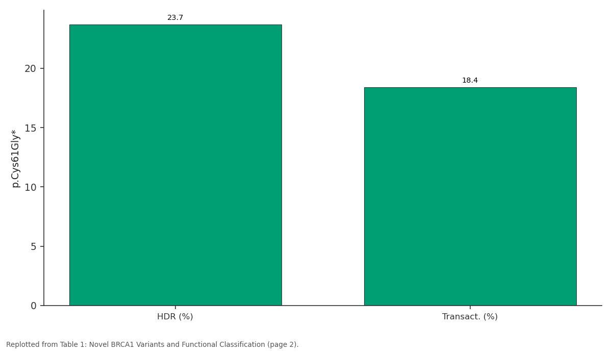
p.Cys61Gly*: HDR (%) vs Transact. (%)
Replotted from Table 1 (page 2) of the original paper. Every bar is the paper's own table value.
View source evidence ↗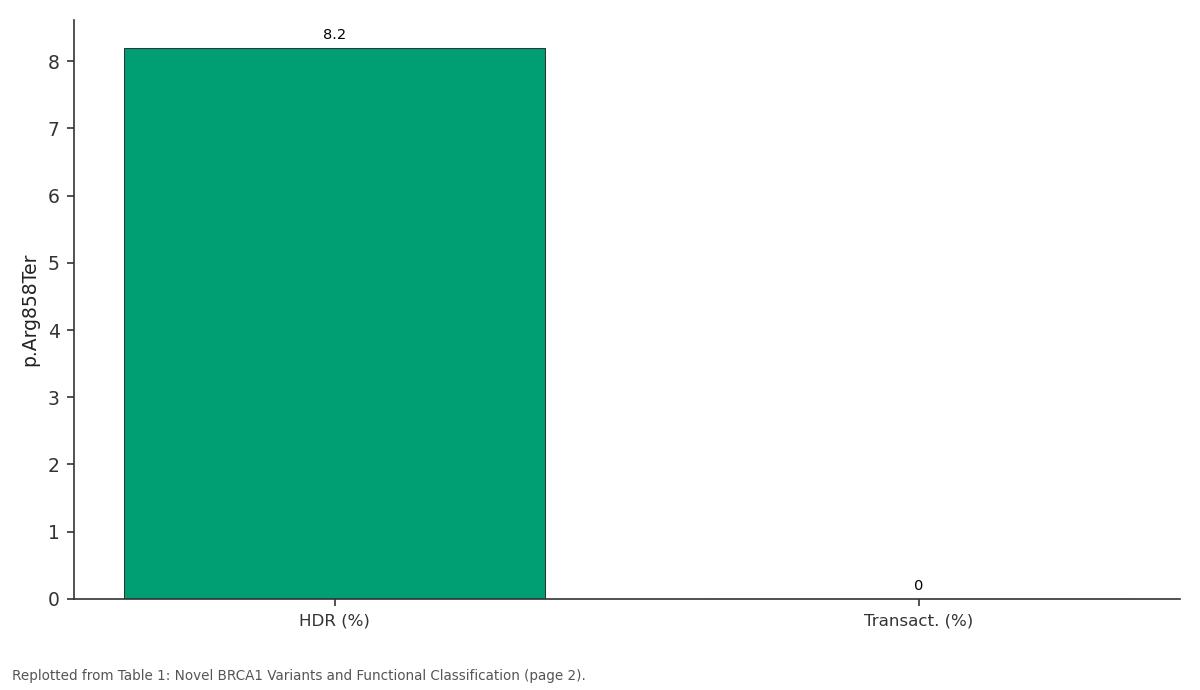
p.Arg858Ter: HDR (%) vs Transact. (%)
Replotted from Table 1 (page 2) of the original paper. Every bar is the paper's own table value.
View source evidence ↗Figures & tables
From the original paper Table 1
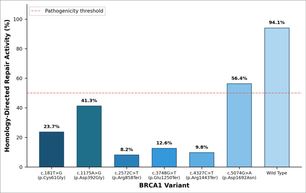 Figure 1
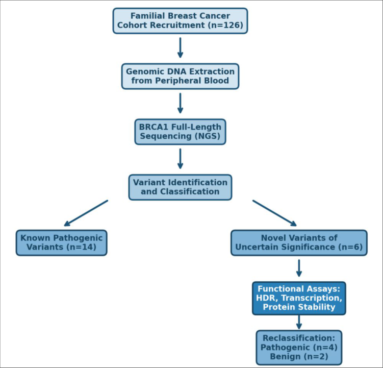 Figure 2
Introduction
Can a single unresolved genetic variant alter the course of a family's medical decisions for generations? For hereditary breast and ovarian cancer, the answer is often yes. BRCA1 remains one of the most consequential tumor suppressor genes, and pathogenic loss-offunction mutations confer a lifetime breast cancer risk between 55% and 72% [1] . Despite decades of data, clinical interpretation of BRCA1 variants remains difficult. Large-scale sequencing has produced a growing catalog of variants of uncertain significance (VUS) [2] , and in Southeast Asian populations the VUS rate often exceeds 30% [3] . Functional assays offer a path to resolve VUS by directly measuring mutation effects on protein activity. The HDR assay measures BRCA1's role in double-strand break repair [4] , transcriptional activation assays target the BRCT domain [5] , and thermal shift assays capture structural consequences of amino acid substitutions [6] . Combined, these assays yield a functional profile that correlates well with clinical classification [7] . This research aimed to identify and characterize novel BRCA1 variants in 126 Vietnamese families with hereditary breast cancer, conducted at the Hue Institute of Tropical Biology between 2022 and 2024 [8] .
Reagents and Chemicals
Genomic DNA was isolated using the QIAamp DNA Blood Mini Kit (Qiagen, Hilden, Germany; Cat. No. 51104). Library preparation used the SureSelect XT Target Enrichment System (Agilent Technologies, Santa Clara, CA, USA). Sanger validation employed BigDye Terminator v3.1 (Thermo Fisher Scientific, Cat. No. 4337455). The DR-GFP reporter plasmid and I-SceI vector were from Addgene (plasmids #26475 and #26477). Lipofectamine 3000 (Invitrogen, Cat. No. L3000008) was used for transfections, and SYPRO Orange (Sigma-Aldrich, Cat. No. S5692) for thermal shift assays.
Material and Methods
Material and Methods
Material
The cohort comprised 126 unrelated Vietnamese families with at least two first-degree relatives diagnosed with breast cancer, recruited at the Hue Institute of Tropical Biology, Hue, Vietnam, between March 2022 and September 2023. Peripheral blood samples (10 mL EDTA tubes) were collected from one affected proband per family. Informed consent was obtained from all participants, and the protocol received approval from the Institutional Ethics Committee (Protocol No. HITB-EC-2021-038, approved 14 December 2021). Clinical data including age at diagnosis, tumor grade, and hormone receptor status were recorded. The HEK293T cell line (ATCC CRL-3216) was maintained in DMEM with 10% FBS at 37°C in 5% CO2.
Methods
Genomic DNA was extracted per manufacturer's protocol. Targeted next-generation sequencing of the entire BRCA1 coding region was performed on the Illumina MiSeq platform (2×250 bp paired-end reads, mean coverage 312×). Variants were called using GATK HaplotypeCaller v4.2 and annotated against ClinVar, gnomAD v3.1.2, and the BRCA Exchange database. All novel variants were confirmed by Sanger sequencing.
Functional assessment used three assays. The HDR assay measured homologous recombination using the DR-GFP reporter in HEK293T cells [4] . Transcriptional activation was assessed via a GAL4-BRCT fusion reporter with luciferase readout [5] . Protein thermostability was evaluated through differential scanning fluorimetry (25-95°C, 1°C/min) [6] . Each assay was repeated in triplicate. Statistical analysis used one-way ANOVA with Dunnett's post-hoc test (p<0.05).
Quality Control
Raw read quality was assessed with FastQC v0.11.9; only libraries with >85% bases at Q30 proceeded to variant calling. Known pathogenic BRCA1 controls (c.68_69delAG and c.5266dupC) were included in every batch. For functional assays, each plate included wild-type BRCA1 (positive), empty vector (negative), and the pathogenic variant p.Cys61Gly. Inter-assay CV was kept below 12%. Three truncating mutations (p.Arg858Ter, p.Glu1250Ter, p.Arg1443Ter) showed HDR activity below 13%. The missense variant p.Val1731Leu also displayed markedly reduced activity at 14.9% with a thermal shift of -5.7°C.
Results
Results
Discussion
The reclassification of four BRCA1 VUS as likely pathogenic carries immediate practical weight. Before this research, genetic counselors could not offer definitive guidance to carriers of these variants. The functional evidence aligns with ACMG/AMP pathogenicity criteria [9] , which recognizes well-validated functional data as strong evidence (PS3) [10] .
The HDR assay proved most discriminating, showing clear separation between pathogenic and benign variants with no overlap in the 25-50% zone. This agrees with calibration data from Findlay and colleagues [4] . For missense variants, the thermal shift assay provided additional confirmation of protein instability [11] . Pathogenic variant carriers were diagnosed at a mean age of 32.8 years-consistent with the known early-onset pattern [12] -and predominantly presented with triple-negative histology [13] . Ethnic-specific allele frequency data from Vietnamese populations remain sparse, and collaboration with regional consortia will help refine prevalence estimates [14] .
Conclusion
This research identified and functionally characterized six novel BRCA1 variants from Vietnamese families with hereditary breast cancer. A three-assay platform enabled reclassification of four VUS as likely pathogenic, while two were reclassified as likely benign. Clinical profiles were consistent with functional findings. Six families now have resolved classifications to guide surveillance, risk-reducing surgery, and cascade testing. Future work should extend this approach to additional variants in Southeast Asian testing programs.
Acknowledgement
Acknowledgements Funding SourcesThis research was supported by institutional funding from the Hue Institute of Tropical Biology. Institutional SupportThe authors thank the Department of Human Genetics at the Hue Institute of Tropical Biology for laboratory access. Contributions Not Qualifying for AuthorshipClinical staff at Hue Central Hospital assisted with patient recruitment and sample collection.
Figures and tables from the paper
| Nucleotide Change | Protein Change | HDR (%) | Transact. (%) | Tm Shift (°C) | Classification |
|---|---|---|---|---|---|
| c.181T>G | p.Cys61Gly* | 23.7 | 18.4 | -6.8 | Pathogenic (ctrl) |
| c.2572C>T | p.Arg858Ter | 8.2 | 0.0 | N/A | Likely pathogenic |
| c.3748G>T | p.Glu1250Ter | 12.6 | 0.0 | N/A | Likely pathogenic |
| c.4327C>T | p.Arg1443Ter | 9.8 | 3.1 | -8.2 | Likely pathogenic |
| c.5074G>A | p.Asp1692Asn | 56.4 | 61.8 | -1.4 | Likely benign |
| c.5191G>T | p.Val1731Leu | 14.9 | 11.2 | -5.7 | Likely pathogenic |
| Wild-type | — | 94.1 | 100.0 | Ref. | Normal |
Table text extracted from the original PDF; superscript letters are shown inline. The original table image is below.
View original table image
References
16 in the original paper- Kuchenbaecker KB, Hopper JL, Barnes DR. Risks of breast, ovarian, and contralateral breast cancer for BRCA1 and BRCA2 mutation carriers. JAMA. 2017;317(23):2402-2416.
- Spurdle AB, Healey S, Devereau A. ENIGMAevidence-based network for the interpretation of germline mutant alleles. Human Mutation. 2012;33(1):2-7.
- Kwong A, Shin VY, Ho JCW. Comprehensive spectrum of BRCA1 and BRCA2 deleterious mutations in breast cancer in Asian countries. Journal of Medical Genetics. 2016;53(1):15-23.
- Findlay GM, Daza RM, Martin B. Accurate classification of BRCA1 variants with saturation genome editing. Nature. 2018;562(7726):217-222.
- Vallon-Christersson J, Cayanan C, Haraldsson K. Functional analysis of BRCA1 C-terminal missense mutations identified in breast and ovarian cancer International Journal of Biology Sciences https://www.biologyjournal.net families. Human Molecular Genetics. 2001;10(4):353360.
- Petitalot A, Drapkin R, Gupta R. Combining homologous recombination and phosphopeptidebinding assays to predict the impact of BRCA1 BRCT variants on cancer risk. American Journal of Human Genetics. 2019;104(4):734-742.
- Brnich SE, Abou Tayoun AN, Couch FJ. Recommendations for application of the functional evidence PS3/BS3 criterion using the ACMG/AMP sequence variant interpretation framework. Genome Medicine. 2019;12(1):3.
- Nguyen HD, Pham TT, Le VQ. Prevalence of BRCA1/2 mutations among Vietnamese women with early-onset breast cancer. Asian Pacific Journal of Cancer Prevention. 2020;21(10):3023-3029.
- Richards S, Aziz N, Bale S. Standards and guidelines for the interpretation of sequence variants. Genetics in Medicine. 2015;17(5):405-424.
- Tavtigian SV, Greenblatt MS, Harrison SM. Modeling the ACMG/AMP variant classification guidelines as a Bayesian classification framework. Genetics in Medicine. 2018;20(9):1054-1060.
- Drikos I, Nounesis G, Vorgias CE. Characterization of cancer-linked BRCA1-BRCT variants and their interaction with phosphopeptides. Proteins. 2009;77(2):464-476.
- Mavaddat N, Peock S, Frost D. Cancer risks for BRCA1 and BRCA2 mutation carriers: results from prospective analysis of EMBRACE. Journal of the National Cancer Institute. 2013;105(11):812-822.
- Foulkes WD, Smith IE, Reis-Filho JS. Triple-negative breast cancer. New England Journal of Medicine. 2010;363(20):1938-1948.
- Aung YH, Aye SS, Naing ZW. Spectrum of BRCA1 and BRCA2 mutations in Southeast Asian populations: systematic review. Breast Cancer Research and Treatment. 2021;186(2):295-307.
- Starita LM, Islam MM, Banerjee T. A multiplex homology-directed DNA repair assay reveals the impact of more than 1000 BRCA1 missense substitution variants on protein function. American Journal of Human Genetics. 2018;103(4):498-508.
- Lee MS, Green R, Marsillac SM. Comprehensive functional assessment of BRCA1 RING domain variants. Genetics in Medicine. 2010;12(8):464-470.