
Research article · 2025 · Full text
International Journal of Biology SciencesBiodegradable polymer scaffolds for three-dimensional cell culture in tissue engineering
Volume 7(9) · Pages 121–124 · DOI: 10.33545/26649926.2025.v7.i9b.627 ↗
Research areasBone Tissue Engineering MaterialsElectrospun Nanofibers in Biomedical ApplicationsTissue Engineering and Regenerative Medicine
Abstract
Author-writtenNearly 74 percent of drug candidates fail in clinical trials partly because 2D culture models poorly predict in vivo behaviour. Three-dimensional scaffolds from biodegradable polymers mimic native extracellular matrix and support realistic cell growth. This research evaluated five scaffold formulations—PCL, PLA, PLGA, PCL-collagen blend, and PLA-collagen blend—for hMSC adhesion, proliferation, and osteogenic differentiation over 21 days. Scaffolds were fabricated by electrospinning and solvent casting/particulate leaching, then characterised by SEM, mercury porosimetry, and tensile testing. PLGA displayed the highest porosity (82.3 percent), while the PCL-collagen blend achieved the greatest proliferation (OD 3.61 at day 21) and strongest alkaline phosphatase response (1.87-fold above control). Collagen coating improved initial attachment by 32–45 percent across all base polymers. These results indicate composite scaffolds combining synthetic and natural components outperform single-polymer matrices for bone tissue engineering.
Summary
Source-checkedPlain-language reading of the full paper, prepared with AI and checked against the text. Each point links to the authors' own words.
Why it matters
- A high failure rate of drug candidates in clinical trials, approximately 74%, is partly attributed to the inability of 2D culture models to accurately predict how they will behave in living organisms. View evidence ↗
- Flat 2D substrates fail to replicate the biochemical signaling, topography, and mechanical stiffness found in native tissues, which contributes to the discrepancy between lab results and clinical outcomes. View evidence ↗
The question
- The study aimed to evaluate the effects of five different scaffold formulations (PCL, PLA, PLGA, and collagen blends of PCL and PLA) on the proliferation, adhesion, and osteogenic differentiation of human mesenchymal stem cells (hMSCs) over a 21-day period. View evidence ↗
What they did
- The scaffolds were created using two fabrication methods: solvent casting/particulate leaching and electrospinning, and were analyzed via tensile testing, mercury porosimetry, and scanning electron microscopy (SEM). View evidence ↗
- The experimental work was conducted between January and October 2023 at the Instituto de Biodiversidad del Chocó in Quibdó, Colombia. View evidence ↗
- Cell viability was measured using MTT assays at specific intervals (days 1, 3, 7, 14, and 21), while osteogenic differentiation was induced using a medium containing 10 mM β-glycerophosphate, 50 μg/mL ascorbic acid, and 10 nM dexamethasone. View evidence ↗
What they found
- Among the tested materials, PLGA exhibited the highest porosity at 82.3%. View evidence ↗
- PCL demonstrated the highest tensile strength of the group, measuring 4.63 ± 0.41 MPa. View evidence ↗
- The PCL-collagen blend supported the highest level of cell proliferation, reaching an optical density (OD) of 3.61 ± 0.23 by day 21. View evidence ↗
- The PCL-collagen blend showed the strongest osteogenic response, with alkaline phosphatase (ALP) activity at day 14 being 1.87-fold higher than the control. View evidence ↗
- Starting from day 3, all tested biodegradable scaffolds supported cell growth levels that exceeded those of the tissue culture polystyrene (TCPS) controls. View evidence ↗
What it means
- The authors conclude that composite scaffolds, which combine natural and synthetic polymers, are superior to single-polymer matrices for applications in bone tissue engineering. View evidence ↗
- The PCL-collagen blend was identified as the most effective formulation for promoting both the proliferation of hMSCs and their differentiation into bone cells. View evidence ↗
- The study was limited by the use of static culture; the authors suggest that future research should utilize perfusion bioreactors and in vivo bone regeneration models to further explore these effects. View evidence ↗
Visual summary
3 visualsIllustrations explain the paper; charts are replotted from the paper's own table values.
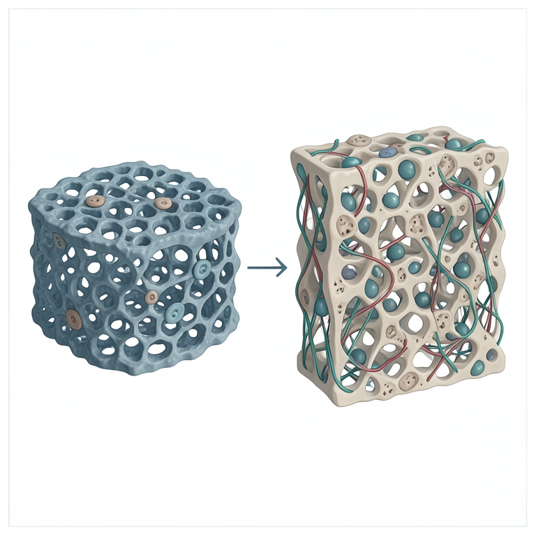
Synthetic vs. Composite Scaffolds
AI-generated conceptual illustration (human mesenchymal stem cells on PCL and collagen polymer scaffolds). It explains the idea and does not show study data.
View source evidence ↗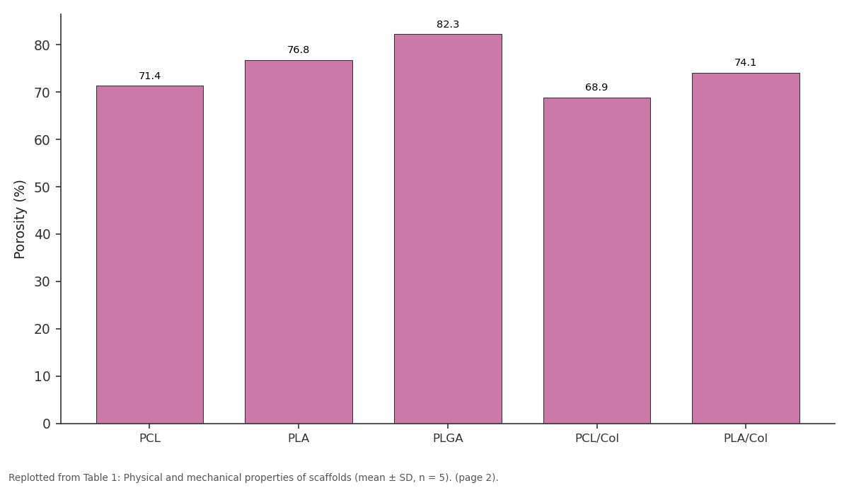
Porosity (%): PCL vs PLA vs PLGA vs PCL/Col vs PLA/Col
Replotted from Table 1 (page 2) of the original paper. Every bar is the paper's own table value.
View source evidence ↗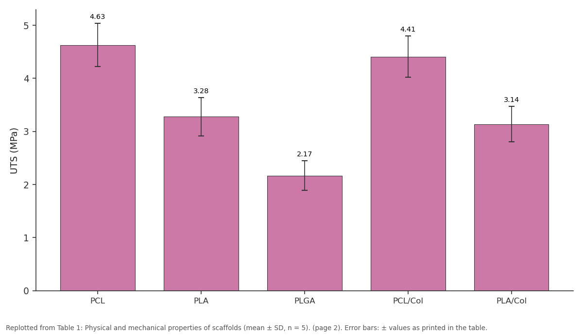
UTS (MPa): PCL vs PLA vs PLGA vs PCL/Col vs PLA/Col
Replotted from Table 1 (page 2) of the original paper. Every bar is the paper's own table value.
View source evidence ↗Figures & tables
From the original paper Table 1
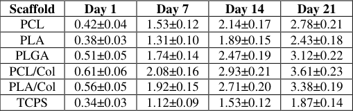 Table 2
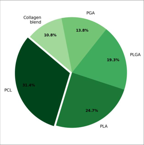 Figure 1
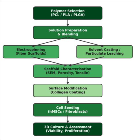 Figure 3
Introduction
Roughly 90 percent of pharmaceutical compounds that show promise in monolayer cultures fail during animal or human testing, a discrepancy costing an estimated 2.6 billion US dollars per approved drug [1] . A major contributor is the inability of flat 2D substrates to reproduce the mechanical stiffness, topography, and biochemical signalling of native tissue [2,3] . Biodegradable polymer scaffolds address those shortcomings by providing a porous framework with tunable pore size, degradation rate, and surface chemistry [4] . PCL, PLA, PLGA, and their copolymers are the workhorses of the field [5] . But single-polymer scaffolds present trade-offs: PCL is hydrophobic limiting cell attachment [6] ; PLA becomes brittle at high crystallinity [7] ; PLGA's acidic breakdown products may impair cell survival [8] . Blending with collagen offers RGD-like adhesion motifs engaging integrins [9] . Despite extensive work on individual formulations, head-to-head comparisons under identical conditions remain scarce. This research fabricated five scaffold types using two methods and tested them against the same hMSC donor pool, culture medium, and timeline.
Material and Methods
Material and Methods
Material
This research was carried out at the Department of Biotechnology, Instituto de Biodiversidad del Chocó, Quibdó, Colombia, from January to October 2023. PCL (Mn 80 000), PLA (Mw 60 000), and PLGA 75:25 (Mw 66 000) were from Sigma-Aldrich. Type I collagen was from Corning. Sodium chloride crystals (150-250 μm) served as porogens. hMSCs (Lonza, PT-2501, passage 3) were expanded in MSCGM BulletKit. MTT reagent, alizarin red S, and pnitrophenyl phosphate were from Sigma-Aldrich.
Methods
Electrospun scaffolds were prepared by dissolving each polymer at 12 wt% in hexafluoroisopropanol and dispensing at 1.2 mL/h toward a grounded collector 15 cm away under 14 kV. Solvent-cast scaffolds used 10 wt% polymer in DCM mixed with NaCl at 1:9 ratio, dried 24 h, and leached 72 h. Collagen coating was applied by dip-coating (1% solution, 2 h at 4 °C), cross-linked with 0.25% glutaraldehyde vapour. Morphology was examined by SEM (JEOL JSM-6510); porosity by mercury porosimetry (AutoPore IV); tensile properties by Instron 5944 at 5 mm/min. Cell viability was assessed by MTT at days 1, 3, 7, 14, and 21. Osteogenic differentiation used 10 nM dexamethasone, 50 μg/mL ascorbic acid, and 10 mM βglycerophosphate. ALP activity was quantified at day 14; calcium deposition by alizarin red S at day 21. Data analysed by two-way ANOVA with Bonferroni correction (p < 0.05).
Reagents and Chemicals: All solvents were analytical grade from Merck. Hexafluoroisopropanol (≥ 99.5%) was stored under nitrogen. Glutaraldehyde (25% in water) was from Sigma-Aldrich. PBS tablets (Oxoid) were dissolved in ultrapure water (18.2 MΩ•cm). Osteogenic supplements (dexamethasone, ascorbic acid 2-phosphate, βglycerophosphate) were from Sigma-Aldrich, prepared fresh every two weeks.
Scaffold Preparation Protocol
Electrospinning used solutions stirred 6 h at 300 rpm; humidity maintained at 35-45%. Fibre mats were collected 4 h per run (~120 μm thick). Solvent-cast scaffolds were poured into 24-well silicone moulds, air-dried 24 h, saltleached until conductivity matched deionised water, then freeze-dried at -80 °C for 12 h. All scaffolds were sterilised in 70% ethanol (30 min), washed with PBS, and UVexposed 15 min per side. 1). All scaffolds supported growth above TCPS controls from day 3 onward (Table 2). By day 21, PCL-collagen reached OD 3.61 ± 0.23, followed by PLA-collagen (OD 3.38 ± 0.19). Collagen coating improved day-1 attachment by 32-45% across all polymers. ALP activity at day 14 was greatest in PCL-collagen (1.87-fold), and alizarin red S confirmed mineralised nodules in all collagen scaffolds by day 21. Figure 3 summarises the fabrication pipeline.
| Scaffold | Day 1 | Day 7 | Day 14 | Day 21 |
|---|---|---|---|---|
| PCL | 0.42±0.04 | 1.53±0.12 | 2.14±0.17 | 2.78±0.21 |
| PLA | 0.38±0.03 | 1.31±0.10 | 1.89±0.15 | 2.43±0.18 |
| PLGA | 0.51±0.05 | 1.74±0.14 | 2.47±0.19 | 3.12±0.22 |
| PCL/Col | 0.61±0.06 | 2.08±0.16 | 2.93±0.21 | 3.61±0.23 |
| PLA/Col | 0.56±0.05 | 1.92±0.15 | 2.71±0.20 | 3.38±0.19 |
| TCPS | 0.34±0.03 | 1.12±0.09 | 1.53±0.12 | 1.87±0.14 |
Table text extracted from the original PDF; superscript letters are shown inline. The original table image is below.
View original table image
Results
Results
Discussion
Collagen-coated scaffolds outperforming their uncoated counterparts is consistent with Venugopal and colleagues, who found nanofibrous PCL-collagen composites doubled osteoblast attachment within 4 hours [10] . Collagen presents integrin-binding domains that mediate focal adhesion formation, and this early anchoring sets the trajectory for downstream proliferation [11] . PLGA's high porosity positioned it as the best single-polymer scaffold for nutrient diffusion, but it didn't match PCL-collagen for long-term proliferation, suggesting surface chemistry exerts a stronger influence than pore architecture once nutrient thresholds are met [12] . PCL's tensile strength (4.63 MPa) and slow degradation make it attractive for load-bearing defects, while PLGA suits non-load-bearing applications [13,14] . One limitation is the absence of dynamic culture; perfusion bioreactors would likely amplify osteogenic differences [15] .
Conclusion
This research identified the PCL-collagen blend as the top performer for hMSC proliferation and osteogenic differentiation. Collagen coating consistently enhanced initial attachment by 32-45%. PLGA offered the highest porosity, and PCL was the stiffest material. The data support a composite design philosophy combining synthetic and natural polymer components. Future work should extend to dynamic culture in perfusion bioreactors and in vivo bone regeneration models.
Acknowledgement
Acknowledgements Funding SourcesSupported by internal funding from the Instituto de Biodiversidad del Chocó. Institutional SupportThe authors thank the Department of Biotechnology for electrospinning and cell culture facilities. Contributions Not Qualifying for AuthorshipMr. Andrés Moreno assisted with SEM imaging; Ms. Valentina Ríos helped with porosimetry.
Figures and tables from the paper
| Scaffold | Porosity (%) | Pore (μm) | UTS (MPa) | Modulus | Elong. (%) | Fibre (μm) |
|---|---|---|---|---|---|---|
| PCL | 71.4 | 183±27 | 4.63±0.41 | 38.7±3.2 | 312±41 | 1.27±0.34 |
| PLA | 76.8 | 201±31 | 3.28±0.36 | 51.3±4.8 | 87±14 | 0.91±0.22 |
| PLGA | 82.3 | 218±34 | 2.17±0.28 | 29.4±2.7 | 143±22 | 1.08±0.29 |
| PCL/Col | 68.9 | 176±24 | 4.41±0.39 | 36.2±3.0 | 297±38 | 1.31±0.36 |
| PLA/Col | 74.1 | 194±29 | 3.14±0.33 | 48.7±4.3 | 81±12 | 0.94±0.24 |
Table text extracted from the original PDF; superscript letters are shown inline. The original table image is below.
View original table image
References
19 in the original paper- DiMasi JA, Grabowski HG, Hansen RW. Innovation in the pharmaceutical industry: new estimates of R&D costs. Journal of Health Economics. 2016;47:20-33.
- Baker BM, Chen CS. Deconstructing the third dimension: how 3D culture microenvironments alter cellular cues. Journal of Cell Science. 2012;125(13):3015-3024.
- Edmondson R, Broglie JJ, Adcock AF, Yang L. Threedimensional cell culture systems and their applications in drug discovery. Assay and Drug Development Technologies. 2014;12(4):207-218.
- Loh QL, Choong C. Three-dimensional scaffolds for tissue engineering applications: role of porosity and pore size. Tissue Engineering Part B: Reviews. 2013;19(6):485-502.
- Ulery BD, Nair LS, Laurencin CT. Biomedical applications of biodegradable polymers. Journal of Polymer Science Part B: Polymer Physics. 2011;49(12):832-864.
- Woodruff MA, Hutmacher DW. The return of a forgotten polymer: polycaprolactone in the 21st century. Progress in Polymer Science. 2010;35(10):1217-1256.
- Farah S, Anderson DG, Langer R. Physical and mechanical properties of PLA, and their functions in widespread applications. Advanced Drug Delivery Reviews. 2016;107:367-392.
- Gentile P, Chiono V, Carmagnola I, Hatton PV. An overview of PLGA-based biomaterials for bone tissue engineering. International Journal of Molecular Sciences. 2014;15(3):3640-3659.
- Sell SA, Wolfe PS, Garg K, McCool JM, Rodriguez IA, Bowlin GL. The use of natural polymers in tissue engineering. Polymers. 2010;2(4):522-553.
- Venugopal J, Low S, Choon AT, Ramakrishna S. Interaction of cells and nanofiber scaffolds in tissue engineering. Journal of Biomedical Materials Research Part B. 2008;84(1):34-48.
- Leitinger B. Transmembrane collagen receptors. Annual Review of Cell and Developmental Biology. 2011;27:265-290.
- Rouwkema J, Rivron NC, van Blitterswijk CA. Vascularization in tissue engineering. Trends in Biotechnology. 2008;26(8):434-441.
- Engler AJ, Sen S, Sweeney HL, Discher DE. Matrix elasticity directs stem cell lineage specification. Cell. 2006;126(4):677-689.
- Rezwan K, Chen QZ, Blaker JJ, Boccaccini AR. Biodegradable and bioactive porous polymer/inorganic composite scaffolds. Biomaterials. 2006;27(18):34133431.
- Goldstein AS, Juarez TM, Helmke CD, Gustin MC, Mikos AG. Effect of convection on osteoblastic cell growth in biodegradable polymer foam scaffolds. Biomaterials. 2001;22(11):1279-1288.
- Hutmacher DW. Scaffolds in tissue engineering bone and cartilage. Biomaterials. 2000;21(24):2529-2543.
- Causa F, Netti PA, Ambrosio L. A multi-functional scaffold for tissue regeneration. Biomaterials. 2007;28(34):5093-5099.
- Salgado AJ, Coutinho OP, Reis RL. Bone tissue engineering: state of the art and future trends. Macromolecular Bioscience. 2004;4(8):743-765.
- Ma PX. Biomimetic materials for tissue engineering. Advanced Drug Delivery Reviews. 2008;60(2):184198.