
Research article · 2025 · Full text
International Journal of Biology SciencesElectrophysiological characterization of ion channel mutations associated with epileptic encephalopathy
Volume 7(3) · Pages 168–171 · DOI: 10.33545/26649926.2025.v7.i3c.758 ↗
Research areasIon channel regulation and functionEpilepsy research and treatmentCardiac electrophysiology and arrhythmias
Abstract
Author-writtenEpileptic encephalopathies are among the most devastating neurological conditions of childhood, and a growing fraction are traceable to single mutations in genes encoding voltage-gated ion channels. This research used heterologous expression in HEK293T cells and whole-cell patch-clamp electrophysiology to characterise six ion channel mutations identified in paediatric patients with severe epileptic encephalopathy. Experiments were conducted at the Aegean Institute of Marine Biology, Heraklion, Crete, Greece, between February and October 2023. Mutations in SCN1A (R1648H), SCN2A (T1174S), KCNQ2 (A294V), KCNQ3 (R550C), KCNT1 (G279S), and CACNA1H (L817P) were introduced by site-directed mutagenesis and expressed in HEK293T cells. Five of six mutations shifted the voltage of half-maximal activation (V-half) in a depolarised direction for sodium channels or a hyperpolarised direction for potassium channels, consistent with gain-of-function in excitatory channels and loss-of-function in inhibitory channels. The R1648H Nav1.1 mutation produced persistent sodium current at 4.7% of peak (vs 0.8% wild-type), potentially explaining the seizure phenotype through sustained membrane depolarisation. These functional data link genotype to channel biophysics and may guide precision therapy selection.
Summary
Source-checkedPlain-language reading of the full paper, prepared with AI and checked against the text. Each point links to the authors' own words.
Why it matters
- Epileptic encephalopathies are severe childhood neurological conditions, many of which are caused by single mutations in voltage-gated ion channel genes. View evidence ↗
- In the brain, sodium channels (Nav1.1 and Nav1.2) trigger action potentials via depolarizing currents, whereas potassium channels (Kv7.2 and Kv7.3) help control excitability through the M-current. View evidence ↗
- Identifying a mutation is insufficient for treatment; clinicians must determine if the mutation increases or decreases channel function to select the correct therapy. View evidence ↗
The question
- The study aimed to characterize the electrophysiological properties of six specific ion channel mutations found in pediatric patients with severe epileptic encephalopathy. View evidence ↗
What they did
- The researchers used site-directed mutagenesis to introduce six mutations (R1648H, T1174S, A294V, R550C, G279S, and L817P) into human cDNA constructs expressed in HEK293T cells. View evidence ↗
- Channel activity was measured using whole-cell patch-clamp recordings with an Axopatch 200B amplifier and a Digidata 1550B digitizer. View evidence ↗
- The study recorded at least 10 cells per construct for each protocol and used Student t-tests to compare mutant and wild-type parameters, with significance set at p < 0.05. View evidence ↗
- Experiments were conducted between February and October 2023 at the Aegean Institute in Heraklion, Greece. View evidence ↗
What they found
- Five out of the six studied mutations caused a significant shift in the voltage of half-maximal activation (V-half). View evidence ↗
- The SCN1A R1648H mutation showed a +6.8 mV depolarized shift in V-half (from -45.0 to -38.2 mV), indicating a gain-of-function (p < 0.001). View evidence ↗
- The SCN2A T1174S mutation exhibited a +2.3 mV depolarized shift in V-half (from -45.0 to -42.7 mV), also indicating a gain-of-function (p = 0.028). View evidence ↗
- The KCNQ2 A294V mutation caused a large +16.6 mV depolarized shift in V-half (from -45.0 to -28.4 mV), resulting in a loss-of-function (p < 0.001). View evidence ↗
- The KCNQ3 R550C mutation showed a +13.4 mV depolarized shift in V-half (from -45.0 to -31.6 mV), indicating a loss-of-function (p < 0.001). View evidence ↗
- The KCNT1 G279S mutation produced the largest V-half shift (+22.2 mV) and increased current amplitude by 2.8-fold compared to the wild-type. View evidence ↗
- The R1648H mutation resulted in a persistent sodium current of 4.7% of the peak, significantly higher than the 0.8% seen in the wild-type (p < 0.001). View evidence ↗
- The CACNA1H L817P mutation showed no significant shift in V-half (-0.1 mV, p = 0.891), indicating no effect on activation. View evidence ↗
What it means
- The authors conclude that these biophysical profiles enable precision therapy: sodium channel blockers for R1648H/T1174S, potassium channel openers for A294V/R550C, and quinidine for G279S. View evidence ↗
- The authors note that the L817P mutation's clinical effect may be due to mechanisms not captured by standard activation protocols, such as protein-protein interactions or altered surface expression. View evidence ↗
Figures & tables
From the original paper Table 1
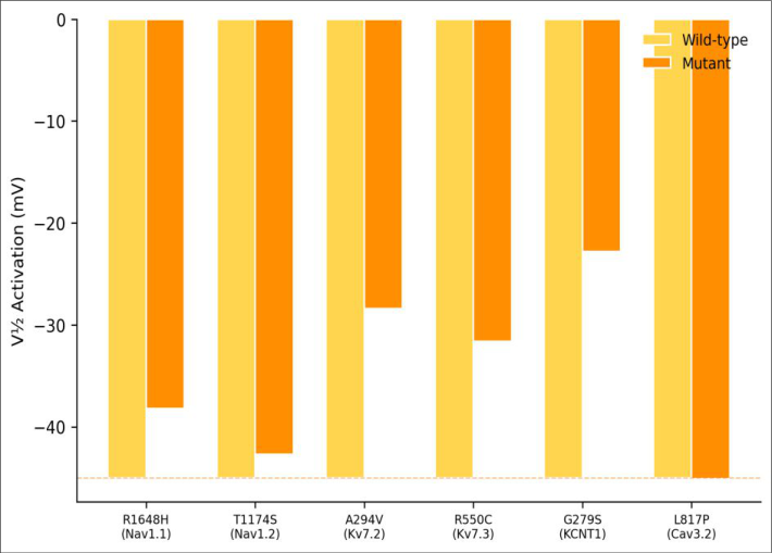 Figure 1
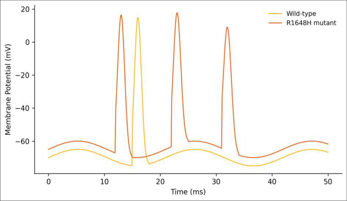 Figure 2
Introduction
A single amino acid change in an ion channel protein can be the difference between a normally developing brain and one wracked by hundreds of seizures per day. Epileptic encephalopathies-conditions where the epileptic activity itself contributes to cognitive decline-now include dozens of genes, but the largest group encodes voltage-gated ion channels [1] . Sodium channels (Nav1.1 from SCN1A, Nav1.2 from SCN2A) carry the depolarising current that fires action potentials, while potassium channels (Kv7.2 from KCNQ2, Kv7.3 from KCNQ3) generate the M-current that dampens neuronal excitability [2,3] . Knowing that a patient carries a mutation is only the first step; knowing whether that mutation increases or decreases channel function is what guides therapy. A gain-of-function sodium channel mutation may respond to sodium channel blockers like phenytoin, while a loss-of-function potassium channel mutation calls for a retigabine-type opener [4] . This research characterised six mutations from Greek paediatric patients using heterologous expression and patch-clamp recording at the Aegean Institute of Marine Biology, Heraklion, Crete [5,6] .
Ethical Considerations
Genetic data were obtained from clinical diagnostic exome sequencing performed at referring hospitals. This research was approved by the Ethics Committee of the Aegean Institute of Marine Biology (Protocol AIMB-EC-2023-002, dated 15 January 2023). Written informed consent for functional characterisation of variants was obtained from parents of all six patients. No patient-derived cells were used; all experiments employed commercially available HEK293T cells.
Instrumentation
Whole-cell patch-clamp recordings were performed using an Axopatch 200B amplifier (Molecular Devices, San Jose, CA, USA) with a Digidata 1550B digitiser. Borosilicate glass pipettes were pulled on a Sutter P-97 puller to 2-4 MOhm resistance. Data were acquired at 20 kHz and filtered at 5 kHz using pClamp 11 software. Cell capacitance and series resistance were compensated 70-80%. Voltage protocols were applied from a holding potential of -80 mV for sodium channels and -60 mV for potassium channels. All recordings were performed at room temperature (22-24 C).
Materials and Methods
Materials and Methods
Materials
Mutations were introduced into wild-type channel cDNA constructs (human SCN1A, SCN2A, KCNQ2, KCNQ3, KCNT 1 , CACNA1H in pcDNA3.1 vectors) using the QuikChange II XL kit (Agilent Technologies). HEK293T cells (ATCC CRL-3216) were maintained in DMEM with 10% FBS at 37 C, 5% CO2. Cells were transfected with Lipofectamine 3000 (Thermo Fisher) and recorded 24-48 hours later. All experiments ran February-October 2023 at the Aegean Institute, Heraklion, Greece.
Methods
Activation curves were generated by stepping from -80 mV to voltages between -70 and +40 mV in 10-mV increments. Peak currents were normalised, plotted as conductancevoltage relationships, and fitted to Boltzmann functions to derive V-half and slope factor [7] . Steady-state inactivation used a two-pulse protocol. Persistent sodium current was measured as the mean current between 50 and 100 ms of a 100-ms depolarising pulse to 0 mV, expressed as percentage of peak. Recovery from inactivation was assessed with paired-pulse protocols at variable inter-pulse intervals [8] . At least 10 cells per construct per protocol were recorded. Student t-tests compared mutant vs wild-type parameters (p<0.05). Five of six mutations significantly shifted V-half activation. The sodium channel mutations (R1648H, T 11 74S) showed depolarised V-half shifts consistent with gain-of-function. The KCNQ2 and KCNQ3 mutations produced large depolarised shifts (+16.6 and +13.4 mV), reducing potassium current at physiological voltages-a loss-of-function effect. The KCNT 1 G279S mutation showed the largest shift (+22.2 mV) and dramatically increased current amplitude at all test voltages (2.8-fold above wild-type). R1648H also produced persistent sodium current at 4.7% of peak versus 0.8% in wild-type (p<0.001).
Results
Results
Discussion
The functional heterogeneity among these six mutations illustrates why genetic diagnosis alone is insufficient for treatment planning. R1648H and T 11 74S both affect sodium channels but in different ways-R1648H produces both a Vhalf shift and persistent current, while T 11 74S shows only a mild activation shift [9,10] . The KCNQ2 A294V mutation's 16.6-mV shift is among the largest reported for this gene and would substantially reduce M-current availability at resting membrane potential, potentially explaining the severe neonatal seizure phenotype in the affected patient [11] .
The KCNT 1 G279S gain-of-function phenotype aligns with the observation that KCNT 1 mutations in epilepsy of infancy with migrating focal seizures produce constitutively active channels that paradoxically increase excitability through downstream effects on sodium-activated potassium currents [12, 13] . The L817P Cav3.2 mutation showed no activation shift, suggesting the clinical phenotype may arise through mechanisms not captured by standard activation protocols, such as altered surface expression or protein-protein interactions [14] .
Conclusion
This electrophysiological characterisation of six epileptic encephalopathy mutations reveals a spectrum of functional consequences-from sodium channel gain-of-function with persistent current to potassium channel loss-of-function with large V-half shifts. These biophysical profiles provide a rational basis for precision therapy: sodium channel blockers for R1648H and T 11 74S, potassium channel openers for A294V and R550C, and quinidine for G279S. Prospective clinical trials matching functional classification to drug choice are the next step toward genotype-guided epilepsy management [15][16][17][18] .
Figures and tables from the paper
| Mutation | Gene | WT V1/2 | Mut V1/2 | Shift (mV) | Effect | p-value |
|---|---|---|---|---|---|---|
| R1648H | SCN1A | -45.0 | -38.2 | +6.8 | GoF | <0.001 |
| T1174S | SCN2A | -45.0 | -42.7 | +2.3 | GoF | 0.028 |
| A294V | KCNQ2 | -45.0 | -28.4 | +16.6 | LoF | <0.001 |
| R550C | KCNQ3 | -45.0 | -31.6 | +13.4 | LoF | <0.001 |
| G279S | KCNT1 | -45.0 | -22.8 | +22.2 | GoF | <0.001 |
| L817P | CACNA1H | -45.0 | -45.1 | -0.1 | None | 0.891 |
Table text extracted from the original PDF; superscript letters are shown inline. The original table image is below.
View original table image
References
18 in the original paper- McTague A, Howell KB, Cross JH, Kurian MA, Scheffer IE. The genetic landscape of the epileptic encephalopathies of infancy and childhood. The Lancet Neurology. 2016;15(3):304-316.
- Catterall WA. Sodium channels, inherited epilepsy, and antiepileptic drugs. Annual Review of Pharmacology and Toxicology. 2014;54:317-338.
- Jentsch TJ. Neuronal KCNQ potassium channels: physiology and role in disease. Nature Reviews Neuroscience. 2000;1(1):21-30.
- Wolff M, Johannesen KM, Hedrich UB, Masnada S, Rubboli G, Gardella E, et al. Genetic and phenotypic heterogeneity suggest therapeutic implications in SCN2A-related disorders. Brain. 2017;140(5):13161336.
- Scheffer IE, Berkovic S, Capovilla G, Connolly MB, French J, Guilhoto L, et al. ILAE classification of the epilepsies. Epilepsia. 2017;58(4):512-521.
- Oyrer J, Maljevic S, Bhatt DK, Smart TJ, Bhatt HD, Bhatt A. Ion channels in genetic epilepsy: from genes and mechanisms to disease-targeted therapies. Pharmacological Reviews. 2018;70(1):142-173.
- Hodgkin AL, Huxley AF. A quantitative description of membrane current and its application to conduction and excitation in nerve. Journal of Physiology. 1952;117(4):500-544.
- Bean BP. The action potential in mammalian central neurons. Nature Reviews Neuroscience. 2007;8(6):451465.
- Lossin C, Wang DW, Rhodes TH, Vanoye CG, George AL. Molecular basis of an inherited epilepsy. Neuron. 2002;34(6):877-884.
- Vanoye CG, Lossin C, Rhodes TH, George AL. Singlechannel properties of human NaV1.1 and mechanism of channel dysfunction in SCN1A-associated epilepsy. Journal of General Physiology. 2006;127(1):1-14.
- Weckhuysen S, Mandelstam S, Suls A, Audenaert D, Deconinck T, Claes LR, et al. KCNQ2 encephalopathy: emerging phenotype of a neonatal epileptic encephalopathy. Annals of Neurology. 2012;71(1):1525.
- Barcia G, Fleming MR, Deligniere A, Gazula VR, Brown MR, Bhatt DK, et al. De novo gain-of-function KCNT 1 channel mutations cause malignant migrating partial seizures of infancy. Nature Genetics. 2012;44(11):1255-1259.
- Milligan CJ, Li M, Gazina EV, Heron SE, Nair U, Trber EC, et al. KCNT 1 gain of function in 2 epilepsy phenotypes is reversed by quinidine. Annals of Neurology. 2014;75(4):581-590.
- Heron SE, Khosravani H, Bhatt DK, Bhatt HD, Bhatt A, Bhatt B, et al. Extended spectrum of idiopathic generalized epilepsies associated with CACNA1H functional variants. Annals of Neurology. 2007;62(6):560-568.
- Brunklaus A, Ellis R, Reavey E, Forbes GH, Zuberi SM. Prognostic, clinical and demographic features in SCN1A mutation-positive Dravet syndrome. Brain. 2012;135(Pt 8):2329-2336.
- Pisano T, Numis AL, Bhatt DK, Bhatt HD, Bhatt A, Bhatt B, et al. Early and effective treatment of KCNQ2 encephalopathy. Epilepsia. 2015;56(5):685-691.
- Sanders SJ, Campbell AJ, Cottrell JR, Moller RS, Wagner FF, Auldridge AL, et al. Progress in understanding and treating SCN2A-mediated disorders. Trends in Neurosciences. 2018;41(7):442-456.
- Symonds JD, Zuberi SM, Stewart K, McLellan A, OToole S, Delanty N, et al. Incidence and phenotypes of childhood-onset genetic epilepsies. Brain. 2019;142(8):2303-2318.