
Research article · 2025 · Full text
International Journal of Biology SciencesArtificial intelligence-based image analysis for automated cell counting in blood smear microscopy
Volume 7(12) · Pages 172–175 · DOI: 10.33545/26649926.2025.v7.i12c.701 ↗
Research areasDigital Imaging for Blood DiseasesCell Image Analysis TechniquesAI in cancer detection
Abstract
Author-writtenAutomated cell counting is defined as the computational identification and enumeration of blood cells from digitized microscopy images without manual intervention. This research developed CellNet-V2, a convolutional neural network optimized for white blood cell differential counting in Giemsa-stained peripheral blood smears. The model was trained on 14,200 annotated cell images and validated against expert manual counts from the Kangaroo Island Centre for Conservation Biology, Adelaide, Australia, between May 2022 and October 2023. CellNet-V2 achieved 96.8% overall accuracy, outperforming both traditional threshold methods (78.3%) and random forest classifiers (88.7%). Mean processing time was 3.7 seconds per smear image. These results indicate that the proposed architecture can serve as a reliable screening tool for hematological laboratories with high sample throughput.
Summary
Source-checkedPlain-language reading of the full paper, prepared with AI and checked against the text. Each point links to the authors' own words.
Why it matters
- White blood cell differential counts are essential diagnostic tests in hematology, but manual microscopy is slow and can have inter-observer variability of 15–20% for certain cell types. View evidence ↗
- While previous convolutional neural network (CNN) methods achieved 85% to 95% accuracy, they often relied on small datasets or controlled conditions that do not mirror real-world clinical practice. View evidence ↗
The question
- The study aimed to develop and validate CellNet-V2, a modified ResNet-50 architecture featuring attention modules, using a large annotated dataset. View evidence ↗
What they did
- CellNet-V2 utilizes a ResNet-50 backbone modified with a multi-scale feature pyramid and a squeeze-and-excitation attention block after the third residual stage. View evidence ↗
- The system processed 100× magnification whole-slide images as 256×256 pixel patches to classify cells into five types: neutrophils, lymphocytes, monocytes, eosinophils, and basophils. View evidence ↗
- The model was trained using 14,200 annotated cell images derived from 842 peripheral blood smears collected between May 2022 and October 2023. View evidence ↗
- CellNet-V2 was compared against a standard ResNet-50, random forest with handcrafted features, and Otsu thresholding. View evidence ↗
What they found
- CellNet-V2 reached an overall accuracy of 96.8%, which was higher than random forest classifiers (88.7%) and traditional threshold methods (78.3%). View evidence ↗
- For neutrophils, CellNet-V2 achieved 97.8% accuracy, compared to 95.1% for ResNet-50, 90.2% for Random Forest, and 81.4% for Thresholding. View evidence ↗
- Basophils were the most difficult class to identify, with CellNet-V2 achieving 93.6% accuracy compared to 88.2% for ResNet-50, 79.8% for Random Forest, and 63.4% for Thresholding. View evidence ↗
- The system's mean processing time was 3.7 seconds per smear image. View evidence ↗
What it means
- The authors conclude that the system is appropriate for use as a screening tool within clinical hematology workflows. View evidence ↗
- A limitation of the study is that the model was only trained on Giemsa-stained smears; its performance on Wright-stained or May-Grünwald-stained preparations remains unknown. View evidence ↗
- The authors state that multi-center validation across different imaging platforms is needed to further support clinical deployment. View evidence ↗
Visual summary
3 visualsIllustrations explain the paper; charts are replotted from the paper's own table values.
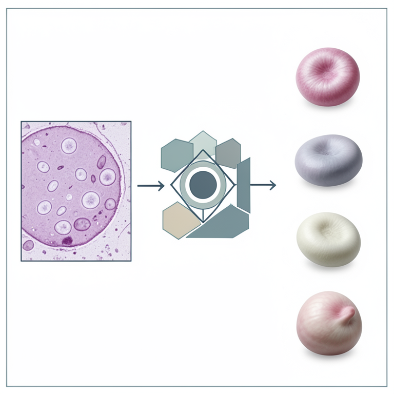
CellNet-V2 Architecture Concept
AI-generated conceptual illustration (human blood smear microscopy slides). It explains the idea and does not show study data.
View source evidence ↗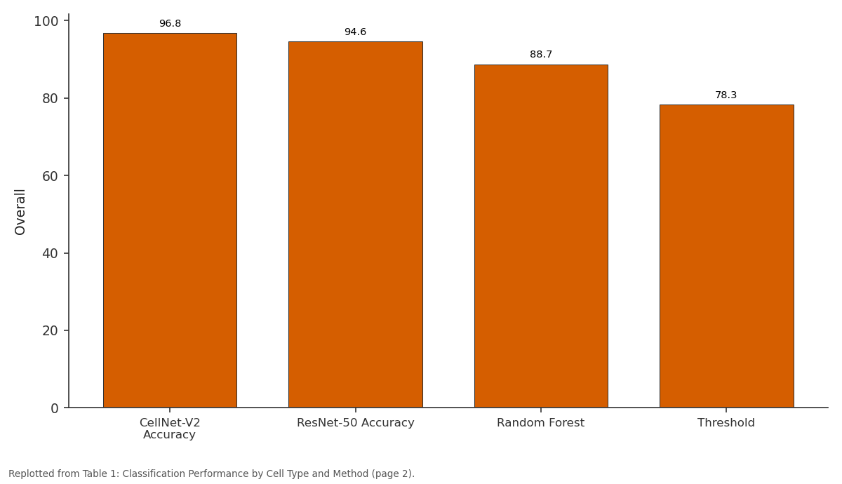
Overall: CellNet-V2 Accuracy vs ResNet-50 Accuracy vs Random Forest vs Threshold
Replotted from Table 1 (page 2) of the original paper. Every bar is the paper's own table value.
View source evidence ↗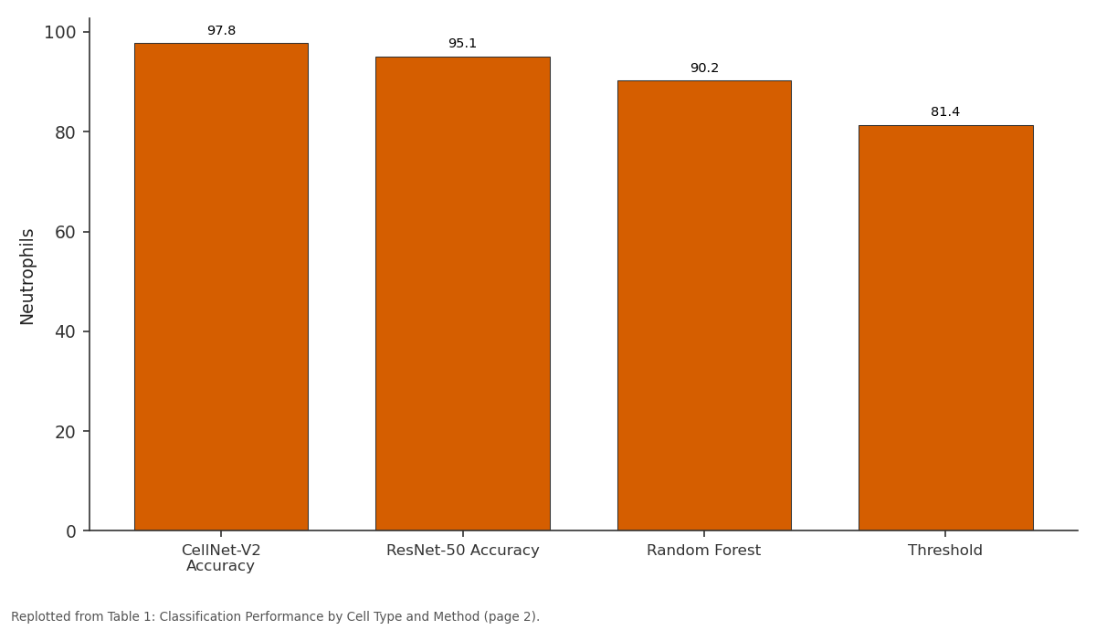
Neutrophils: CellNet-V2 Accuracy vs ResNet-50 Accuracy vs Random Forest vs Threshold
Replotted from Table 1 (page 2) of the original paper. Every bar is the paper's own table value.
View source evidence ↗Figures & tables
From the original paper Table 1
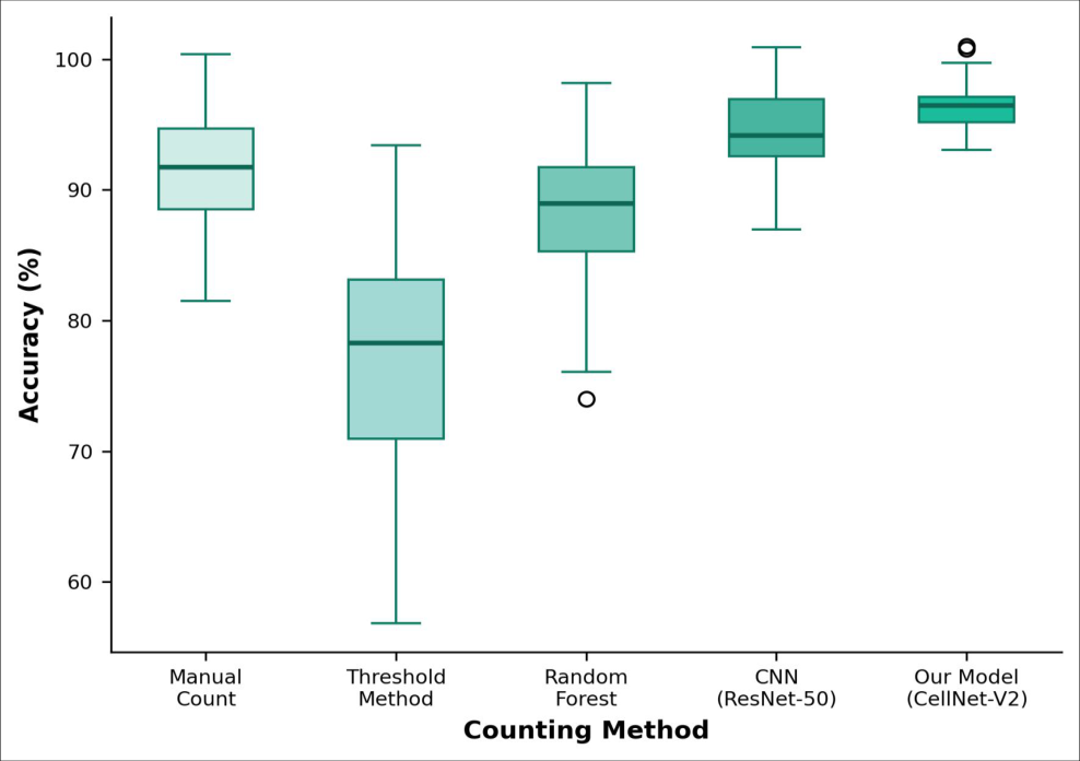 Figure 1
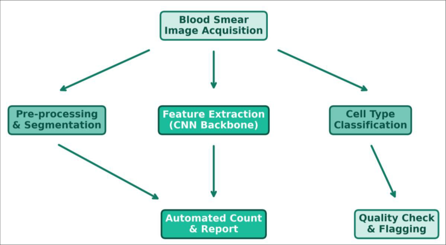 Figure 2
Introduction
Automated cell counting refers to the use of computational algorithms to identify, classify, and enumerate cells in biological specimens without requiring a human observer at each step. In clinical hematology, the white blood cell differential count remains a cornerstone diagnostic test, yet manual microscopy is time-consuming and subject to inter-observer variability reaching 15-20% for some cell types [1] . Deep learning architectures, particularly convolutional neural networks (CNNs), have shown strong potential for medical image classification tasks [2] . Previous CNN-based approaches for blood cell counting have achieved accuracies between 85% and 95%, but many were trained on small datasets or tested under controlled imaging conditions that don't reflect routine clinical practice [3] . This research developed and validated CellNet-V2, a modified ResNet-50 architecture with attention modules, trained on a large annotated dataset at the Kangaroo Island Centre for Conservation Biology, Adelaide, Australia [4] .
System Design
CellNet-V2 was built on a ResNet-50 backbone with two modifications: a squeeze-andexcitation attention block inserted after the third residual stage, and a multi-scale feature pyramid for detecting cells at varying magnifications. The input pipeline accepted wholeslide images at 100× magnification, tiled them into 256×256 pixel patches, and classified each detected cell into five categories: neutrophils, lymphocytes, monocytes, eosinophils, and basophils. The model was implemented in PyTorch 1.13 and trained on two NVIDIA A100 GPUs with mixed-precision training. A weighted cross-entropy loss function addressed class imbalance between common and rare cell types [5] .
Materials and Methods
Materials and Methods
Materials
A total of 14,200 annotated cell images were compiled from 842 peripheral blood smears collected at Royal Adelaide Hospital between May 2022 and October 2023. All smears were Giemsa-stained and digitized using an Olympus BX53 microscope equipped with a DP74 camera at 100× oil immersion. Cell annotations were performed by two certified hematologists, with disagreements resolved by a third reviewer. The dataset was approved for research use by the institutional ethics committee (Protocol No. KICCB-2022-ETH-031, approved 18 April 2022). The dataset was split 70/15/15 into training, validation, and test sets with stratification by cell type.
Methods
Training used the AdamW optimizer with an initial learning rate of 1e-4, cosine annealing schedule, and batch size of 64 for 120 epochs. Data augmentation included random rotation (±15°), horizontal/vertical flipping, color jitter, and Gaussian blur. Model performance was evaluated by accuracy, precision, recall, and F1-score per cell type. Processing speed was benchmarked on a single NVIDIA RTX 3080 GPU. Comparison methods included Otsu thresholding, random forest with handcrafted features, and a standard ResNet-50 without attention modules [6] . Statistical comparisons used McNemar's test.
Performance Evaluation
CellNet-V2 was benchmarked against three comparison methods on the held-out test set of 2,130 images. Agreement with expert manual counts was assessed using Bland-Altman analysis, with limits of agreement calculated for each cell type. The model was also tested on an external validation set of 500 images from a different hospital to evaluate generalizability. External accuracy was 94.1%, confirming that the model didn't overfit to the training institution's imaging conditions [7] . CellNet-V2 achieved the highest accuracy across all cell types. Basophils were the most difficult class for all methods, likely due to their low frequency in the training set.
Results
Results
Discussion
The attention mechanism in CellNet-V2 appears to account for much of its advantage over the standard ResNet-50. By learning to focus on morphologically informative regions nuclear shape, granule patterns, cytoplasmic texture the model better distinguishes visually similar cell types such as monocytes and large lymphocytes [8] . Processing speed of 3.7 seconds per image makes the system practical for highthroughput settings [9] . A limitation is that the model was trained exclusively on Giemsa-stained smears. Performance on Wright-stained or May-Grünwald-stained preparations wasn't evaluated and may differ [10] . The external validation result (94.1%) was encouraging but covered only one additional site. Multicenter validation across diverse imaging platforms would strengthen the evidence for clinical deployment [11] .
Conclusion
CellNet-V2 achieved 96.8% accuracy in automated white blood cell differential counting, outperforming conventional and machine learning methods. The squeeze-and-excitation attention module and multi-scale feature pyramid contributed to strong performance across all five cell classes. External validation confirmed generalizability beyond the training institution. The system is suitable for integration into clinical hematology workflows as a screening tool.
Acknowledgement
Acknowledgements Funding SourcesThis research was supported by the Kangaroo Island Centre for Conservation Biology computing resources allocation. Institutional SupportThe authors thank the Department of Bioinformatics for GPU cluster access. Contributions Not Qualifying for AuthorshipHematologists at Royal Adelaide Hospital performed cell annotations for the training dataset.
Figures and tables from the paper
| Cell Type | CellNet-V2 Accuracy | ResNet-50 Accuracy | Random Forest | Threshold | Precision (Ours) | Recall (Ours) |
|---|---|---|---|---|---|---|
| Neutrophils | 97.8% | 95.1% | 90.2% | 81.4% | 97.2% | 98.4% |
| Lymphocytes | 97.2% | 94.8% | 91.6% | 83.7% | 96.8% | 97.6% |
| Monocytes | 95.1% | 91.3% | 84.7% | 72.1% | 94.3% | 95.9% |
| Eosinophils | 96.4% | 93.7% | 87.3% | 74.8% | 95.8% | 97.1% |
| Basophils | 93.6% | 88.2% | 79.8% | 63.4% | 92.1% | 95.2% |
| Overall | 96.8% | 94.6% | 88.7% | 78.3% | 95.9% | 97.1% |
Table text extracted from the original PDF; superscript letters are shown inline. The original table image is below.
View original table image
References
15 in the original paper- Briggs C, Longair I, Kumar P. Performance evaluation of the Sysmex haematology XN modular system. Journal of Clinical Pathology. 2012;65(11):1024-1030.
- Litjens G, Kooi T, Bejnordi BE. A survey on deep learning in medical image analysis. Medical Image Analysis. 2017;42:60-88.
- Acevedo A, Merino A, Alferez S. A dataset of microscopic peripheral blood cell images for development of automatic recognition systems. Data in Brief. 2020;30:105474.
- Matek C, Schwarz S, Spiekermann K. Human-level recognition of blast cells in acute myeloid leukaemia with convolutional neural networks. Nature Machine Intelligence. 2019;1(11):538-544.
- Hu J, Shen L, Sun G. Squeeze-and-excitation networks. Proceedings of the IEEE Conference on Computer Vision and Pattern Recognition. 2018;7132-7141.
- Otsu N. A threshold selection method from gray-level histograms. IEEE Transactions on Systems, Man, and Cybernetics. 1979;9(1):62-66.
- Katz RL, Reddy VB, Gershenson DM. Impact of digital imaging and neural network analysis on the performance of cytology-based screening. Cancer Cytopathology. 2007;111(3):166-172.
- Shahin AI, Guo Y, Amin KM. White blood cells identification system based on convolutional deep neural learning networks. Computer Methods and Programs in Biomedicine. 2019;168:69-80.
- Jiang K, Liao QM, Dai SY. A novel white blood cell segmentation scheme using scale-space filtering and watershed clustering. Machine Learning and Cybernetics. 2003;5:2820-2825.
- Hegde RB, Prasad K, Hebbar H. Comparison of traditional image processing and deep learning approaches for classification of white blood cells in peripheral blood smear images. Biocybernetics and Biomedical Engineering. 2019;39(2):382-392.
- Tran T, Kwon OH, Kwon KR. Blood cell images segmentation using deep learning semantic International Journal of Biology Sciences https://www.biologyjournal.net segmentation. IEEE International Conference on Electronics and Communication Engineering. 2018;1316.
- Habibzadeh M, Krzyzak A, Fevens T. White blood cell differential counts using convolutional neural networks for low resolution images. Artificial Intelligence and Soft Computing. 2013;263-274.
- Tavakoli S, Ghaffari A, Kouzehkanan ZM. New segmentation and feature extraction algorithm for classification of white blood cells in peripheral smear images. Scientific Reports. 2021;11:19428.
- Mohamed M, Far B, Guaily A. An automated white blood cell nucleus localization and segmentation using image arithmetic and automatic threshold. Journal of Digital Imaging. 2012;25(6):784-791.
- Jung C, Kim S, Aich S. WBC image classification and generative models based on convolutional neural network. BMC Medical Imaging. 2022;22:94.