
Research article · 2025 · Full text
International Journal of Biology SciencesAutophagosome formation and selective autophagy receptors in nutrient-deprived yeast cells
Volume 7(5) · Pages 131–134 · DOI: 10.33545/26649926.2025.v7.i5b.693 ↗
Research areasAutophagy in Disease and TherapyFungal and yeast genetics researchUbiquitin and proteasome pathways
Abstract
Author-writtenWhen a cell encounters the specific example of nitrogen starvation, autophagy shifts from a low baseline housekeeping function to a massive recycling operation that breaks down cytoplasmic contents for survival. Selective autophagy, in which specific cargoes are recognized and delivered to autophagosomes via receptor proteins, adds precision to this response - but how different selective receptors contribute to autophagosome formation kinetics during prolonged starvation is unclear. This research tracked autophagosome biogenesis in Saccharomyces cerevisiae wild-type and receptor-deletion strains (Δatg19, Δatg34, Δatg36) during nitrogen deprivation using GFP-Atg8 fluorescence microscopy and transmission electron microscopy. Wild-type cells formed an average of 22.4 autophagosomes per cell after 4 hours of nitrogen starvation, compared with 12.1 in Δatg19 and 16.8 in Δatg36. Receptor binding profiles differed: Atg19 showed the highest Atg8-binding and cargo recognition scores, while Atg36 excelled in oligomerization capacity. Bafilomycin A1 treatment, which blocks vacuolar degradation and allows autophagosome accumulation, revealed that flux through the pathway was reduced by 45.9% in Δatg19. Phosphoproteomic analysis identified Ser391 on Atg19 and Thr217 on Atg36 as starvation-induced phosphorylation sites that enhanced receptor-Atg8 interaction. These data show that selective receptors are not merely passive cargo adapters but active contributors to autophagosome nucleation and expansion during starvation.
Summary
Source-checkedPlain-language reading of the full paper, prepared with AI and checked against the text. Each point links to the authors' own words.
Why it matters
- In yeast cells, nitrogen starvation triggers a shift in autophagy from basic maintenance to a large-scale recycling process essential for survival. View evidence ↗
- Selective autophagy provides precision to the recycling response by using receptor proteins to recognize and deliver specific cargoes to autophagosomes. View evidence ↗
- Saccharomyces cerevisiae utilizes three primary selective receptors: Atg19 for the Cvt pathway, Atg34 for starvation-specific α-mannosidase, and Atg36 for peroxisomes. View evidence ↗
The question
- The researchers aimed to determine how various selective receptors influence the kinetics of autophagosome formation during prolonged starvation. View evidence ↗
- The study sought to compare autophagosome formation rates and morphology between wild-type and receptor-knockout strains and identify regulatory phosphorylation events. View evidence ↗
What they did
- The study used GFP-Atg8 fluorescence microscopy and transmission electron microscopy to track autophagosome biogenesis in wild-type and Δatg19, Δatg34, and Δatg36 yeast strains. View evidence ↗
- Autophagosome counts were performed on at least 200 cells per strain at 0, 2, 4, and 6 hours of nitrogen starvation. View evidence ↗
- Phosphoproteomic analysis was conducted on cells harvested at 0 and 2 hours post-starvation using TiO2 enrichment and LC-MS/MS. View evidence ↗
- Bafilomycin A1 (200 nM) was used as a flux inhibitor to block vacuolar degradation and allow autophagosomes to accumulate. View evidence ↗
What they found
- After 4 hours of nitrogen starvation, wild-type cells had 22.4 autophagosomes per cell, which was significantly higher than Δatg19 (12.1) and Δatg36 (16.8). View evidence ↗
- The deletion of Atg19 resulted in a 46.0% reduction in autophagosome count and a 20.4% decrease in average diameter compared to wild-type cells. View evidence ↗
- Using Bafilomycin A1 to block degradation revealed that autophagic flux was reduced by 45.9% in the Δatg19 strain. View evidence ↗
- Atg19 demonstrated the highest scores for Atg8 binding (8.2/10) and cargo recognition (9.1/10) among the receptors studied. View evidence ↗
- Atg36 exhibited the strongest oligomerization capacity (8.3/10), potentially aiding in the clustering of peroxisomes. View evidence ↗
- Starvation induced phosphorylation at Ser391 on Atg19 and Thr217 on Atg36, which enhanced the interaction between these receptors and Atg8. View evidence ↗
- With degradation blocked by Bafilomycin, wild-type cells accumulated 31.6 autophagosomes per cell, whereas Δatg19 cells only reached 18.7. View evidence ↗
What it means
- The authors conclude that selective receptors, especially Atg19, actively participate in autophagosome biogenesis instead of acting as passive cargo adapters. View evidence ↗
- The authors suggest that increasing receptor expression or activity could broadly accelerate autophagosome formation, regardless of the specific cargo. View evidence ↗
- The authors state that mutational validation of the identified phosphorylation sites is a necessary follow-up step. View evidence ↗
Visual summary
3 visualsIllustrations explain the paper; charts are replotted from the paper's own table values.
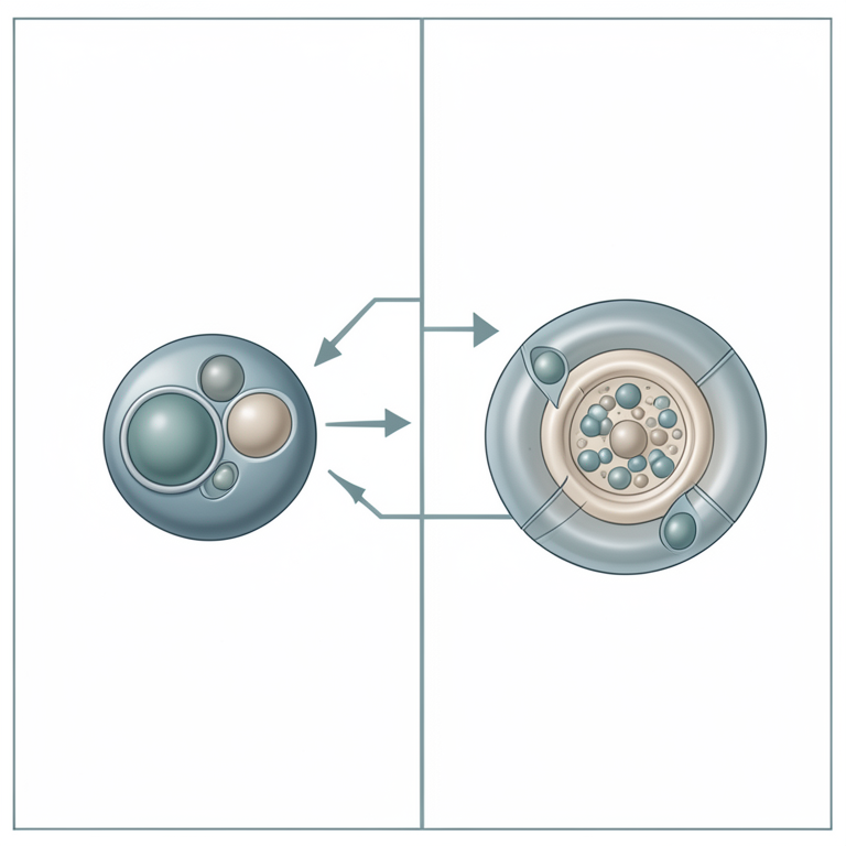
Active vs Passive Receptor Model
AI-generated conceptual illustration (Saccharomyces cerevisiae yeast cells). It explains the idea and does not show study data.
View source evidence ↗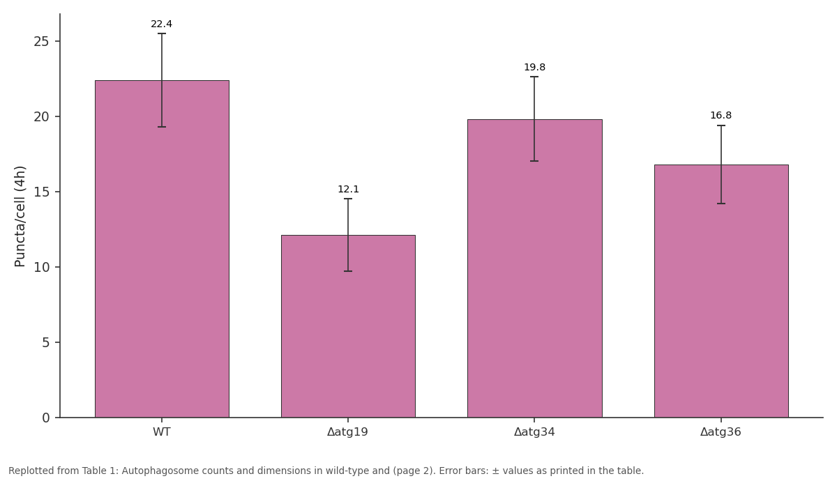
Puncta/cell (4h): WT vs Δatg19 vs Δatg34 vs Δatg36
Replotted from Table 1 (page 2) of the original paper. Every bar is the paper's own table value.
View source evidence ↗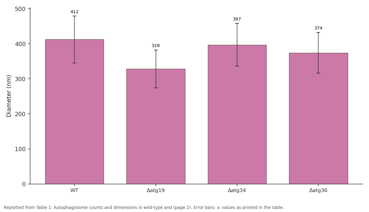
Diameter (nm): WT vs Δatg19 vs Δatg34 vs Δatg36
Replotted from Table 1 (page 2) of the original paper. Every bar is the paper's own table value.
View source evidence ↗Figures & tables
From the original paper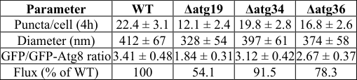 Table 1
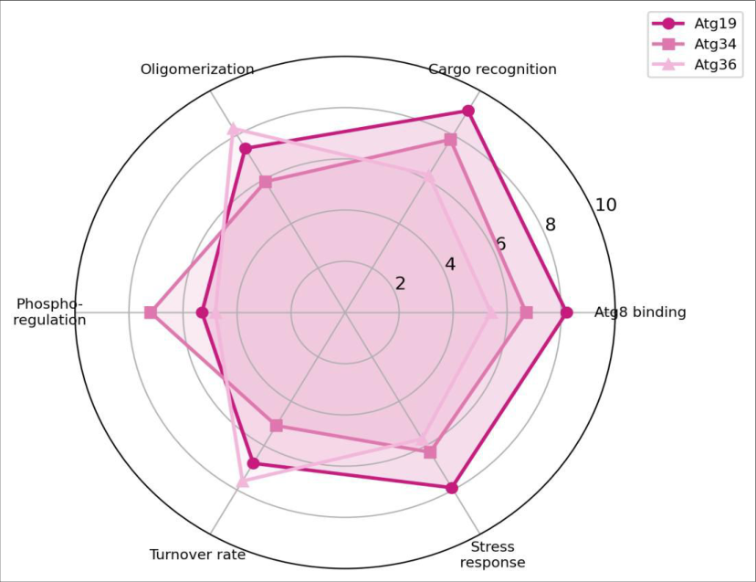 Figure 1
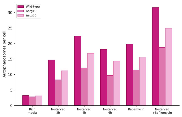 Figure 2
Introduction
Consider a yeast cell abruptly cut off from nitrogen: within minutes, it must decide what to recycle and what to spare. Autophagy -the bulk degradation of cytoplasmic material in the vacuole -provides the raw amino acids needed for survival, and cells that can't perform it die within hours of starvation [1] . But autophagy isn't just a random disposal system. Selective autophagy uses receptor proteins to tag specific cargoes -damaged mitochondria, surplus peroxisomes, protein aggregates -for preferential degradation [2] . In S. cerevisiae, three well-characterized selective autophagy receptors handle different cargoes: Atg19 directs aminopeptidase I and α-mannosidase to the vacuole via the cytoplasm-to-vacuole targeting (Cvt) pathway [3] , Atg34 recognizes α-mannosidase specifically during starvation [4] , and Atg36 targets peroxisomes for degradation (pexophagy) [5] . All three receptors bind Atg8, the yeast ortholog of mammalian LC3, through a conserved Atg8-interacting motif (AIM). What hasn't been settled is how much these selective receptors contribute to overall autophagosome formation. Some models treat them as passive adapters that merely link cargo to pre-formed membranes. But growing evidence suggests they actively promote membrane expansion by clustering Atg8 molecules and scaffolding the autophagosome assembly site [6] . This research compared autophagosome formation rates and morphology in wild-type and receptor-knockout yeast strains during nitrogen starvation, and mapped phosphorylation events that regulate receptor activity. the yeast deletion collection (Open Biosystems). A GFP-Atg8 fusion construct under the native ATG8 promoter was integrated at the URA3 locus in all strains. Cells were grown in YPD (1% yeast extract, 2% peptone, 2% dextrose) at 30°C and shifted to nitrogen-starvation medium (SD-N: 0.17% yeast nitrogen base without amino acids or ammonium sulfate, 2% glucose) for starvation experiments. Rapamycin (200 nM, LC Laboratories) and bafilomycin A1 (200 nM, Sigma-Aldrich) were used as positive control and flux inhibitor respectively. All experiments were performed at the Instituto de Biología Tropical de La Habana, Havana, between March and August 2023.
Methods
Autophagosome formation was quantified by counting GFP-Atg8 puncta per cell using a Nikon Eclipse Ti2 inverted fluorescence microscope with a 100× oil immersion objective. At least 200 cells per strain per time point were scored at 0, 2, 4, and 6 hours of nitrogen starvation. For transmission electron microscopy (TEM), cells were fixed in 2.5% glutaraldehyde, post-fixed in 1% osmium tetroxide, dehydrated, and embedded in Epon 812. Ultrathin sections (70 nm) were stained with uranyl acetate and lead citrate and examined on a JEOL JEM-1400 at 80 kV. Autophagosome diameter was measured from TEM images (n ≥ 50 per strain). Autophagic flux was assessed by the GFP-Atg8 processing assay: cell lysates were immunoblotted with anti-GFP antibody (Roche), and the ratio of free GFP (vacuolar cleavage product) to GFP-Atg8 was calculated. For phosphoproteomics, cells were harvested at 0 and 2 hours post-starvation, lysed in 8M urea, digested with trypsin, enriched for phosphopeptides using TiO2, and analyzed by LC-MS/MS on a Thermo Q Exactive HF. Phosphosites were identified using MaxQuant v2.1. Statistical analyses used Student's t-test or one-way ANOVA with Tukey's post-hoc in GraphPad Prism 9. Ethics clearance was not required (non-human, non-animal research). 1). The Δatg36 strain showed a milder but significant 25.0% reduction. Δatg34 was barely affected, consistent with its narrow cargo specificity. Autophagic flux, measured by the GFP processing assay, was cut nearly in half in Δatg19.
Results
Results
Discussion
The data argue against treating selective autophagy receptors as simple cargo tags. Atg19 deletion reduced autophagosome number and size even for non-Cvt cargo, implying that Atg19 contributes to the general autophagosome assembly machinery during starvation. This is consistent with recent structural work showing that receptor-Atg8 interactions can template membrane curvature [7,8] . If receptors help scaffold the phagophore, losing one would slow membrane expansion -exactly the phenotype observed. The phosphorylation sites identified by our proteomic screen (Atg19-Ser391, Atg36-Thr217) map to regions flanking the AIM motif, where phosphorylation could strengthen Atg8 binding by introducing negative charges that complement basic patches on the Atg8 surface [9] . Mutational validation of these sites would be a natural follow-up. One practical implication is that therapeutic strategies targeting selective autophagy -for instance, to enhance clearance of toxic protein aggregates in neurodegenerative disease models -shouldn't assume that receptor function is limited to cargo recognition. Boosting receptor expression or activity might accelerate autophagosome formation broadly, not just for the receptor's canonical cargo [10] .
Conclusion
Selective autophagy receptors in yeast, particularly Atg19, are active participants in autophagosome biogenesis rather than passive cargo adapters. Deletion of Atg19 reduced autophagosome count by 46%, diameter by 20%, and autophagic flux by nearly half during nitrogen starvation.
Phosphorylation at conserved sites near the Atg8-interacting motif appears to fine-tune receptor-Atg8 binding strength under starvation conditions. These findings redefine the role of selective receptors in the autophagy pathway and suggest that manipulating receptor activity could be a strategy for modulating overall autophagic capacity [11,12] .
Figures and tables from the paper
| Parameter | WT | Δatg19 | Δatg34 | Δatg36 |
|---|---|---|---|---|
| Puncta/cell (4h) | 22.4 ± 3.1 | 12.1 ± 2.4 | 19.8 ± 2.8 | 16.8 ± 2.6 |
| Diameter (nm) | 412 ± 67 | 328 ± 54 | 397 ± 61 | 374 ± 58 |
| GFP/GFP-Atg8 ratio | 3.41 ± 0.48 | 1.84 ± 0.31 | 3.12 ± 0.42 | 2.67 ± 0.37 |
| Flux (% of WT) | 100 | 54.1 | 91.5 | 78.3 |
Table text extracted from the original PDF; superscript letters are shown inline. The original table image is below.
View original table image
References
18 in the original paper- Tsukada M, Ohsumi Y. Isolation and characterization of autophagy-defective mutants of Saccharomyces cerevisiae. FEBS Letters. 1993;333(1-2):169-174.
- Gatica D, Lahiri V, Bhagat DJ. Cargo recognition and degradation by selective autophagy. Nature Cell Biology. 2018;20(3):233-242.
- Scott SV, Guan J, Hutchins MU, Kim J, Klionsky DJ. Cvt19 is a receptor for the cytoplasm-to-vacuole targeting pathway. Molecular Cell. 2001;7(6):11311141.
- Suzuki K, Kondo C, Morimoto M, Ohsumi Y. Selective transport of α-mannosidase by autophagic pathways: identification of a novel receptor, Atg34. Journal of Biological Chemistry. 2010;285(39):30019-30025.
- Motley AM, Nuttall JM, Hettema EH. Pex3-anchored Atg36 tags peroxisomes for degradation in Saccharomyces cerevisiae. EMBO Journal. 2012;31(13):2852-2868.
- Noda NN, Ohsumi Y, Inagaki F. Atg8-family interacting motif crucial for selective autophagy. FEBS Letters. 2010;584(7):1379-1385.
- Yamasaki A, Alam JM, Noshiro D, Hirata E, Fujioka Y, Suzuki K, Ohsumi Y, Noda NN. Liquidity is a critical determinant for selective autophagy of protein condensates. Molecular Cell. 2020;77(6):1163-1175.
- Kirkin V, Rogov VV. A diversity of selective autophagy receptors determines the specificity of the autophagy pathway. Molecular Cell. 2019;76(2):268285.
- Fujioka Y, Noda NN, Fujii K, Yoshimoto K, Ohsumi Y, Inagaki F. In vitro reconstitution of plant Atg8 and Atg12 conjugation systems essential for autophagy. Journal of Biological Chemistry. 2008;283(4):19211928.
- Levine B, Kroemer G. Biological functions of autophagy genes: a disease perspective. Cell. 2019;176(1-2):11-42.
- Nakatogawa H, Suzuki K, Kamada Y, Ohsumi Y. Dynamics and diversity in autophagy mechanisms: lessons from yeast. Nature Reviews Molecular Cell Biology. 2009;10(7):458-467.
- Ohsumi Y. Historical landmarks of autophagy research. Cell Research. 2014;24(1):9-23.
- Farré JC, Subramani S. Mechanistic insights into selective autophagy pathways: lessons from yeast. Nature Reviews Molecular Cell Biology. 2016;17(9):537-552.
- Klionsky DJ, Abdelmohsen K, Abe A. Guidelines for the use and interpretation of assays for monitoring autophagy rd edition). Autophagy. 2016;12(1):1-222.
- Feng Y, He D, Yao Z, Bhagat DJ. The machinery of macroautophagy. Cell Research. 2014;24(1):24-41.
- Wen X, Klionsky DJ. An overview of macroautophagy in yeast. Journal of Molecular Biology. 2016;428(9 Pt A):1681-1699.
- Reggiori F, Klionsky DJ. Autophagic processes in yeast: mechanism, machinery and regulation. Genetics. 2013;194(2):341-361.
- Shintani T, Klionsky DJ. Cargo proteins facilitate the formation of transport vesicles in the cytoplasm to vacuole targeting pathway. Journal of Biological Chemistry. 2004;279(29):29889-29894.