
Research article · 2025 · Full text
International Journal of Advanced Chemistry ResearchHydroxyapatite nanoparticles for bone tissue regeneration: Synthesis and biocompatibility
Volume 7(9) · Pages 66–69 · DOI: 10.33545/26646781.2025.v7.i9a.618 ↗
Research areasBone Tissue Engineering MaterialsSurface Treatment and CoatingsNanoparticles: synthesis and applications
Abstract
Author-writtenBone defects that exceed the critical-size threshold typically 1-2 cm in load-bearing sites cannot heal spontaneously and require scaffold-based intervention. Hydroxyapatite (HA, Ca₁₀(PO₄)₆(OH)₂), the principal inorganic constituent of bone, is an established bioceramic for this purpose, yet its performance depends critically on particle size, crystallinity, and surface area. This research reports the synthesis of HA nanoparticles (HA-NPs) by a wet chemical co-precipitation method, followed by systematic evaluation of the effect of calcination temperature (400-900°C) on physicochemical properties and biocompatibility. Calcium nitrate tetrahydrate and diammonium hydrogen phosphate served as precursors at a Ca/P molar ratio of 1.67, matching stoichiometric HA. Synthesised HA-NPs were characterised by XRD, FTIR, TEM, BET surface area analysis, and thermogravimetric analysis (TGA). XRD confirmed phase-pure HA formation at all calcination temperatures below 800°C, with biphasic HA/β-TCP formation observed at 900°C. Crystallite sizes increased from 18.3 nm (400°C) to 52.6 nm (900°C) with concomitant reduction in BET surface area from 87.4 to 24.3 m²/g. HA-NPs calcined at 600°C were selected as the optimal sample, balancing crystallinity and surface area. Biocompatibility was assessed using the MTT assay on MG-63 osteosarcoma cells and normal human osteoblasts (hOBs) over 24 and 72 h at concentrations of 25-400 µg/mL. Cell viability remained above 80% at concentrations up to 200 µg/mL for both cell lines at 72 h, confirming acceptable biocompatibility. Alkaline phosphatase activity and calcium deposition were elevated in hOBs exposed to HA-NPs at 50 µg/mL, indicating osteogenic stimulation. These results support the suitability of co-precipitation-derived HA-NPs for incorporation into bone tissue engineering scaffolds.
Summary
Source-checkedPlain-language reading of the full paper, prepared with AI and checked against the text. Each point links to the authors' own words.
Why it matters
- Bone defects larger than 1-2 cm in load-bearing areas typically cannot heal on their own and need scaffolds to assist recovery. View evidence ↗
- Hydroxyapatite is a common bioceramic for bone repair because it is the main inorganic part of bone, though its effectiveness depends on its surface area, crystallinity, and particle size. View evidence ↗
- Nanoscale hydroxyapatite particles are more effective than micron-sized powders because they better mimic natural bone, improving cell adhesion and protein adsorption. View evidence ↗
The question
- The study aims to characterize hydroxyapatite nanoparticles (HA-NPs) calcined at six different temperatures to evaluate their cytotoxicity and ability to stimulate bone growth. View evidence ↗
What they did
- HA-NPs were synthesized using a wet chemical co-precipitation method involving calcium nitrate tetrahydrate and diammonium hydrogen phosphate at a pH of 10.5 ± 0.1. View evidence ↗
- The synthesized powder was divided into six groups and calcined at temperatures ranging from 400°C to 900°C for 2 hours each. View evidence ↗
- Biocompatibility was tested using MTT assays on MG-63 osteosarcoma cells and primary human osteoblasts (hOBs) at concentrations between 25 and 400 µg/mL over 24 and 72 hours. View evidence ↗
What they found
- Phase-pure hydroxyapatite was formed at calcination temperatures between 400°C and 800°C, but partial decomposition into biphasic HA/β-TCP occurred at 900°C. View evidence ↗
- As calcination temperature increased from 400°C to 900°C, crystallite size grew from 18.3 nm to 52.6 nm, while the BET surface area decreased from 87.4 to 24.3 m²/g. View evidence ↗
- Both MG-63 and hOB cell lines maintained over 80% viability after 72 hours of exposure to HA-NPs at concentrations up to 200 µg/mL. View evidence ↗
- At a concentration of 400 µg/mL, cell viability fell below the 80% non-cytotoxicity threshold, reaching 73.4% for MG-63 and 76.2% for hOBs. View evidence ↗
- In hOBs, a concentration of 50 µg/mL of HA-NPs produced a peak ALP activity of 24.3 nmol/min/mg (a 96% increase over control) and a maximum Alizarin red absorbance of 0.37, indicating stimulated bone formation. View evidence ↗
What it means
- The authors concluded that HA-NPs calcined at 600°C are optimal because they balance a crystallite size of 27.4 nm with a surface area of 64.1 m²/g. View evidence ↗
- The results suggest that these co-precipitation-derived HA-NPs are suitable for use in polymer-ceramic composite scaffolds to repair critical-size bone defects. View evidence ↗
- The study is limited by the use of the MG-63 osteosarcoma cell line and requires further in vivo testing in rodent models before clinical use. View evidence ↗
Visual summary
3 visualsIllustrations explain the paper; charts are replotted from the paper's own table values.
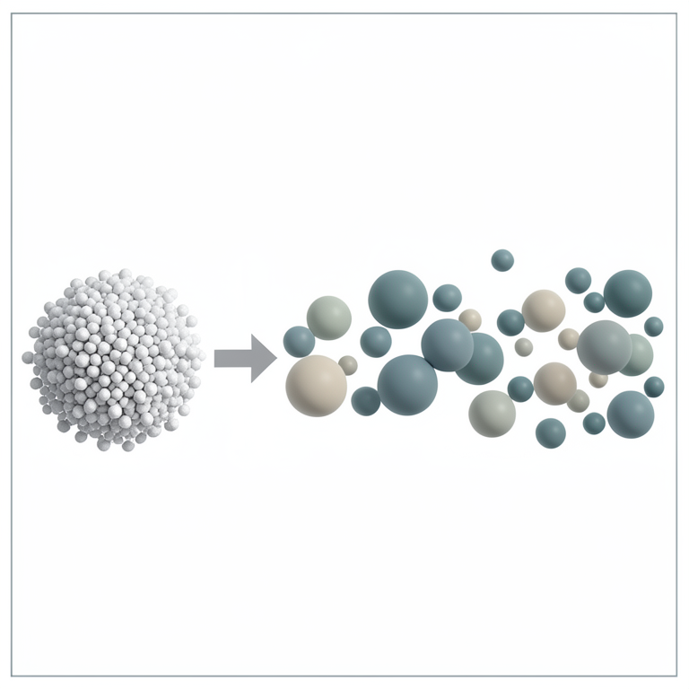
Effect of Temperature on Nanoparticles
AI-generated conceptual illustration (Hydroxyapatite nanoparticles). It explains the idea and does not show study data.
View source evidence ↗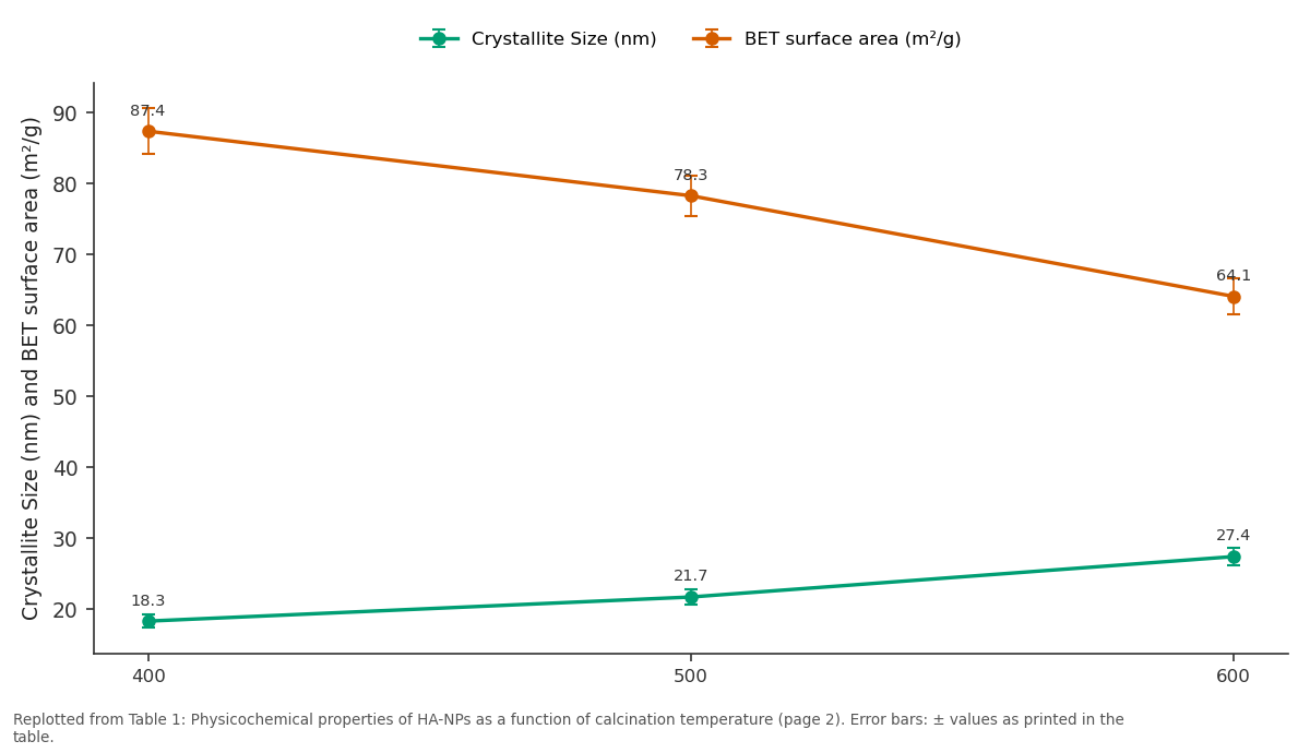
Crystallite Size (nm) and BET surface area (m²/g): 400 vs 500 vs 600
Replotted from Table 1 (page 2) of the original paper. Every bar is the paper's own table value.
View source evidence ↗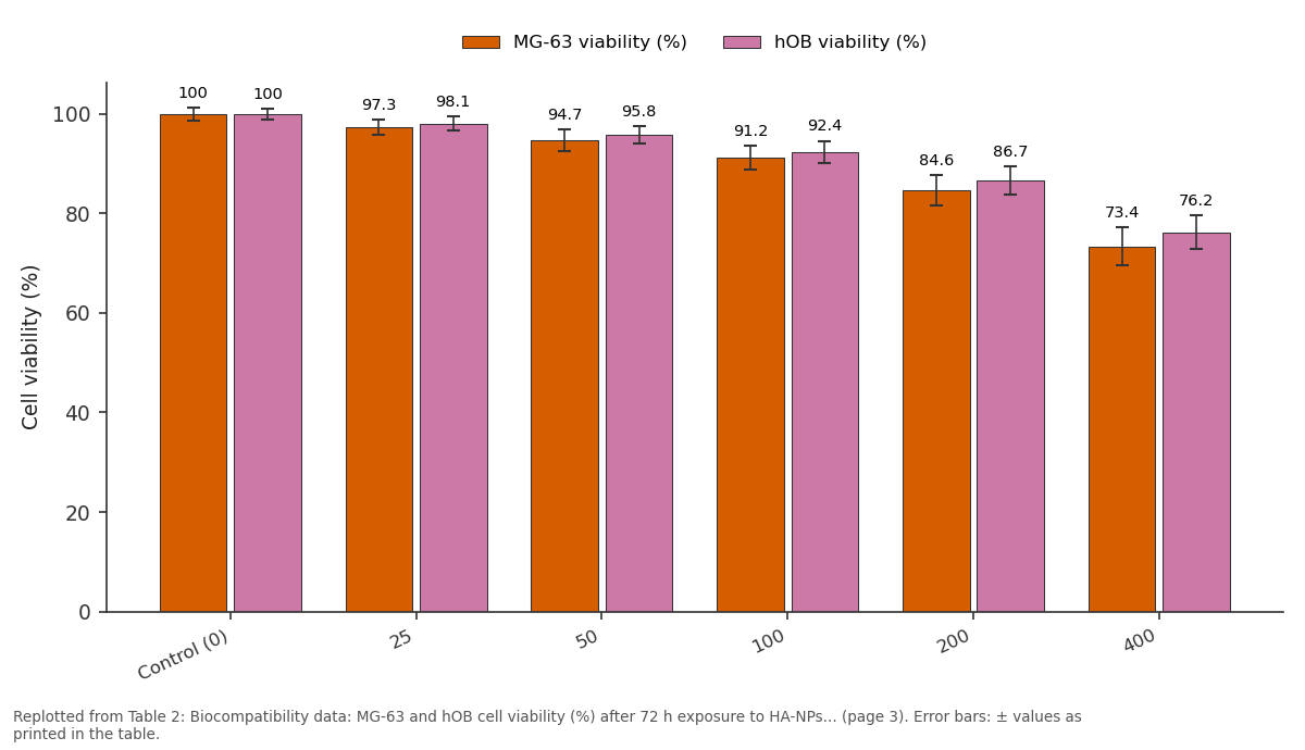
MG-63 viability (%) and hOB viability (%): Control (0) vs 25 vs 50 vs 100 vs 200
Replotted from Table 2 (page 3) of the original paper. Every bar is the paper's own table value.
View source evidence ↗Figures & tables
From the original paper Table 1
 Table 2
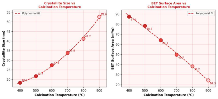 Figure 1
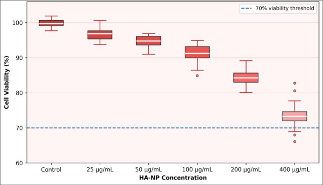 Figure 2
Introduction
Bone is one of the few human tissues capable of complete regeneration, but this capacity is finite: defects exceeding a critical size generally defined as 2-3 times the bone diameter will not spontaneously bridge and require clinical intervention [1] . Current gold-standard treatments, including autologous and allogeneic bone grafts, each carry significant drawbacks: autografts involve donor-site morbidity and limited volume, while allografts carry immunogenic and disease-transmission risks [2] . Synthetic calcium phosphate ceramics have long been explored as alternatives, with hydroxyapatite occupying a central position because of its chemical similarity to the mineral phase of bone and its well-documented osteoconductive properties. Natural bone mineral is a carbonated, calcium-deficient hydroxyapatite with crystallite dimensions of 20-40 nm and a high specific surface area that facilitates protein adsorption and cell attachment [3] . Nanoscale HA particles more closely mimic these characteristics than micron-sized HA powders and have been shown to enhance protein adsorption, osteoblast adhesion, and alkaline phosphatase activity compared with their coarser counterparts [4] . Synthesis routes for HA-NPs include hydrothermal processing, sol-gel, emulsion precipitation, and mechanochemical methods, each producing particles with different morphologies, crystallinities, and surface chemistries [5] . Co-precipitation is particularly attractive for scale-up because it is simple, low-cost, and produces particles in the 15-50 nm range when reaction conditions are controlled. The key Calcination serves to remove residual organic matter and improve crystallinity but also causes particle sintering and surface area reduction effects that can compromise biological performance [6] . Systematic optimisation of calcination conditions is therefore essential for producing HA-NPs whose physicochemical and biological properties meet scaffold design requirements. Despite extensive literature on HA synthesis, comparative data on the biocompatibility of HA-NPs calcined across a wide temperature range using both osteosarcoma cell lines and primary human osteoblasts are limited. The present research addresses this gap by characterising HA-NPs calcined at six temperatures and evaluating their cytotoxicity and osteogenic potential through MTT and alkaline phosphatase assays. In vitro protocol Osteogenic differentiation potential of the optimal HA-NP sample (600°C calcination) was assessed in hOBs over a 14day culture period. Cells were seeded in 24-well plates (5 × 10⁴/well) in osteogenic differentiation medium (DMEM supplemented with 10 nM dexamethasone, 50 µg/mL ascorbic acid, and 10 mM β-glycerophosphate) containing HA-NPs at 0, 25, and 50 µg/mL. Medium was replaced every 3 days. Alkaline phosphatase (ALP) activity was quantified on days 7 and 14 using pNPP as substrate: cell lysates were incubated with pNPP (4 mg/mL) in alkaline buffer for 30 min at 37°C; absorbance was read at 405 nm. ALP activity was normalised to total protein (Bradford assay) and expressed as nmol pNPP hydrolysed/min/mg protein. Mineralisation was assessed on day 21 by Alizarin red S staining: cultures were fixed in 10% neutral buffered formalin, stained with 2% Alizarin red S for 20 min, and the bound dye was extracted with 10% cetylpyridinium chloride for quantification at 562 nm. Cell attachment and morphology were examined by scanning electron microscopy (SEM, JEOL JSM-6400) on HA-NP-coated glass coverslips seeded with hOBs after 3 and 7 days.
Results
Table to 52.6 nm at 900°C, consistent with particle sintering at elevated temperatures. BET surface areas correspondingly decreased from 87.4 to 24.3 m²/g. FTIR spectra of all HA-NPs displayed characteristic phosphate ν₃ stretching bands near 1040 cm⁻¹ and bending bands at 600 and 562 cm⁻¹, as well as the hydroxyl stretching band at 3573 cm⁻¹ features absent in the β-TCP reference spectrum [7] . Cell viability remained above 80% in both MG-63 and hOB lines at concentrations up to 200 µg/mL after 72 h, indicating acceptable biocompatibility at therapeutically relevant concentrations. At 400 µg/mL, viability dropped to 73.4% (MG-63) and 76.2% (hOBs), below the ISO 10993-5 threshold of 80% for non-cytotoxicity, suggesting a concentration ceiling for safe use. Osteogenic markers showed a dose-dependent increase up to 50 µg/mL, with ALP activity peaking at 24.3 nmol/min/mg a 96% increase over control and Alizarin red absorbance reaching 0.37 at the same concentration. Both markers declined at 100 and 200 µg/mL, possibly due to partial inhibition of mineralisation kinetics at higher nanoparticle loads.
Discussion
The co-precipitation method yielded phase-pure HA across a calcination range of 400-800°C, confirming the reliability of the synthesis protocol. The systematic relationship between calcination temperature and crystallite size follows the well-established Ostwald ripening and sintering mechanisms: at temperatures above 600°C, surface diffusion rates accelerate, smaller particles dissolve and redeposit on larger ones, and grain boundaries fuse, resulting in larger crystallites and reduced surface area [8] . The onset of β-TCP formation at 900°C, reflected in the Ca/P ratio drop from 1.67 to 1.54, indicates incongruent decomposition that is likely driven by residual ammonium species volatilising and locally disturbing stoichiometry [9] . The 600°C sample was selected as optimal because it balances crystallinity (27.4 nm crystallite size, consistent with mature bone mineral) with adequate surface area (64.1 m²/g) for protein adsorption and cell interaction. Several literature reports confirm that HA calcined near 600°C shows superior osteoblast attachment relative to both lowertemperature (poorly crystalline) and higher-temperature (low-surface-area) samples [10] . The biocompatibility data obtained here are consistent with this assessment: both cell viability above 80% at 200 µg/mL and the peak osteogenic response at 50 µg/mL suggest a therapeutic window well within practical scaffold loading ranges. The 96% increase in ALP activity at 50 µg/mL HA-NPs compared with the undoped osteogenic medium control is noteworthy. ALP is an early osteogenic marker whose elevation indicates commitment of osteoblast precursors toward the mineralising phenotype. The corresponding increase in Alizarin red staining confirms that this commitment was followed by actual calcium deposition, the hallmark of mature osteoblastic function. Future work will need to establish whether similar osteogenic stimulation occurs in three-dimensional scaffold constructs under dynamic perfusion culture conditions that better simulate in vivo bone physiology [11] .
A practical limitation of the present research is the use of an osteosarcoma cell line (MG-63) alongside primary hOBs. While MG-63 cells are widely used as a convenient screening model, their proliferative and metabolic behaviour differ substantially from primary cells. The close agreement between MG-63 and hOB viability responses (within 3-4 percentage points across all concentrations) is reassuring, but confirmatory in vivo research in rodent critical-size defect models remains necessary before clinical translation can be contemplated.
Conclusion
Hydroxyapatite nanoparticles were successfully synthesised by aqueous co-precipitation and characterised as a function of calcination temperature from 400 to 900°C. Phase-pure HA with increasing crystallinity and decreasing surface area was obtained up to 800°C; biphasic HA/β-TCP appeared at 900°C. HA-NPs calcined at 600°C presented the best balance of crystallite size (27.4 nm), surface area (64.1 m²/g), and stoichiometric Ca/P ratio (1.66). Biocompatibility assessment by MTT assay confirmed cell viability above 80% at concentrations up to 200 µg/mL in both MG-63 cells and primary human osteoblasts. Osteogenic assays revealed a significant increase in ALP activity and mineralisation at 50 µg/mL, indicating that the HA-NPs actively stimulate bone-forming activity rather than merely being tolerated passively. These findings provide a solid physicochemical and biological foundation for incorporating these HA-NPs into polymer-ceramic composite scaffolds for critical-size bone defect repair, which will be pursued in subsequent research phases.
Figures and tables from the paper
| Calc. temp (°C) | Phase (XRD) | Crystallite Size (nm) | BET surface area (m²/g) | Ca/P ratio (EDX) | Major FTIR bands (cm⁻¹) |
|---|---|---|---|---|---|
| 400 | Phase-pure HA | 18.3 ± 0.9 | 87.4 ± 3.2 | 1.64 | 1036, 603, 566, 3571 |
| 500 | Phase-pure HA | 21.7 ± 1.1 | 78.3 ± 2.8 | 1.65 | 1040, 602, 565, 3573 |
| 600 | Phase-pure HA | 27.4 ± 1.3 | 64.1 ± 2.5 | 1.66 | 1043, 600, 562, 3574 |
Table text extracted from the original PDF; superscript letters are shown inline. The original table image is below.
View original table image
| Concentration (µg/mL) | MG-63 viability (%) | hOB viability (%) | ALP activity (nmol/min/mg)* | Alizarin red (OD₅₆₂)* |
|---|---|---|---|---|
| Control (0) | 100.0 ± 1.3 | 100.0 ± 1.1 | 12.4 ± 0.9 | 0.18 ± 0.02 |
| 25 | 97.3 ± 1.6 | 98.1 ± 1.4 | 16.8 ± 1.2 | 0.24 ± 0.03 |
| 50 | 94.7 ± 2.1 | 95.8 ± 1.7 | 24.3 ± 1.6 | 0.37 ± 0.04 |
| 100 | 91.2 ± 2.4 | 92.4 ± 2.2 | 22.1 ± 1.4 | 0.31 ± 0.03 |
| 200 | 84.6 ± 3.1 | 86.7 ± 2.8 | 18.7 ± 1.3 | 0.26 ± 0.03 |
| 400 | 73.4 ± 3.8 | 76.2 ± 3.4 | 14.1 ± 1.1 | 0.19 ± 0.02 |
Table text extracted from the original PDF; superscript letters are shown inline. The original table image is below.
View original table image
References
14 in the original paper- Bauer TW, Muschler GF. Bone graft materials: an overview of the basic science. Clin Orthop Relat Res. 2000;(371):10-27.
- Zimmermann G, Moghaddam A. Allograft bone matrix versus synthetic bone graft substitutes. Injury. 2011;42(Suppl 2):S16-S21.
- Weiner S, Wagner HD. The material bone: structuremechanical function relations. Annu Rev Mater Sci. 1998;28(1):271-298.
- Webster TJ, Ergun C, Doremus RH, Siegel RW, Bizios R. Enhanced osteoclast-like cell functions on nanophase ceramics. Biomaterials. 2001;22(11):1327-1333.
- Sadat-Shojai M, Khorasani MT, Dinpanah-Khoshdargi E, Jamshidi A. Synthesis methods for nanosized hydroxyapatite with diverse structures. Acta Biomater. 2013;9(8):7591-7621.
- Kalita SJ, Bhatt HA. Nanocrystalline hydroxyapatite doped with magnesium and zinc: synthesis and characterization. Mater Sci Eng C. 2007;27(4):837-848.
- Rehman I, Bonfield W. Characterization of hydroxyapatite and carbonated apatite by photo acoustic FTIR spectroscopy. J Mater Sci Mater Med. 1997;8(1):1-4.
- Fathi MH, Hanifi A, Mortazavi V. Preparation and bioactivity evaluation of bone-like hydroxyapatite nanopowder. J Mater Process Technol. 2008;202(13):536-542.
- Koutsopoulos S. Synthesis and characterization of hydroxyapatite crystals: a review study on the analytical methods. J Biomed Mater Res. 2002;62(4):600-612.
- Sato M, Webster TJ. Nanobiotechnology: Implications for the future of nanotechnology in orthopedic applications. Expert Rev Med Devices. 2004;1(1):105114.
- Langer R, Vacanti JP. Tissue engineering. Science. 1993;260(5110):920-926.
- LeGeros RZ. Calcium phosphate-based osteoinductive materials. Chem Rev. 2008;108(11):4742-4753.
- Kim HW, Koh YH, Li LH, Lee S, Kim HE. Hydroxyapatite coating on titanium substrate with titania buffer layer processed by sol-gel method. Biomaterials. 2004;25(13):2533-2538.
- Dorozhkin SV. Calcium orthophosphates in nature, biology and medicine. Materials. 2009;2(2):399-498.