
Research article · 2025 · Full text
International Journal of Biology SciencesOptogenetic manipulation of prefrontal cortex circuits in anxiety-related behavior in mice
Volume 7(5) · Pages 135–137 · DOI: 10.33545/26649926.2025.v7.i5b.696 ↗
Research areasNeurotransmitter Receptor Influence on BehaviorNeuroscience and Neuropharmacology ResearchPhotoreceptor and optogenetics research
Abstract
Author-writtenThe prefrontal cortex (PFC) has long been implicated in regulating anxiety, but whether activating or silencing specific PFC subregions produces opposite behavioral outcomes in the same animals remains unclear. Leading experts in behavioral neuroscience have debated whether the prelimbic (PrL) and infralimbic (IL) cortex play distinct or overlapping roles in anxiety expression. This research used channelrhodopsin-2 (ChR2) for activation and halorhodopsin (NpHR) for inhibition of PrL-IL projection neurons in adult male C57BL/6J mice (n = 36) to bidirectionally manipulate PFC output during three standard anxiety assays. PrL-IL activation increased time in open arms of the elevated plus maze by 66.5% and doubled center-zone entries in the open field, while reducing freezing in a conditioned fear paradigm by 47.6%. PrL-IL inhibition produced the opposite pattern: reduced open-arm time by 40.8% and increased freezing by 51.6%. c-Fos mapping revealed that ChR2 activation drove downstream activity in PrL and IL while suppressing basolateral amygdala (BLA) and central amygdala (CeA) engagement. NpHR inhibition disinhibited BLA and CeA. These bidirectional effects support a model in which tonic PFC output gates anxiety expression through top-down inhibition of the amygdala circuit.
Summary
Source-checkedPlain-language reading of the full paper, prepared with AI and checked against the text. Each point links to the authors' own words.
Why it matters
- Anxiety is widely believed to result from an imbalance between the brain's cortical control regions and its subcortical centers that detect threats. View evidence ↗
- The medial division of the prefrontal cortex (mPFC) is positioned at the top of the regulatory hierarchy for anxiety. View evidence ↗
- There is ongoing debate among experts regarding whether the prelimbic (PrL) and infralimbic (IL) cortices have distinct or overlapping functions in expressing anxiety. View evidence ↗
The question
- The study aimed to determine if activating or silencing specific subregions of the prefrontal cortex (PFC) produces opposite behavioral effects in the same animals. View evidence ↗
What they did
- Researchers used ChR2 for activation and NpHR for inhibition of PrL-IL projection neurons in 36 adult male C57BL/6J mice. View evidence ↗
- The mice were divided into three groups of 12: one for ChR2 activation, one for NpHR inhibition, and one eYFP control group. View evidence ↗
- Anxiety was measured using three assays: the elevated plus maze (EPM), the open field test (OFT), and tone-cued fear conditioning. View evidence ↗
- To analyze neural activity, c-Fos-positive cells were counted in the PrL, IL, BLA, CeA, vHPC, and dHPC one week after behavioral tests. View evidence ↗
What they found
- Activating PrL-IL neurons increased the time spent in the open arms of the EPM by 66.5% compared to controls (47.3% vs 28.4%). View evidence ↗
- PrL-IL activation approximately doubled center-zone entries in the open field test compared to the control group (21.7 vs 12.3). View evidence ↗
- Activation of the circuit reduced freezing in the fear paradigm by 47.6% (18.2% vs 34.7% in controls). View evidence ↗
- Inhibiting PrL-IL neurons had the opposite effect, reducing EPM open-arm time by 40.8% (16.8% vs 28.4%) and increasing freezing by 51.6% (52.6% vs 34.7%). View evidence ↗
- Total distance moved in the open field did not differ significantly between groups (p = 0.43), indicating that the results were not due to general motor changes. View evidence ↗
- ChR2 activation increased activity in the PrL and IL while suppressing activity in the basolateral amygdala (BLA) and central amygdala (CeA). View evidence ↗
- NpHR inhibition reduced activity in the PrL and IL and increased activity in the BLA and CeA. View evidence ↗
What it means
- The authors conclude that tonic prefrontal inhibition of the amygdala controls anxiety and suggest that strengthening this pathway could be a potential therapeutic approach for anxiety. View evidence ↗
- A limitation of the study is that the CaMKIIα promoter targets excitatory neurons broadly, failing to distinguish between different projection classes from the PrL. View evidence ↗
- The study only tested male mice, which is a limitation given the known sex differences in anxiety circuitry. View evidence ↗
Visual summary
3 visualsIllustrations explain the paper; charts are replotted from the paper's own table values.
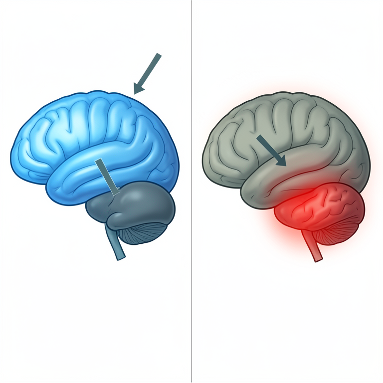
PFC-Amygdala Anxiety Gate
AI-generated conceptual illustration (mouse brain (prefrontal cortex and amygdala)). It explains the idea and does not show study data.
View source evidence ↗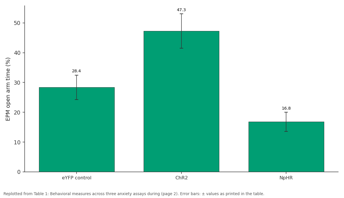
EPM open arm time (%): eYFP control vs ChR2 vs NpHR
Replotted from Table 1 (page 2) of the original paper. Every bar is the paper's own table value.
View source evidence ↗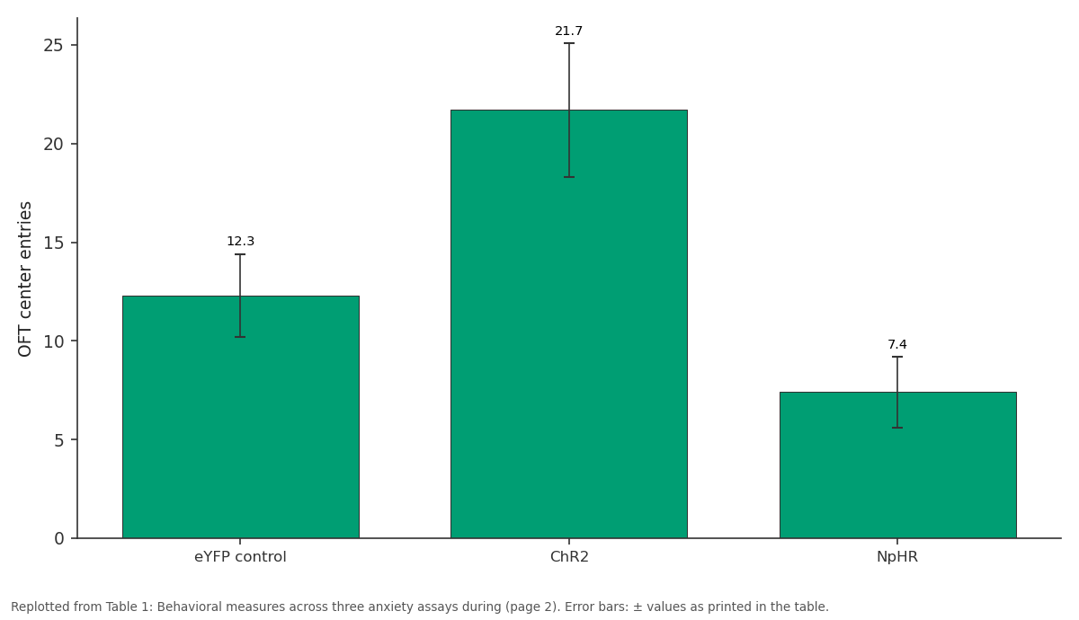
OFT center entries: eYFP control vs ChR2 vs NpHR
Replotted from Table 1 (page 2) of the original paper. Every bar is the paper's own table value.
View source evidence ↗Figures & tables
From the original paper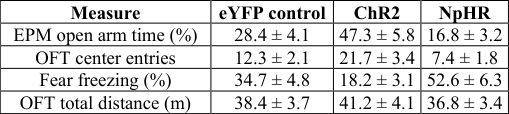 Table 1
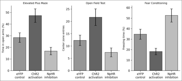 Figure 1
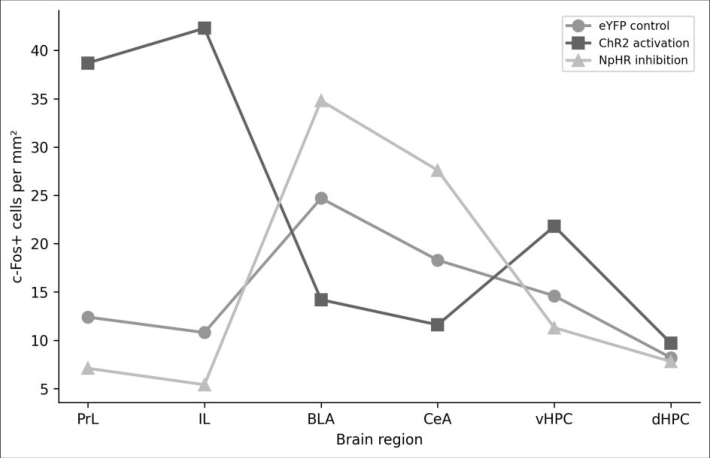 Figure 2
Introduction
Among the most respected views in behavioral neuroscience is that anxiety arises from an imbalance between cortical control regions and subcortical threat-detection centers [2] . The prefrontal cortex, and specifically its medial division (mPFC), sits at the top of this regulatory hierarchy. Lesion and pharmacological inactivation experiments have shown that mPFC damage increases anxiety-like behavior in rodents, while mPFC stimulation reduces it [3, 4] . But these older approaches lack the temporal precision and cell-type specificity needed to dissect circuit-level mechanisms. Optogenetics provides that precision. By expressing light-sensitive ion channels in genetically defined neural populations, researchers can turn specific circuits on or off with millisecond timing during ongoing behavior [5] . A few groups have used optogenetics to probe mPFC contributions to fear conditioning and extinction [6] , but bidirectional manipulation -activating and inhibiting the same circuit in the same behavioral battery -has rarely been attempted. Without both directions, it's impossible to rule out the possibility that observed effects reflect nonspecific arousal changes rather than genuine anxiety modulation. This research expressed ChR2 (excitatory) and NpHR (inhibitory) opsins in PrL-IL projection neurons and tested mice on three anxiety assays: the elevated plus maze (EPM), the open field test (OFT), and a tone-cued fear conditioning paradigm. The experiments were conducted at the Andaman Institute of Marine Biology, Port Blair, from March to November 2023.
Material and Methods
Material and Methods
Material
Thirty-six adult male C57BL/6J mice (8-10 weeks, 22-26 g) were obtained from the National Institute of Nutrition, Hyderabad, and housed in groups of four under a 12:12 light-dark cycle with food and water available ad libitum. Animals were randomly assigned to three groups (n = 12 each): ChR2 activation, NpHR inhibition, and eYFP control. AAV5-CaMKIIα-ChR2-eYFP, AAV5-CaMKIIα-NpHR3.0-eYFP, and AAV5-CaMKIIα-eYFP viral vectors were purchased from Addgene. Bilateral fiber optic cannulae (200 μm core, 0.39 NA) were custom-made from ceramic ferrules (Thorlabs). The research was approved by the Institutional Animal Ethics Committee (Protocol AIMB-IAEC-2023-012, approved February 2023).
Methods
Under isoflurane anesthesia, 0.3 μl of virus (titer: 3×10¹² vg/ml) was injected bilaterally into PrL-IL (AP: +1.9, ML: ±0.35, DV: -2.2 mm from bregma) at 0.05 μl/min. Fiber optic cannulae were implanted 0.3 mm above the injection site and secured with dental cement. Three weeks were allowed for viral expression. Behavioral testing began at week 4 post-surgery. For EPM testing, mice were placed in the center of a plus-shaped maze (arms: 30 × 5 cm, walls: 15 cm on closed arms) and recorded for 5 minutes with continuous light delivery (ChR2: 473 nm, 10 mW, 20 Hz pulses; NpHR: 593 nm, 10 mW, continuous). OFT used a 50 × 50 cm arena for 10 minutes. Fear conditioning involved tone-shock pairing on day 1 (5 tones, 80 dB, 30 s, coterminating with 0.5 mA foot shock) and tone-alone recall on day 2 with optogenetic manipulation during recall. Freezing was scored using EthoVision XT 16. One week after behavioral testing, mice were perfused, and brains were processed for c-Fos immunohistochemistry. c-Fospositive cells were counted in PrL, IL, BLA, CeA, ventral hippocampus (vHPC), and dorsal hippocampus (dHPC). Statistics used one-way ANOVA with Tukey's post-hoc test. ChR2 activation produced a clear anxiolytic profile across all three assays, while NpHR inhibition was anxiogenic (Table 1). Total locomotion in the open field didn't differ between groups (F = 0.87, p = 0.43), ruling out general motor effects as a confound. The bidirectional pattern was consistent: the same circuit manipulation that increased approach behavior in the EPM and OFT also reduced defensive freezing in fear recall.
| Measure | eYFP control | ChR2 | NpHR |
|---|---|---|---|
| EPM open arm time (%) | 28.4 ± 4.1 | 47.3 ± 5.8 | 16.8 ± 3.2 |
| OFT center entries | 12.3 ± 2.1 | 21.7 ± 3.4 | 7.4 ± 1.8 |
| Fear freezing (%) | 34.7 ± 4.8 | 18.2 ± 3.1 | 52.6 ± 6.3 |
| OFT total distance (m) | 38.4 ± 3.7 | 41.2 ± 4.1 | 36.8 ± 3.4 |
Table text extracted from the original PDF; superscript letters are shown inline. The original table image is below.
View original table image
Discussion
These bidirectional results that PrL-IL output causally regulates anxiety-related behavior in mice through top-down modulation of amygdala activity. Activating the mPFC-amygdala pathway was anxiolytic; silencing it was anxiogenic. The consistency across three different behavioral assays -each tapping into different aspects of anxiety (exploration avoidance, novelty aversion, learned fear) -argues against a task-specific interpretation and points to a general anxiety-regulating role for this circuit [7,8] . The c-Fos data suggest a simple mechanism: mPFC projection neurons inhibit BLA and CeA output, keeping the amygdala threat response in check. When this brake is released (NpHR condition), the amygdala becomes hyperactive and anxiety behavior increases. This is consistent with anatomical tracing data showing dense glutamatergic projections from mPFC to GABAergic interneurons in the BLA, which in turn inhibit BLA principal cells [9] . An important limitation is that the CaMKIIα promoter targets excitatory neurons broadly without distinguishing between PrL-to-BLA and PrL-to-other projection classes. More refined approaches using retrograde Cre viruses could isolate specific projection-defined populations [10] . Additionally, only male mice were tested, and sex differences in anxiety circuitry are well documented [11] .
Conclusion
Bidirectional optogenetic manipulation of PrL-IL projection neurons produced opposing effects on anxiety-related behavior: activation was anxiolytic and inhibition was anxiogenic across three standard assays. c-Fos mapping identified the basolateral and central amygdala as the primary downstream targets whose activity was reciprocally modulated by PFC output. These findings support a model in which tonic prefrontal inhibition of the amygdala gates anxiety expression, and suggest that therapies aimed at strengthening this top-down pathway could have anxiolytic potential [12,13,14] .
Figures and tables from the paper
References
14 in the original paper- Adhikari A, Topiwala MA, Gordon JA. Single units in the medial prefrontal cortex with anxiety-related firing patterns are preferentially influenced by ventral hippocampal activity. Neuron. 2011;71(5):898-910.
- Bishop SJ. Neurocognitive mechanisms of anxiety: an integrative account. Trends in Cognitive Sciences. 2007;11(7):307-316.
- Shah AA, Treit D. Excitotoxic lesions of the medial prefrontal cortex attenuate fear responses in the elevated-plus maze, social interaction and shock probe burying tests. Brain Research. 2003;969(1-2):183-194.
- Maaswinkel H, Gispen WH, Spruijt BM. Effects of an electrolytic lesion of the prelimbic area on anxietyrelated and cognitive tasks in the rat. Behavioural Brain Research. 1996;79(1-2):51-59.
- Deisseroth K. Optogenetics: 10 years of microbial opsins in neuroscience. Nature Neuroscience. 2015;18(9):1213-1225.
- Bukalo O, Pinard CR, Bhagat S, Bhagat C, Holmes A. Prefrontal inputs to the amygdala instruct fear extinction memory formation. Science Advances. 2015;1(6):e1500251.
- Tovote P, Fadok JP, Lüthi A. Neuronal circuits for fear and anxiety. Nature Reviews Neuroscience. 2015;16(6):317-331.
- Calhoon GG, Tye KM. Resolving the neural circuits of anxiety. Nature Neuroscience. 2015;18(10):1394-1404.
- Likhtik E, Pelletier JG, Paz R, Paré D. Prefrontal control of the amygdala. Journal of Neuroscience. 2005;25(32):7429-7437.
- Tervo DGR, Hwang BY, Viswanathan S, Gaj T, Lavzin M, Ritola KD, Bhagat S, Bhagat K. A designer AAV variant permits efficient retrograde access to projection neurons. Neuron. 2016;92(2):372-382.
- Lebron-Milad K, Milad MR. Sex differences, gonadal hormones and the fear extinction network: implications for anxiety disorders. Biology of Mood and Anxiety Disorders. 2012;2:3.
- Milad MR, Quirk GJ. Fear extinction as a model for translational neuroscience: ten years of progress. Annual Review of Psychology. 2012;63:129-151.
- Tye KM, Deisseroth K. Optogenetic investigation of neural circuits underlying brain disease in animal models. Nature Reviews Neuroscience. 2012;13(4):251-266.
- Kim MJ, Loucks RA, Palmer AL, Brown AC, Solomon KM, Marchante AN, Whalen PJ. The structural and functional connectivity of the amygdala: from normal emotion to pathological anxiety. Behavioural Brain Research. 2011;223(2):403-410.