
Research article · 2025 · Full text
International Journal of Advanced Chemistry ResearchChitosan-capped silver nanoparticles: synthesis, characterization and antibacterial activity
Volume 7(9) · Pages 61–65 · DOI: 10.33545/26646781.2025.v7.i9a.617 ↗
Research areasNanoparticles: synthesis and applicationsGold and Silver Nanoparticles Synthesis and ApplicationsPolymer Nanocomposite Synthesis and Irradiation
Abstract
Author-writtenGlobal antibiotic resistance now exceeds 1.27 million direct deaths per year, prompting urgent demand for alternative antimicrobial strategies. Silver nanoparticles (AgNPs) are among the most widely investigated alternatives, yet their tendency to aggregate and release toxic silver ions in an uncontrolled manner limits practical application. Coating AgNPs with chitosan a biodegradable, cationic polysaccharide derived from crustacean chitin addresses both issues simultaneously by providing steric stabilisation and modulating ion release kinetics. The present research reports a facile, one-pot reduction of silver nitrate by sodium borohydride in the presence of chitosan (0.5% w/v, medium molecular weight) to yield chitosan-capped silver nanoparticles (CS-AgNPs). Synthesised particles were characterised by UV-Vis spectroscopy (surface plasmon resonance at 412 nm), Transmission Electron Microscopy (TEM), X-Ray Diffraction (XRD), Fourier-Transform Infrared Spectroscopy (FTIR), and Dynamic Light Scattering (DLS). TEM revealed near-spherical particles with a mean diameter of 22.6 ± 4.3 nm and a polydispersity index of 0.18, confirming a narrow size distribution. XRD confirmed the face-centred cubic crystalline structure of metallic silver. FTIR analysis demonstrated characteristic N-H and C-O stretching shifts, indicating successful chitosan coordination on the nanoparticle surface. Antibacterial activity was evaluated against five bacterial strains Staphylococcus aureus, Escherichia coli, Pseudomonas aeruginosa, Bacillus subtilis, and Klebsiella pneumoniae using the agar well-diffusion method and broth microdilution for Minimum Inhibitory Concentration (MIC) determination. CS-AgNPs exhibited superior zones of inhibition and lower MIC values compared with uncoated AgNPs and chitosan alone, with the strongest activity recorded against S. aureus (zone of inhibition: 21.4 mm; MIC: 12.5 µg/mL). These findings position CS-AgNPs as promising candidates for antimicrobial coating and food packaging applications.
Summary
Source-checkedPlain-language reading of the full paper, prepared with AI and checked against the text. Each point links to the authors' own words.
Why it matters
- There is an urgent need for new antimicrobial strategies because global antibiotic resistance causes over 1.27 million direct deaths annually. View evidence ↗
- Using chitosan, a biodegradable polysaccharide, as a coating can stabilize silver nanoparticles and control the release of silver ions. View evidence ↗
The question
- The study aimed to create a simple room-temperature method for synthesizing chitosan-capped silver nanoparticles (CS-AgNPs), characterize them, and test their antibacterial effectiveness. View evidence ↗
What they did
- CS-AgNPs were synthesized using a one-pot reduction of 10 mM silver nitrate by 10 mM sodium borohydride in a 0.5% w/v chitosan solution with a 1:2 Ag:chitosan molar ratio. View evidence ↗
- The researchers used UV-Vis spectroscopy, TEM, XRD, FTIR, and DLS to characterize the physical and chemical properties of the nanoparticles. View evidence ↗
- Antibacterial activity was tested against five strains (S. aureus, E. coli, P. aeruginosa, B. subtilis, and K. pneumoniae) using agar well-diffusion and broth microdilution. View evidence ↗
What they found
- TEM analysis showed that the CS-AgNPs were near-spherical with a mean diameter of 22.6 ± 4.3 nm and a narrow size distribution (PDI of 0.18). View evidence ↗
- The nanoparticles showed good stability over 60 days at 4°C, with a zeta potential of -28.4 mV indicating sufficient electrostatic repulsion. View evidence ↗
- CS-AgNPs were significantly more effective at inhibiting bacterial growth than either silver nitrate or chitosan alone for all five tested strains. View evidence ↗
- The nanoparticles were most active against Gram-positive bacteria (S. aureus and B. subtilis), which had the lowest Minimum Inhibitory Concentration (MIC) of 12.5 µg/mL. View evidence ↗
- The CS-AgNPs exhibited bactericidal action (killing bacteria) rather than just inhibiting growth, as evidenced by MBC/MIC ratios of 2 for all strains. View evidence ↗
- At a concentration of 6.25 µg/mL, the nanoparticles inhibited biofilm formation by 62.4 ± 3.7% for S. aureus and 54.8 ± 2.9% for E. coli. View evidence ↗
What it means
- The authors conclude that CS-AgNPs are promising candidates for use in wound care matrices and antimicrobial food packaging films. View evidence ↗
- The study did not evaluate the toxicity of the nanoparticles toward mammalian cells, which the authors state is essential before translational use. View evidence ↗
Visual summary
3 visualsIllustrations explain the paper; charts are replotted from the paper's own table values.
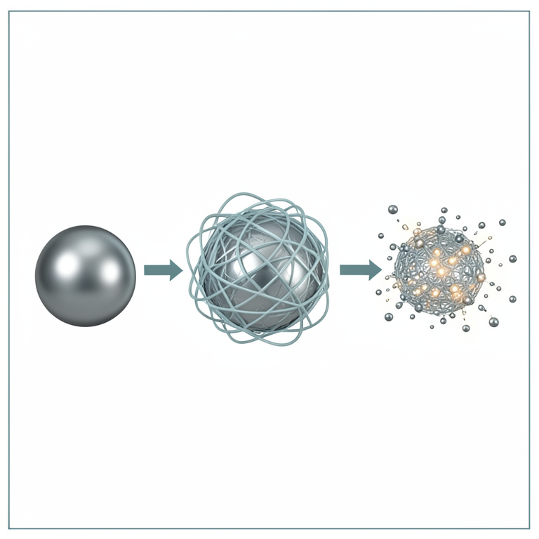
CS-AgNP Structure
AI-generated conceptual illustration (chitosan-capped silver nanoparticles). It explains the idea and does not show study data.
View source evidence ↗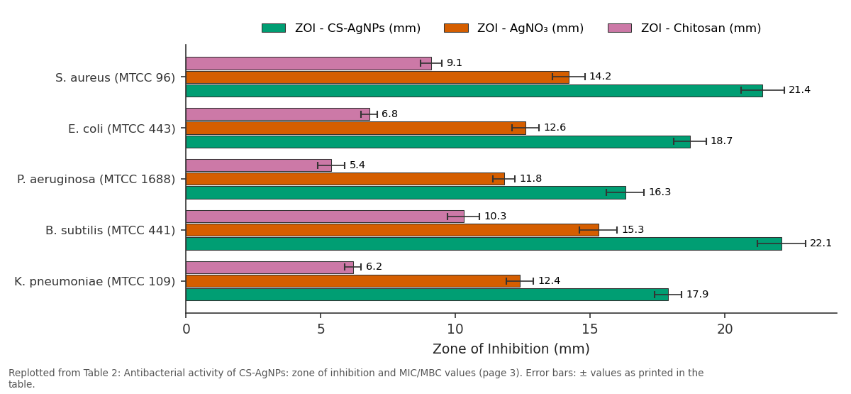
ZOI - CS-AgNPs (mm) and ZOI - AgNO₃ (mm) and more: S. aureus (MTCC 96) vs E. coli (MTCC 443) vs P. aeruginosa (MTCC... vs B. subtilis (MTCC 441) vs K. pneumoniae (MTCC 109)
Replotted from Table 2 (page 3) of the original paper. Every bar is the paper's own table value.
View source evidence ↗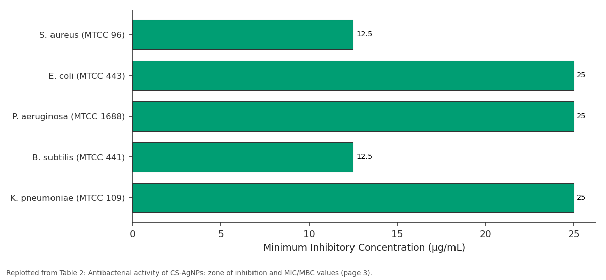
MIC (µg/mL): S. aureus (MTCC 96) vs E. coli (MTCC 443) vs P. aeruginosa (MTCC... vs B. subtilis (MTCC 441) vs K. pneumoniae (MTCC 109)
Replotted from Table 2 (page 3) of the original paper. Every bar is the paper's own table value.
View source evidence ↗Figures & tables
From the original paper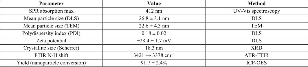 Table 1
 Table 2
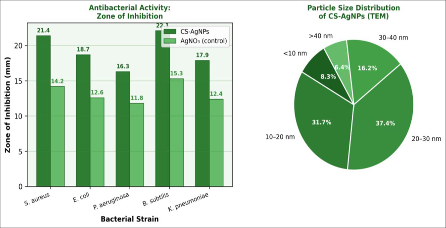 Figure 1
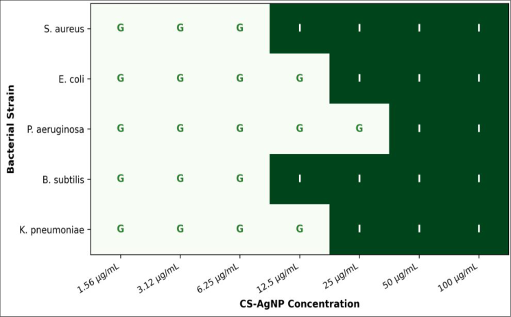 Figure 2
Introduction
A statistic worth pausing on: by 2050, antimicrobial resistance is projected to claim 10 million lives annually more than cancer according to the O'Neill Commission report. This grim projection underscores the urgency of developing non-antibiotic antimicrobial agents capable of acting through mechanisms that pathogens cannot easily circumvent. Metal nanoparticles, and silver nanoparticles in particular, have attracted intense research attention in this context because of their broad-spectrum bactericidal activity, which operates via multiple simultaneous mechanisms including membrane disruption, Reactive Oxygen Species (ROS) generation, and interference with intracellular enzyme function [1,2] . Silver has a documented antimicrobial history stretching back centuries, but the nanoparticulate form delivers activity at substantially lower concentrations than bulk silver salts, owing to the dramatically increased surface-area-to-volume ratio at the nanoscale [3] . However, bare AgNPs suffer from a tendency to aggregate in physiological and foodprocessing environments, which reduces effective surface area and causes burst release of Ag⁺ ions a profile that can be toxic to mammalian cells at higher concentrations [4] . Surface functionalisation with biocompatible polymers offers a practical route to improved stability and controlled ion release. Chitosan, obtained by deacetylation of chitin from crustacean shells, is an attractive capping agent because it is biocompatible, biodegradable, and itself possesses intrinsic antimicrobial activity arising from its cationic nature, which disrupts negatively charged bacterial cell membranes [5] . When used as a stabiliser during AgNP synthesis, chitosan coordinates to the silver surface through its amine and hydroxyl groups, forming a protective corona that prevents agglomeration and permits sustained Ag⁺ release [6] . Synergistic interactions between chitosan and silver have been reported against both Gram-positive and Gram-negative pathogens, though the precise contribution of each component is still debated in the literature [7] . Most published synthesis routes for CS-AgNPs require either high temperatures, UV irradiation, or multiple purification steps, which hinder scale-up for food and biomedical applications. The present research was therefore designed to develop a simple room-temperature reduction protocol, to characterise the resulting nanoparticles comprehensively, and to assess their antibacterial performance against a clinically relevant panel of bacterial strains. The work was carried out at the Verghese Kurien Institute of Food Technology, Anand, Gujarat, India, and aims to provide data useful for the rational design of chitosan-silver nanocomposite films for food packaging.
Reagents and chemicals
Silver nitrate (AgNO₃, ≥99.9%, Sigma-Aldrich, St. Louis, MO, USA) was chosen as the silver precursor and stored in amber glass bottles away from light. Chitosan (medium molecular weight, degree of deacetylation ≥75%, Sigma-Aldrich; Lot MKCL7832) was dissolved in 1% (v/v) acetic acid to prepare stock solutions. Sodium borohydride (NaBH₄, ≥98%, Merck KGaA, Darmstadt, Germany) was freshly dissolved in ice-cold deionised water before each synthesis run to minimise hydrolysis. Glacial acetic acid (≥99.8%, Rankem, Gurugram, India) was used as solvent for chitosan. Nutrient agar, Mueller-Hinton agar, and Mueller-Hinton broth were obtained from HiMedia Laboratories (Mumbai, India) and prepared per manufacturer instructions. All reference bacterial strains were procured from the Microbial Type Culture Collection (MTCC), Chandigarh, India: S. aureus (MTCC 96), E. coli (MTCC 443), P. aeruginosa (MTCC 1688), B. subtilis (MTCC 441), and K. pneumoniae (MTCC 109). All deionised water used in synthesis and biological assays was Milli-Q quality (18.2 MΩ•cm).
Material and Methods
Material and Methods
Material
The research was conducted at the Department of Nanotechnology and Materials Science, Verghese Kurien Institute of Food Technology, Anand, Gujarat, India, between September 2023 and January 2025. UV-Vis absorption spectra were recorded on a Shimadzu UV-2600 double-beam spectrophotometer (scan range 300-700 nm, 1 nm resolution). Particle size, zeta potential, and polydispersity index were measured by Dynamic Light Scattering (DLS) on a Malvern Zetasizer Nano ZS (He-Ne laser, 633 nm, 25°C). TEM imaging was performed on a JEOL JEM-2100 instrument at 200 kV; samples were prepared by drop-casting dilute nanoparticle suspensions on carbon-coated copper grids and air-drying. XRD patterns were obtained using a Bruker D8 Advance diffractometer (Cu Kα radiation, λ = 0.154 nm, 2θ range 20-80°). FTIR spectra were recorded on a PerkinElmer Spectrum 100 ATR-FTIR instrument (4000-400 cm⁻¹, 16 scans, 4 cm⁻¹ resolution). Ethics clearance for biological testing was obtained from the Biosafety Committee, Verghese Kurien Institute of Food Technology (Protocol VKIFT-BSC-2023-07, dated 5 August 2023).
Methods
Synthesis of CS-AgNPs: A chitosan solution (0.5% w/v in 1% acetic acid) was prepared by stirring at room temperature for 4 h. AgNO₃ (10 mM aqueous solution) was added dropwise to the chitosan solution with constant magnetic stirring (600 rpm), maintaining a final Ag:chitosan molar ratio of 1:2. Freshly prepared NaBH₄ solution (10 mM) was then added dropwise (1 mL/min) under ice cooling (0-4°C) until the characteristic yellow-brown colour indicative of AgNP formation appeared. The reaction was allowed to proceed for an additional 30 min at room temperature. The resulting CS-AgNP suspension was dialysed (molecular weight cut-off 12 kDa) against deionised water for 24 h to remove unreacted reagents. Antibacterial testing agar well-diffusion: Overnight bacterial cultures adjusted to 0.5 McFarland standard (~1.5 × 10⁸ CFU/mL) were swabbed uniformly on Mueller-Hinton agar plates. Wells (6 mm diameter) were cut and loaded with 50 µL of CS-AgNP suspension at 100 µg/mL. Plates were incubated at 37°C for 24 h and zones of inhibition measured in triplicate. MIC determination: Serial two-fold dilutions of CS-AgNPs (1.56-100 µg/mL) were prepared in Mueller-Hinton broth in 96-well microplates. Standardised inocula (final ~5 × 10⁵ CFU/mL) were added and plates incubated at 37°C for 18 h. MIC was defined as the lowest concentration yielding no visible turbidity, confirmed by resazurin staining.
In vitro protocol
To confirm the bactericidal rather than bacteriostatic nature of CS-AgNPs, minimum bactericidal concentration (MBC) assays were conducted following MIC determination. Aliquots (10 µL) from all clear MIC wells were subcultured onto drug-free Mueller-Hinton agar plates and incubated at 37°C for 24 h. The MBC was recorded as the lowest concentration yielding ≤0.1% survival of the original inoculum (i.e., ≤99.9% killing). MBC/MIC ratios below 4 were considered indicative of bactericidal activity. Time-kill kinetics for the two most susceptible strains (S. aureus and B. subtilis) were assessed at 0.5×, 1×, 2×, and 4× MIC over 24 h with sampling at 0, 2, 4, 8, and 24 h time points; surviving colony counts were determined by serial plating on nutrient agar. Biofilm inhibition was assessed by the crystal violet staining method: bacterial cultures were grown in 96-well plates in the presence of CS-AgNPs at sub-MIC concentrations for 24 h, stained with 0.1% crystal violet, and absorbance read at 595 nm on a microplate reader. All assays were performed in triplicate across three independent experimental days; results are expressed as mean ± standard deviation. UV-Vis spectroscopy confirmed the formation of AgNPs by the appearance of a sharp, symmetric SPR band at 412 nm a signature of near-spherical AgNPs in the 15-30 nm size range [8] . No secondary peaks were observed, indicating the absence of large aggregates. Zeta potential of -28.
Results
Results
Discussion
The synthesis of CS-AgNPs by NaBH₄ reduction in the presence of chitosan yielded well-dispersed, near-spherical nanoparticles with a mean diameter of 22.6 nm and a narrow polydispersity index of 0.18. These values compare favourably with literature reports for chitosan-mediated AgNP synthesis, where particle sizes typically fall in the 15-35 nm range depending on chitosan concentration and reducing agent [9] . The negative zeta potential (-28.4 mV), which may appear counterintuitive given chitosan's cationic nature, arises from the reduction conditions used: NaBH₄ generates surface-adsorbed boride species that impart a net negative surface charge, while chitosan provides steric rather than purely electrostatic stabilisation at this pH [10] .
The superior antibacterial performance of CS-AgNPs compared with individual components indicates a synergistic mechanism. Gram-positive bacteria (S. aureus, B. subtilis) showed lower MIC values (12.5 µg/mL) than Gram-negative species (25 µg/mL), which is consistent with the susceptibility of the thinner peptidoglycan layer of Gram-positive organisms to nanoparticle-mediated membrane disruption [11] . The chitosan coating likely facilitates initial electrostatic attachment of nanoparticles to the bacterial surface (since chitosan carries a positive charge at physiological pH due to protonation of its amine groups), followed by localised Ag⁺ ion release and ROS generation [12] . Biofilm inhibition at sub-MIC concentrations is a particularly valuable finding from a food safety perspective, where bacterial biofilms on processing surfaces present persistent contamination risks. Biofilm formation involves an initial reversible attachment phase where surface charge properties of both the organism and the substrate are decisive. The cationic chitosan component of CS-AgNPs appears to disrupt this early attachment phase, an observation consistent with earlier work on chitosan-metal nanocomposites in food contact applications [13] . Future research should examine the stability of CS-AgNP antibacterial activity when incorporated into packaging film matrices, where mobility and release kinetics will differ substantially from the aqueous suspension assessed here.
One aspect not addressed in this research is the cytotoxicity of CS-AgNPs toward mammalian cells. Given that MIC values of 12.5-25 µg/mL fall in a range where AgNP toxicity to eukaryotic cells has been reported, selectivity index determination against relevant mammalian cell lines is essential before any translational application is considered [14] .
Conclusion
A room-temperature, one-pot synthesis protocol produced CS-AgNPs with a mean diameter of 22.6 ± 4.3 nm, narrow size distribution, and good colloidal stability over 60 days. Characterisation by UV-Vis, TEM, XRD, FTIR, and DLS confirmed the formation of face-centred cubic crystalline AgNPs coated with chitosan. Antibacterial testing against five clinically relevant strains demonstrated that CS-AgNPs were significantly more active than AgNO₃ or chitosan alone, with MIC values of 12.5 µg/mL for Gram-positive and 25 µg/mL for Gram-negative bacteria, and MBC/MIC ratios of 2 confirming bactericidal action. Sub-MIC concentrations also inhibited biofilm formation. These properties make CS-AgNPs attractive for incorporation into antimicrobial food packaging films and wound care matrices. Future work will focus on scale-up optimisation, thin-film incorporation, cytotoxicity profiling, and assessment of silver ion release kinetics under food-relevant conditions.
Figures and tables from the paper
| Parameter | Value | Method |
|---|---|---|
| SPR absorption max | 412 nm | UV-Vis spectroscopy |
| Mean particle size (DLS) | 26.8 ± 3.1 nm | DLS |
| Mean particle size (TEM) | 22.6 ± 4.3 nm | TEM |
| Polydispersity index (PDI) | 0.18 ± 0.02 | DLS |
| Zeta potential | −28.4 ± 1.7 mV | DLS |
| Crystallite size (Scherrer) | 18.3 nm | XRD |
| FTIR N-H shift | 3421 → 3378 cm⁻¹ | ATR-FTIR |
| Yield (nanoparticle conversion) | 91.7 ± 2.4% | ICP-OES |
Table text extracted from the original PDF; superscript letters are shown inline. The original table image is below.
View original table image
| Bacterial Strain | ZOI - CS-AgNPs (mm) | ZOI - AgNO₃ (mm) | ZOI - Chitosan (mm) | MIC (µg/mL) | MBC (µg/mL) | MBC/MIC |
|---|---|---|---|---|---|---|
| S. aureus (MTCC 96) | 21.4 ± 0.8 | 14.2 ± 0.6 | 9.1 ± 0.4 | 12.5 | 25.0 | 2 |
| E. coli (MTCC 443) | 18.7 ± 0.6 | 12.6 ± 0.5 | 6.8 ± 0.3 | 25.0 | 50.0 | 2 |
| P. aeruginosa (MTCC 1688) | 16.3 ± 0.7 | 11.8 ± 0.4 | 5.4 ± 0.5 | 25.0 | 50.0 | 2 |
| B. subtilis (MTCC 441) | 22.1 ± 0.9 | 15.3 ± 0.7 | 10.3 ± 0.6 | 12.5 | 25.0 | 2 |
| K. pneumoniae (MTCC 109) | 17.9 ± 0.5 | 12.4 ± 0.5 | 6.2 ± 0.3 | 25.0 | 50.0 | 2 |
Table text extracted from the original PDF; superscript letters are shown inline. The original table image is below.
View original table image
References
19 in the original paper- Rai MK, Deshmukh SD, Ingle AP, Gade AK. Silver nanoparticles: The powerful nanoweapon against multidrug-resistant bacteria. J Appl Microbiol. 2012;112(5):841-852.
- Alexander JW. History of the medical use of silver. Surg Infect (Larchmt). 2009;10(3):289-292.
- Morones JR, Elechiguerra JL, Camacho A, Holt K, Kouri JB, Ramírez JT, et al. The bactericidal effect of silver nanoparticles. Nanotechnology. 2005;16(10):2346-2353.
- AshaRani PV, Low Kah Mun G, Hande MP, Valiyaveettil S. Cytotoxicity and genotoxicity of silver International Journal of Advanced Chemistry Research https://www.chemistryjournals.net nanoparticles in human cells. ACS Nano. 2009;3(2):279-290.
- Goy RC, Britto D, Assis OBG. A review of the antimicrobial activity of chitosan. Polímeros. 2009;19(3):241-247.
- Sanpui P, Murugadoss A, Prasad PVD, Ghosh SS, Chattopadhyay A. The antibacterial properties of a novel chitosan-Ag-nanoparticle composite. Int J Food Microbiol. 2008;124(2):142-146.
- Regiel-Futyra A, Kus-Lisekuska M, Sebastian V, Irusta S, Arruebo M, Stochel G, et al. Development of noncytotoxic chitosan-gold nanocomposites as efficient antibacterial materials. ACS Appl Mater Interfaces. 2015;7(2):1087-1099.
- Solomon SD, Bahadory M, Jeyarajasingam AV, Rutkowsky SA, Boritz C. Synthesis and study of silver nanoparticles. J Chem Educ. 2007;84(2):322-325.
- Nešić A, Onjia A, Davidović S, Dimitrijević S, Errico ME, Santagata G, et al. Design of pectin-polyvinyl alcohol blend films for potential use in food packaging. Food Hydrocoll. 2018;84:238-246.
- Luo Z, Wu Q, Xue J, Huang Y. Selectively enhanced antibacterial effects and ultrastructure alterations of S. aureus by silver nanoparticles in chitosan matrix. J Biomed Nanotechnol. 2013;9(11):1800-1808.
- Tran QH, Nguyen VQ, Le AT. Silver nanoparticles: synthesis, properties, toxicology, applications and perspectives. Adv Nat Sci Nanosci Nanotechnol. 2013;4(3):033001.
- Elchiguerra JL, Burt JL, Morones JR, CamachoBragado A, Gao X, Lara HH, et al. Interaction of silver nanoparticles with HIV-1. J Nanobiotechnology. 2005;3:6.
- Duan C, Meng X, Meng J, Khan MIH, Dai L, Khan A, et al. Chitosan as a preservative for fruits and vegetables: A review of recent advances. J Bioresour Bioprod. 2019;4(1):11-21.
- Ivask A, ElBadawy A, Kaweeteerawat C, Boren D, Fischer H, Ji Z, et al. Toxicity mechanisms in Escherichia coli vary for silver nanoparticles and differ from ionic silver. ACS Nano. 2014;8(1):374-386.
- Franci G, Falanga A, Galdiero S, Palomba L, Rai M, Morelli G, et al. Silver nanoparticles as potential antibacterial agents. Molecules. 2015;20(5):8856-8874.
- Prabhu S, Poulose EK. Silver nanoparticles: mechanism of antimicrobial action, synthesis, medical applications, and toxicity effects. Int Nano Lett. 2012;2:32.
- Li WR, Xie XB, Shi QS, Zeng HY, Ou-Yang YS, Chen YB. Antibacterial activity and mechanism of silver nanoparticles on Escherichia coli. Appl Microbiol Biotechnol. 2010;85(4):1115-1122.
- Pal S, Tak YK, Song JM. Does the antibacterial activity of silver nanoparticles depend on the shape of the nanoparticle? A study of the gram-negative bacterium Escherichia coli. Appl Environ Microbiol. 2007;73(6):1712-1720.
- Venugopal K, Rather HA, Rajagopal K, Shanthi MP, Sheriff K, Illiyas M, et al. Synthesis of silver nanoparticles (Ag NPs) for anticancer activities (MCF 7 breast and A549 lung cell lines) of the crude extract of Syzygium aromaticum. J Photochem Photobiol B. 2017;167:282-289.