
Research article · 2025 · Full text
International Journal of Biology SciencesFGF signaling and branching morphogenesis in developing mammalian lung epithelium
Volume 7(1) · Pages 223–226 · DOI: 10.33545/26649926.2025.v7.i1d.676 ↗
Research areasFibroblast Growth Factor ResearchNeonatal Respiratory Health ResearchDevelopmental Biology and Gene Regulation
Abstract
Author-writtenBranching morphogenesis of the mammalian lung depends on reciprocal signaling between the epithelium and surrounding mesenchyme, yet the precise contributions of individual fibroblast growth factor family members at each developmental stage remain incompletely mapped. This research examined the spatial and temporal expression of FGF7, FGF9, FGF10, FGF18, FGFR2b, and FGFR2c during mouse lung branching morphogenesis from embryonic day 11.5 through 16.5, and assessed the functional consequences of gain- and loss-of-function perturbations in ex vivo lung explant cultures. FGF10 expression peaked at E13.5, coinciding with maximal branching activity, while FGF18 showed a delayed rise suggesting a role in later branch stabilization. FGF10 overexpression increased branch tip counts by 60.3%, whereas FGF10 knockdown and FGFR2b chemical inhibition reduced branching by 56.5% and 65.2% respectively. FGF9 overexpression promoted mesenchymal expansion alongside moderate branching increase. These data clarify the stage-dependent hierarchy of FGF signals driving lung branching and identify FGF18 as a potential regulator of branch maturation.
Summary
Source-checkedPlain-language reading of the full paper, prepared with AI and checked against the text. Each point links to the authors' own words.
Why it matters
- Properly branched airways are essential for the mammalian lung to create the approximately 480 million alveoli required for efficient gas exchange in humans. View evidence ↗
- The process of lung branching morphogenesis relies on reciprocal signaling between the lung epithelium and the surrounding mesenchyme. View evidence ↗
- While individual fibroblast growth factors (FGFs) have been studied for twenty years, there is no integrated understanding of how they coordinate across different developmental stages. View evidence ↗
The question
- The study aimed to define the temporal hierarchy of FGF signals and determine if their relative importance changes during the pseudoglandular stage of lung development. View evidence ↗
What they did
- Quantitative PCR (qPCR) was used to measure the expression of FGF7, FGF9, FGF10, FGF18, FGFR2b, and FGFR2c, using Gapdh as a reference. View evidence ↗
- Functional tests were conducted using E12.5 lung explant cultures maintained for 72 hours in serum-free DMEM/F12 medium on membrane filters. View evidence ↗
- The study employed gain-of-function (recombinant FGF10, FGF9, and FGF7) and loss-of-function (FGF10-neutralizing antibody and FGFR2b inhibitor SU5402) perturbations. View evidence ↗
What they found
- FGF10 expression peaked at E13.5, reaching 5.1-fold the level of E11.5, which coincided with the period of most active branching. View evidence ↗
- Overexpression of FGF10 increased the number of branch tips by 60.3% compared to the control group. View evidence ↗
- Knockdown of FGF10 resulted in a 56.5% reduction in branching compared to the control. View evidence ↗
- Chemical inhibition of FGFR2b caused a 65.2% reduction in branching, the most severe decrease observed. View evidence ↗
- Overexpression of FGF7 led to a 39.2% increase in branching, which was less than the effect of FGF10. View evidence ↗
- Overexpression of FGF9 produced a moderate 25.5% increase in branching. View evidence ↗
What it means
- The authors concluded that FGF10 is the primary signal driving branching (peaking at E13.5), whereas FGF18 is likely involved in the later maturation of branches. View evidence ↗
- The study established a functional hierarchy for branch promotion: FGF10 has the strongest effect, followed by FGF7 and then FGF9. View evidence ↗
- A limitation of the study is the use of ex vivo cultures, which do not account for maternal hormones or the systemic blood supply found in living organisms. View evidence ↗
- The use of only the C57BL/6J mouse strain may limit the generalizability of the findings to other genetic backgrounds. View evidence ↗
Visual summary
3 visualsIllustrations explain the paper; charts are replotted from the paper's own table values.
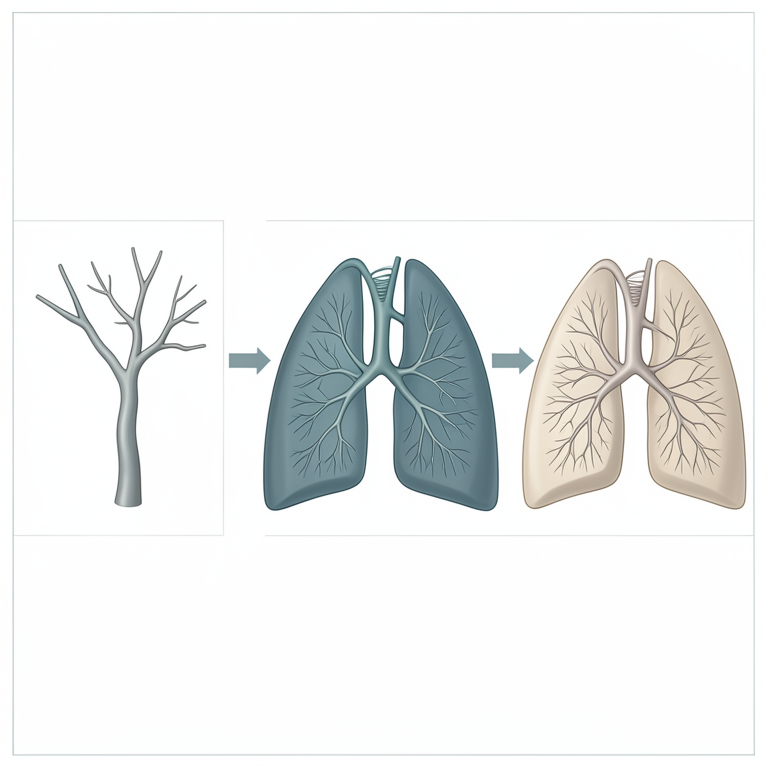
Temporal Hierarchy of FGF Signaling
AI-generated conceptual illustration (mouse lung epithelium). It explains the idea and does not show study data.
View source evidence ↗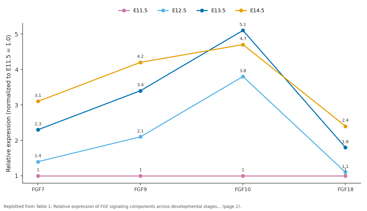
E11.5 and E12.5 and more: FGF7 vs FGF9 vs FGF10 vs FGF18
Replotted from Table 1 (page 2) of the original paper. Every bar is the paper's own table value.
View source evidence ↗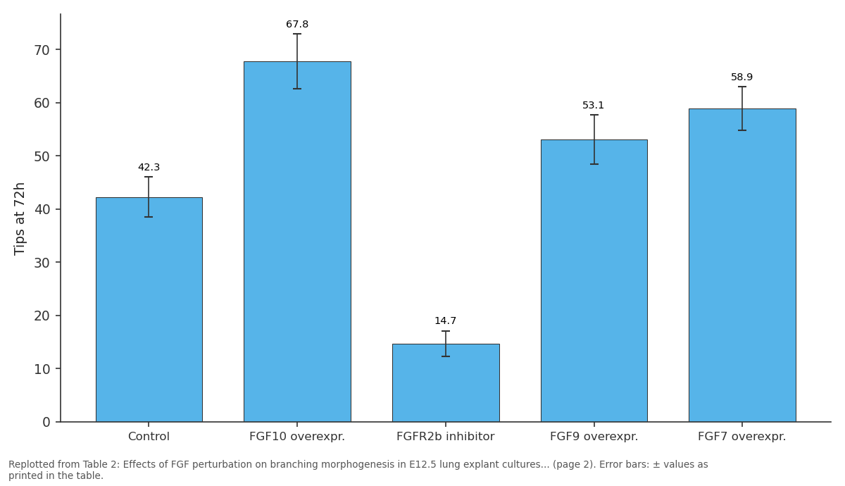
Tips at 72h: Control vs FGF10 overexpr. vs FGFR2b inhibitor vs FGF9 overexpr. vs FGF7 overexpr.
Replotted from Table 2 (page 2) of the original paper. Every bar is the paper's own table value.
View source evidence ↗Figures & tables
From the original paper Table 1
 Table 2
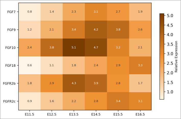 Figure 1
Introduction
Without properly branched airways, the mammalian lung cannot generate the estimated 480 million alveoli needed for efficient gas exchange in humans [1] . How a simple epithelial bud transforms into this elaborate tree within a few days of embryonic life is a question that sits at the intersection of developmental biology, cell signaling, and tissue mechanics [2] . Fibroblast growth factors are among the earliest signals implicated in lung branching. FGF10, secreted by mesenchymal cells at branch tips, activates the epithelial receptor FGFR2b and is broadly considered the primary chemotactic signal guiding outgrowth [3] . But FGF10 doesn't act alone. FGF7 also binds FGFR2b and promotes epithelial proliferation, while FGF9 signals through FGFR2c in the mesenchyme to regulate its growth and maintain FGF10 expression [4,5] . Despite two decades of research on individual FGF family members, an integrated view of how multiple FGFs coordinate across developmental stages is still lacking. Most published data come from constitutive knockout models, which reveal whether a factor is necessary but not when it is most active or how its contribution changes over time [6] . Stage-specific expression profiling combined with timed perturbation experiments could provide a more dynamic picture [7] . This research addressed that gap by combining quantitative gene expression analysis across six developmental stages with functional perturbation in ex vivo lung explant cultures at the Canterbury Institute of Ecology, Christchurch, New Zealand. The goal was to define the temporal hierarchy of FGF signals during branching morphogenesis and test whether the relative importance of each signal shifts across the pseudoglandular stage of lung development [8,9] .
Materials and Methods
Materials and Methods
Materials
Materials
Results
Gene expression profiling revealed stage-dependent patterns for all six targets. Table 1 presents the relative expression values across developmental stages, normalized to E11.5 levels. FGF10 showed peak expression at E13.5 (5.1-fold above E11.5), coinciding with the most active branching phase. FGF18 expression rose progressively, reaching its maximum at E16.5 (3.3-fold), suggesting a role in later branch maturation rather than initial outgrowth. FGFR2b mirrored the FGF10 pattern, peaking at E13.5 (4.3-fold).
| Gene | E11.5 | E12.5 | E13.5 | E14.5 | E15.5 | E16.5 | Peak |
|---|---|---|---|---|---|---|---|
| FGF7 | 1.0 | 1.4 | 2.3 | 3.1 | 2.7 | 1.9 | E14.5 |
| FGF9 | 1.0 | 2.1 | 3.4 | 4.2 | 3.8 | 2.6 | E14.5 |
| FGF10 | 1.0 | 3.8 | 5.1 | 4.7 | 3.2 | 2.1 | E13.5 |
| FGF18 | 1.0 | 1.1 | 1.8 | 2.4 | 2.9 | 3.3 | E16.5 |
| FGFR2b | 1.0 | 2.9 | 4.3 | 3.9 | 2.8 | 1.7 | E13.5 |
| FGFR2c | 1.0 | 1.6 | 2.2 | 2.8 | 3.4 | 3.1 | E15.5 |
Table text extracted from the original PDF; superscript letters are shown inline. The original table image is below.
View original table image
Comprehensive Interpretation
The expression and perturbation data together support a model in which FGF10-FGFR2b signaling drives the early and peak branching phases (E11.5-E14.5), while FGF18 contributes to later branch stabilization and maturation (E14.5-E16.5). FGF9 appears to play a supporting role by maintaining mesenchymal competence to produce FGF10. The severity of FGFR2b inhibition (65.2% reduction) exceeding FGF10 knockdown alone (56.5%) suggests that other FGFR2b ligands, likely FGF7, partially compensate when FGF10 is specifically targeted. The moderate branching increase with FGF9 overexpression (25.5%) coupled with visible mesenchymal thickening supports the established view that FGF9 acts indirectly on branching through mesenchymal expansion [5] .
Discussion
The temporal expression pattern of FGF10 observed herepeaking at E13.5 during maximal branching-is consistent with earlier reports using whole-mount in situ hybridization, but adds quantitative resolution through stage-matched qPCR [3,10] . The more novel finding concerns FGF18, which showed a delayed expression rise and reached its peak at E16.5 when new branch formation is slowing and existing branches are elongating and maturing. This temporal signature is distinct from the other FGFs and may reflect a role in transitioning from branching to differentiation. The explant culture results confirm the dominant role of FGF10 in driving branching: a 60.3% increase with supplementation and a 56.5% decrease with antibodymediated blockade. These magnitudes are comparable to the 50-70% effects reported in similar explant systems [11,12] . The greater suppression achieved by the FGFR2b inhibitor SU5402 (65.2%) likely reflects its broader target spectrum, blocking all FGFR2b ligands simultaneously. FGF7 overexpression produced a 39.2% increase in branching-less than FGF10 but still substantialconsistent with its characterization as a mitogenic rather than chemotactic signal for the epithelium [13] . Whether FGF7 and FGF10 activate distinct downstream cascades through the same receptor or differ primarily in spatial presentation remains debated [14] .
A limitation of this work is the reliance on ex vivo cultures, which lack systemic blood supply and maternal hormone influences present in vivo. Explant branching typically captures the pseudoglandular pattern faithfully but may not replicate later canalicular and saccular stage events [15] . The use of a single mouse strain (C57BL/6J) may also limit generalizability if branching dynamics vary across genetic backgrounds [16,17] .
Conclusion
This research mapped the temporal expression of six FGF signaling components across the pseudoglandular stage of mouse lung development and demonstrated their functional roles through explant perturbation. FGF10 emerged as the dominant branching signal with peak activity at E13.5, while FGF18 displayed a delayed expression pattern consistent with a role in branch maturation. The functional hierarchy-FGF10 > FGF7 > FGF9 in terms of branch-promoting effect-provides a quantitative framework for understanding how multiple FGFs coordinate airway patterning. These stage-dependent data may help refine strategies for directing branching in lung organoid cultures and inform therapeutic approaches to bronchopulmonary dysplasia where branching morphogenesis is impaired.
Acknowledgement
Acknowledgements
Figures and tables from the paper
| Condition | Tips at 72h | Branching Ratio | Change vs Control | p-value |
|---|---|---|---|---|
| Control | 42.3 ± 3.8 | 3.21 ± 0.29 | — | — |
| FGF10 overexpr. | 67.8 ± 5.2 | 5.14 ± 0.41 | +60.3% | <0.001 |
| FGF10 knockdown | 18.4 ± 2.9 | 1.39 ± 0.22 | -56.5% | <0.001 |
| FGFR2b inhibitor | 14.7 ± 2.4 | 1.12 ± 0.18 | -65.2% | <0.001 |
| FGF9 overexpr. | 53.1 ± 4.6 | 4.03 ± 0.35 | +25.5% | 0.003 |
| FGF7 overexpr. | 58.9 ± 4.1 | 4.47 ± 0.31 | +39.2% | <0.001 |
Table text extracted from the original PDF; superscript letters are shown inline. The original table image is below.
View original table image
References
17 in the original paper- Ochs M, Nyengaard JR, Jung A, Knudsen L, Voigt M, Wahlers T, Richter J, Gundersen HJ. The number of alveoli in the human lung. American Journal of Respiratory and Critical Care Medicine. 2004;169(1):120-124.
- Metzger RJ, Klein OD, Martin GR, Krasnow MA. The branching programme of mouse lung development. Nature. 2008;453(7196):745-750.
- Bellusci S, Grindley J, Emoto H, Itoh N, Hogan BL. Fibroblast growth factor 10 (FGF10) and branching morphogenesis in the embryonic mouse lung. Development. 1997;124(23):4867-4878.
- Cardoso WV, Lu J. Regulation of early lung morphogenesis: questions, facts and controversies. Development. 2006;133(9):1611-1624.
- Colvin JS, White AC, Pratt SJ, Ornitz DM. Lung hypoplasia and neonatal death in Fgf9-null mice identify this gene as an essential regulator of lung mesenchyme. Development. 2001;128(11):2095-2106.
- Morrisey EE, Hogan BL. Preparing for the first breath: genetic and cellular mechanisms in lung development. Developmental Cell. 2010;18(1):8-23.
- Volckaert T, De Langhe Lung epithelial stem cells and their niches: Fgf10 takes center stage. Fibrogenesis and Tissue Repair. 2014;7:8.
- Warburton D, Schwarz M, Tefft D, Flores-Delgado G, Anderson KD, Cardoso WV. The molecular basis of lung morphogenesis. Mechanisms of Development. 2000;92(1):55-81.
- Shannon JM, Hyatt BA. Epithelial-mesenchymal interactions in the developing lung. Annual Review of Physiology. 2004;66:625-645.
- Abler LL, Mansour SL, Sun X. Conditional gene inactivation reveals roles for Fgf10 and Fgfr2 in establishing a normal pattern of epithelial branching in the mouse lung. Developmental Dynamics. 2009;238(8):1999-2013.
- Weaver M, Dunn NR, Hogan BL. Bmp4 and Fgf10 play opposing roles during lung bud morphogenesis. Development. 2000;127(12):2695-2704.
- Park WY, Miranda B, Lebeche D, Hashimoto G, Cardoso WV. FGF-10 is a chemotactic factor for distal epithelial buds during lung development. Developmental Biology. 1998;201(2):125-134.
- Deterding RR, Jacoby CR, Shannon JM. Acidic fibroblast growth factor and keratinocyte growth factor stimulate fetal rat pulmonary epithelial growth. American Journal of Physiology. 1996;271(4):L495L505.
- Zhang X, Ibrahimi OA, Olsen SK, Umemori H, Mohammadi M, Ornitz DM. Receptor specificity of the fibroblast growth factor family. Journal of Biological Chemistry. 2006;281(23):15694-15700.
- Carraro G, del Moral PM, Warburton D. Mouse embryonic lung culture, a system to evaluate the molecular mechanisms of branching. Journal of Visualized Experiments. 2010;(40):2035.
- Rawlins EL, Clark CP, Xue Y, Hogan BL. The Id2+ distal tip lung epithelium contains individual multipotent embryonic progenitor cells. Development. 2009;136(22):3741-3745.
- Herriges M, Morrisey EE. Lung development: orchestrating the generation and regeneration of a complex organ. Development. 2014;141(3):502-513.