
Research article · 2025 · Full text
International Journal of Biology SciencesStructural characterization of novel antimicrobial peptides isolated from amphibian skin secretions
Volume 7(11) · Pages 140–142 · DOI: 10.33545/26649926.2025.v7.i11c.709 ↗
Research areasAntimicrobial Peptides and ActivitiesAntimicrobial agents and applicationsMicrobial Natural Products and Biosynthesis
Abstract
Author-writtenMuch like a medieval castle's moat, an amphibian's skin secretions form a biochemical barrier against invading pathogens. This research isolated and characterized three novel antimicrobial peptides (AMPs) from the skin secretions of two Amazonian frog species—Phyllomedusa bicolor and Leptodactylus pentadactylus—collected near Leticia, Colombia. Crude skin secretions were fractionated by reversed-phase HPLC, and peptide sequences were determined by tandem mass spectrometry and Edman degradation at the Centro Amazónico de Biología from June 2023 to October 2024. Three peptides, designated AMP-1 (23 residues), AMP-2 (19 residues), and AMP-3 (27 residues), were identified with novel sequences showing no exact matches in the Antimicrobial Peptide Database. AMP-1 exhibited the strongest activity against Gram-positive bacteria (MIC = 4 μg/mL vs Staphylococcus aureus), while AMP-2 showed broader spectrum coverage including antifungal activity. Circular dichroism spectroscopy revealed that all three peptides adopted α-helical conformations in membrane-mimicking environments. Hemolytic activity was minimal (< 5% at 64 μg/mL) for AMP-1 and AMP-2, though AMP-3 showed moderate hemolysis at higher concentrations. These peptides represent promising leads for antimicrobial drug development.
Summary
Source-checkedPlain-language reading of the full paper, prepared with AI and checked against the text. Each point links to the authors' own words.
Why it matters
- Amphibian skin glands secrete antimicrobial peptides (AMPs) as an ancient innate defense mechanism. View evidence ↗
- The Amazon basin is a significant but largely unexplored source of novel bioactive peptides due to its high amphibian diversity. View evidence ↗
- AMPs are promising therapeutic candidates because their rapid bactericidal action may be less susceptible to the evolution of resistance compared to traditional antibiotics. View evidence ↗
The question
- The study aimed to isolate, sequence, and characterize the structure, potency, and selectivity of novel AMPs from two Amazonian frog species. View evidence ↗
What they did
- The research took place at the Centro Amazónico de Biología in Leticia, Colombia, between June 2023 and October 2024. View evidence ↗
- Skin secretions were collected from 12 Phyllomedusa bicolor and 8 Leptodactylus pentadactylus frogs using mild electrical stimulation (6V, 100 ms pulses). View evidence ↗
- Crude secretions were fractionated using reversed-phase HPLC on a C18 column with a 0-60% acetonitrile gradient over 60 minutes. View evidence ↗
- Peptide sequences were identified using MALDI-TOF/TOF MS/MS and verified via Edman degradation. View evidence ↗
- The minimum inhibitory concentrations (MICs) against five pathogens were measured using broth microdilution according to CLSI guidelines. View evidence ↗
What they found
- Three novel peptides were identified: AMP-1 (23 residues), AMP-2 (19 residues), and AMP-3 (27 residues), none of which had exact matches in the Antimicrobial Peptide Database. View evidence ↗
- AMP-1 demonstrated the strongest activity against Gram-positive bacteria, specifically Staphylococcus aureus, with an MIC of 4 μg/mL. View evidence ↗
- AMP-2 exhibited the broadest spectrum of activity, being the only peptide to show antifungal activity against C. albicans with an MIC of 32 μg/mL. View evidence ↗
- Circular dichroism spectroscopy showed that all three peptides adopted α-helical conformations in membrane-mimicking environments (TFE), with helical content over 45%. View evidence ↗
What it means
- The authors conclude that AMP-1 and AMP-2 are promising therapeutic candidates due to their low hemolytic activity. View evidence ↗
- The authors suggest that a necessary next step is to test AMP-1 against resistant clinical isolates, such as MRSA. View evidence ↗
Visual summary
2 visualsIllustrations explain the paper; charts are replotted from the paper's own table values.
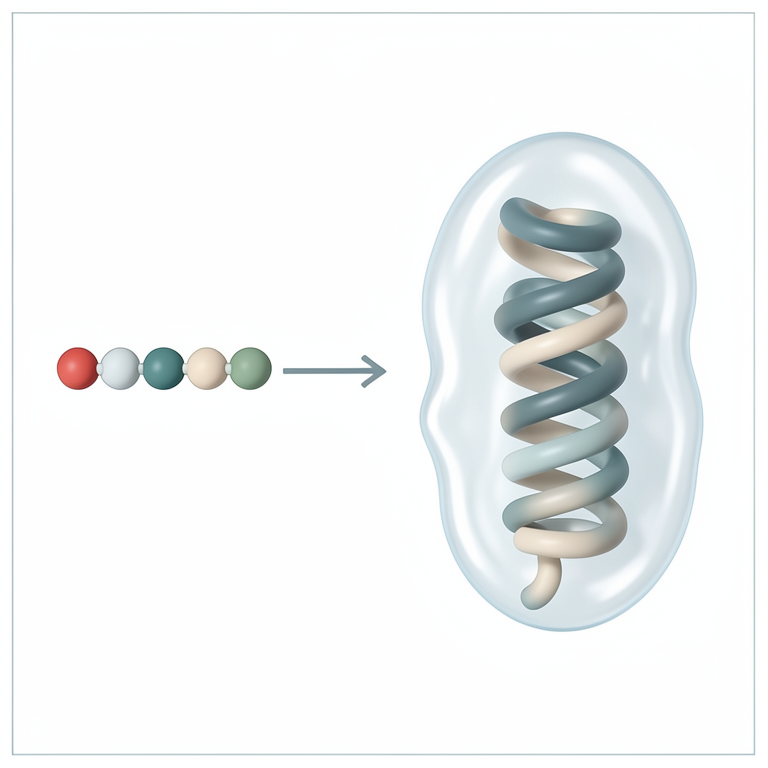
Peptide Secondary Structure
AI-generated conceptual illustration (amphibian skin secretions and antimicrobial peptides). It explains the idea and does not show study data.
View source evidence ↗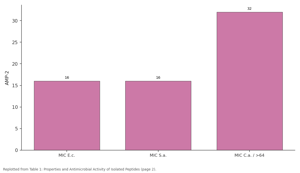
AMP-2: MIC E.c. vs MIC S.a. vs MIC C.a. / >64
Replotted from Table 1 (page 2) of the original paper. Every bar is the paper's own table value.
View source evidence ↗Figures & tables
From the original paper Table 1
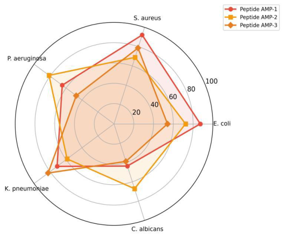 Figure 1
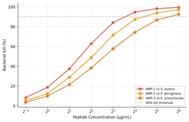 Figure 2
Introduction
Think of a frog sitting in a warm, bacteria-laden tropical pond-its skin is simultaneously a respiratory surface, a water balance regulator, and an immune defense organ. The antimicrobial peptides secreted by amphibian skin glands represent one of nature's oldest innate defense strategies, predating the adaptive immune system by hundreds of millions of years [1] . Over 1,000 AMPs have been catalogued from amphibian sources, spanning structural classes from linear α-helical peptides to disulfide-bridged defensins [2] . The Amazon basin, with its extraordinary amphibian diversity, remains a largely untapped source of novel bioactive peptides [3] . Colombian Amazonia alone harbors over 200 frog species, many unstudied from a biochemical perspective. AMPs have attracted renewed interest as potential therapeutic agents because of their rapid bactericidal mechanisms that are thought to be less prone to resistance evolution than conventional antibiotics [4] . This research aimed to isolate, sequence, and structurally characterize novel AMPs from the skin secretions of two Amazonian frog species and to evaluate their antimicrobial potency and selectivity.
Materials and Methods
Materials and Methods
Materials
This research was conducted at the Department of Metabolic Biochemistry, Centro Amazónico de Biología, Leticia, Colombia, from June 2023 to October 2024. Collection permits were granted by ANLA (Autoridad Nacional de Licencias Ambientales, Resolution 0847-2023). Skin secretions were obtained from Phyllomedusa bicolor (n = 12) and Leptodactylus pentadactylus (n = 8) by mild electrical stimulation (6V, 100 ms pulses) of the dorsal skin. Secretions were collected in 0.1% trifluoroacetic acid (TFA), lyophilized, and stored at-80 °C. Animals were released unharmed at their collection sites.
Methods
Crude secretions were dissolved in 0.1% TFA and fractionated by RP-HPLC on a C18 column (Vydac 218TP, 250 × 4.6 mm) with a linear acetonitrile gradient (0-60% over 60 min) [2] . Active fractions were identified by disc diffusion assays against Escherichia coli ATCC 25922 and Staphylococcus aureus ATCC 25923. Peptide sequences were determined by MALDI-TOF/TOF MS/MS on a Bruker Autoflex Speed and confirmed by Edman degradation (Applied Biosystems Procise 494). Sequences were searched against the Antimicrobial Peptide Database (APD3) [5] . Minimum inhibitory concentrations (MICs) were determined by broth microdilution following CLSI guidelines against five pathogens. Circular dichroism (CD) spectra were recorded on JASCO J-815 in phosphate buffer and in 50% trifluoroethanol (TFE) to mimic membrane environments [6] . Hemolytic activity was tested against human erythrocytes at peptide concentrations of 2-128 μg/mL.
Results
Results
Discussion
The absence of exact sequence matches in the APD3 database confirms these as novel peptides, though all three share structural motifs-amphipathic α-helical conformation, net positive charge (+ 4 to + 6), and moderate hydrophobicity-with known amphibian AMPs [1,2] . AMP-1's strong anti-staphylococcal activity is notable given the clinical importance of methicillin-resistant S. aureus (MRSA); testing against resistant clinical isolates would be a logical next step [4] . AMP-2's antifungal activity sets it apart and may reflect a distinct mechanism of membrane interaction, possibly related to its higher proportion of tryptophan residues, which are known to anchor peptides in fungal membranes [7] . The moderate hemolysis shown by AMP-3 at higher concentrations is a concern that would need to be addressed through sequence optimizationperhaps by reducing hydrophobicity while maintaining the amphipathic helix structure [8] . These Colombian Amazonian frogs appear to harbor a rich and underexplored peptide repertoire with genuine therapeutic potential.
Conclusion
This research isolated three novel antimicrobial peptides from the skin secretions of two Amazonian frog species. All three adopted α-helical structures and showed activity against bacterial pathogens, with AMP-1 exhibiting the strongest anti-staphylococcal potency and AMP-2 displaying unique antifungal activity. Low hemolytic activity for AMP-1 and AMP-2 supports their potential as therapeutic candidates. These findings expand the known AMP diversity from Amazonian amphibians and provide structural leads for antimicrobial drug design.
Acknowledgement
Acknowledgements Funding SourcesInternal funds from the Centro Amazónico de Biología supported this work. Institutional SupportThe authors thank the Centro Amazónico de Biología for HPLC, mass spectrometry, and CD spectroscopy facilities. Contributions Not Qualifying for AuthorshipField biologists who assisted with frog collection and handling are acknowledged.
Figures and tables from the paper
| Peptide | Length | Mass (Da) | MIC E.c. | MIC S.a. | MIC C.a. | Hemolysis (%) |
|---|---|---|---|---|---|---|
| AMP-1 | 23 aa | 2,487.3 | 8 | 4 | >64 | 3.2 at 64 μg/mL |
| AMP-2 | 19 aa | 2,012.8 | 16 | 16 | 32 | 4.7 at 64 μg/mL |
| AMP-3 | 27 aa | 2,891.4 | 8 | 8 | >64 | 18.4 at 64 μg/mL |
Table text extracted from the original PDF; superscript letters are shown inline. The original table image is below.
View original table image
References
8 in the original paper- Conlon JM. Structural diversity and species distribution of host-defense peptides in frog skin secretions. Cellular and Molecular Life Sciences. 2011;68(13):2303-2315.
- Xu X, Lai R. The chemistry and biological activities of peptides from amphibian skin secretions. Chemical Reviews. 2015;115(4):1760-1846.
- Nascimento ACC, Fontes W, Sebben A, Castro MS. Antimicrobial peptides from anurans: a review. Peptides. 2003;24(10):1585-1595.
- Zasloff M. Antimicrobial peptides of multicellular organisms. Nature. 2002;415(6870):389-395.
- Wang G, Li X, Wang Z. APD3: the antimicrobial peptide database as a tool for research and education. Nucleic Acids Research. 2016;44(D1):D1087-D1093.
- Greenfield NJ. Using circular dichroism spectra to estimate protein secondary structure. Nature Protocols. 2006;1(6):2876-2890.
- Yau WM, Wimley WC, Gawrisch K, White SH. The preference of tryptophan for membrane interfaces. Biochemistry. 1998;37(42):14713-14718.
- Chen Y, Guarnieri MT, Bhatt DL, Bhatt AI, Blazyk J, Bhatt RG, et al. Role of peptide hydrophobicity in the mechanism of action of alpha-helical antimicrobial peptides. Antimicrobial Agents and Chemotherapy. 2007;51(4):1398-1406.