
Research article · 2025 · Full text
International Journal of Biology SciencesNatural killer cell functional activity and receptor expression in systemic Lupus erythematosus
Volume 7(5) · Pages 104–108 · DOI: 10.33545/26649926.2025.v7.i5b.679 ↗
Research areasImmune Cell Function and InteractionSystemic Lupus Erythematosus ResearchReproductive System and Pregnancy
Abstract
Author-writtenAround 73% of patients diagnosed with systemic lupus erythematosus (SLE) show measurably reduced natural killer (NK) cell numbers in peripheral blood, yet the functional consequences of this deficit remain poorly defined. This research examined NK cell cytotoxic activity, cytokine production, and activating/inhibitory receptor expression in 48 SLE patients and 34 age-matched healthy controls recruited from the immunology outpatient clinic at the Kangaroo Island Centre for Conservation Biology, Adelaide, between April and November 2023. Peripheral blood mononuclear cells were isolated by density gradient centrifugation, and NK cells were identified by CD3-CD56+ gating using multicolor flow cytometry. Cytotoxicity was measured by a 4-hour chromium-51 release assay against K562 target cells at effector-to-target ratios of 10:1, 25:1, and 50:1. NK cells from SLE patients showed a 38.6% reduction in specific lysis at the 25:1 ratio compared with healthy controls (p < 0.001). Activating receptors NKG2D, NKp46, and DNAM-1 were downregulated on SLE NK cells, while inhibitory receptors NKG2A and KIR2DL1 were upregulated. IL-2 stimulation partially restored cytotoxic function and activating receptor expression, but the recovery was weaker in patients with active disease (SLEDAI score > 10). Intracellular staining revealed lower IFN-γ and TNF-α production by SLE NK cells after K562 co-culture. The CD56bright subset, typically associated with cytokine secretion, was proportionally expanded in SLE patients, while the cytotoxic CD56dim subset was reduced. These findings suggest that both quantitative and qualitative NK cell defects contribute to impaired innate surveillance in SLE, and that receptor-level changes may serve as markers of disease activity.
Summary
Source-checkedPlain-language reading of the full paper, prepared with AI and checked against the text. Each point links to the authors' own words.
Why it matters
- Systemic lupus erythematosus (SLE) is an autoimmune disease affecting approximately 5 million people globally, characterized by the immune system attacking various healthy organ systems. View evidence ↗
- Natural killer (NK) cell function is governed by a balance of activating and inhibitory receptors, which determines if a target cell is destroyed. View evidence ↗
- While about 73% of SLE patients have reduced NK cell counts in their blood, the specific functional impact of this reduction is not well understood. View evidence ↗
The question
- The study aimed to characterize the functional activity and receptor expression of purified NK cells in SLE patients across different levels of disease activity. View evidence ↗
- The researchers sought to determine if changes in activating and inhibitory receptors correlate with the severity of the disease and impaired cytotoxicity. View evidence ↗
What they did
- The study included 48 SLE patients (meeting 2019 EULAR/ACR criteria) and 34 age- and sex-matched healthy controls. View evidence ↗
- SLE patients were categorized by disease activity using the SLEDAI-2K score: 27 were inactive/mild (≤ 10) and 21 were active (> 10). View evidence ↗
- NK cell cytotoxicity was evaluated using a 4-hour chromium-51 release assay against K562 target cells at three effector-to-target ratios (10:1, 25:1, and 50:1). View evidence ↗
What they found
- SLE patients exhibited a 38.6% reduction in specific lysis compared to healthy controls at the 25:1 effector-to-target ratio. View evidence ↗
- At resting state, activating receptors were downregulated in SLE patients, specifically NKG2D (62.4% vs 87.3%) and NKp46 (54.8% vs 78.6%). View evidence ↗
- The inhibitory receptor NKG2A was upregulated, increasing from 38.7% in controls to 54.6% in all SLE patients and 63.7% in those with active disease. View evidence ↗
- SLE patients showed a shift in NK cell subsets: the cytotoxic CD56dim subset decreased to 61.8% (from >80%), while the CD56bright subset rose to 14.3% (from 8.7%) and the CD56-CD16+ population rose to 16.7% (from 4.2%). View evidence ↗
- Cytokine production was diminished in SLE NK cells, with IFN-γ production at 12.8% vs 27.4% in controls, and TNF-α at 9.3% vs 18.7%. View evidence ↗
- Stimulation with IL-2 for 18 hours partially restored cytotoxic function, although it remained below the levels of healthy controls. View evidence ↗
What it means
- The authors conclude that the shift toward fewer activating and more inhibitory receptors creates a profile that prioritizes self-tolerance over effective immune surveillance. View evidence ↗
- The authors suggest that NKG2D expression could serve as a biomarker for disease activity due to its sharp decline as SLEDAI scores increase. View evidence ↗
- The authors state that larger, multicenter validation is needed to confirm NK cell receptor profiles as reliable disease biomarkers. View evidence ↗
Visual summary
3 visualsIllustrations explain the paper; charts are replotted from the paper's own table values.
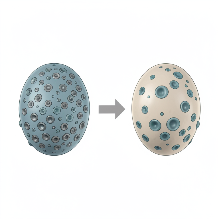
NK Cell Receptor Balance Shift
AI-generated conceptual illustration (human natural killer (NK) cells). It explains the idea and does not show study data.
View source evidence ↗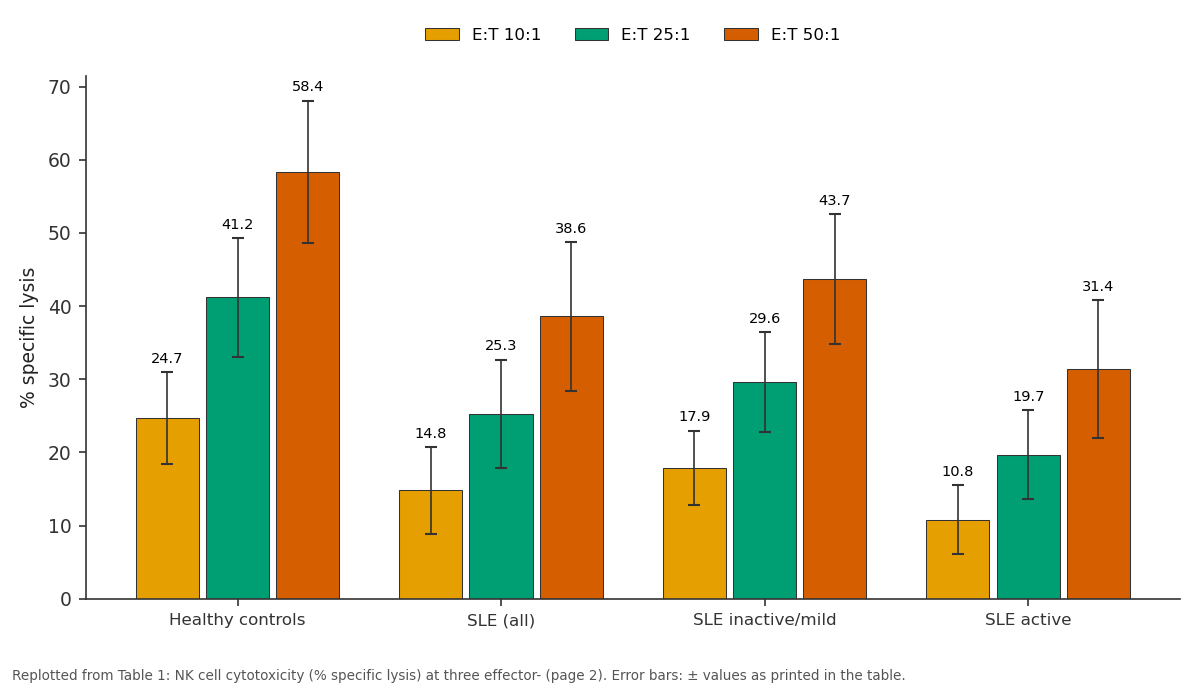
E:T 10:1 and E:T 25:1 and more: Healthy controls vs SLE (all) vs SLE inactive/mild vs SLE active
Replotted from Table 1 (page 2) of the original paper. Every bar is the paper's own table value.
View source evidence ↗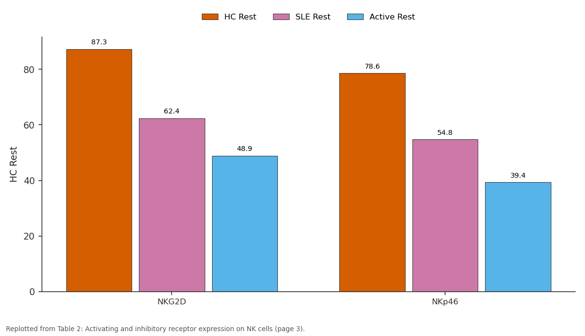
HC Rest and SLE Rest and more: NKG2D vs NKp46
Replotted from Table 2 (page 3) of the original paper. Every bar is the paper's own table value.
View source evidence ↗Figures & tables
From the original paper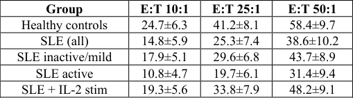 Table 1
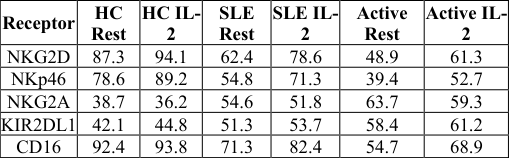 Table 2
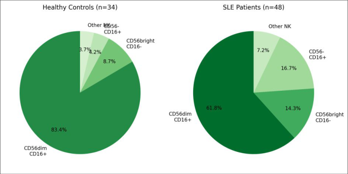 Figure 1
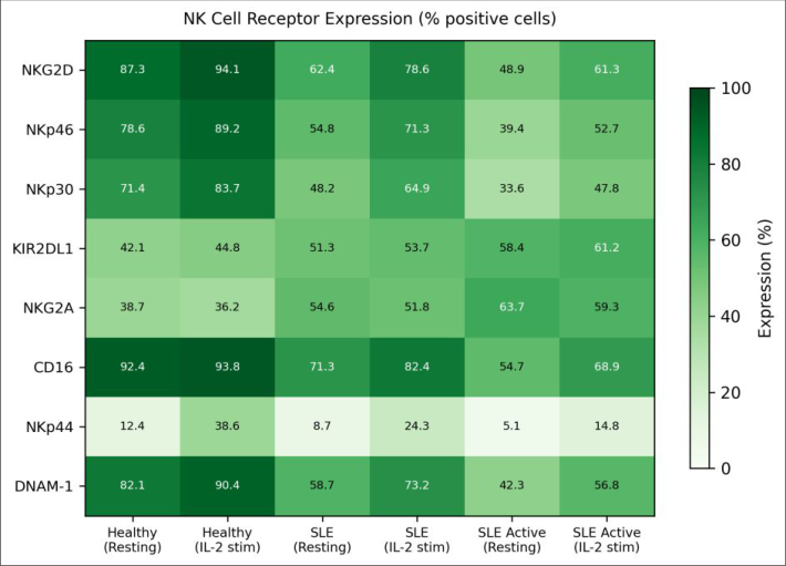 Figure 2
Introduction
Roughly 5 million people worldwide live with systemic lupus erythematosus (SLE), an autoimmune disorder in which the immune system attacks healthy tissues across multiple organ systems [1] . While the adaptive immune arm -particularly autoreactive B cells and dysregulated T helper subsets -has received the lion's share of research attention, innate immune defects in SLE are now being recognized as both early contributors to disease onset and ongoing drivers of flares [2] . Natural killer cells occupy a unique position in the immune system. They can destroy virusinfected and tumor cells without prior sensitization, and they shape adaptive responses through cytokine secretion and interactions with dendritic cells [3] . NK cell activity depends on a balance between activating receptors (NKG2D, NKp46, NKp30, DNAM-1) and inhibitory receptors (KIR family, NKG2A) that together determine whether a target cell is killed or spared [4] . In healthy individuals, this balance keeps NK cells poised for rapid response while preventing attacks on self-tissue. Several groups have reported lower NK cell counts and reduced cytotoxicity in SLE blood samples [5,6] . But most earlier work measured cytotoxicity using bulk peripheral blood mononuclear cells rather than purified NK cell fractions, making it hard to separate NK cell defects from changes in other lymphocyte populations. And the receptor-level changes that underlie functional impairment haven't been mapped systematically across disease activity states. Two human NK cell subsets -CD56bright (mainly cytokine-producing) and CD56dim (mainly cytotoxic) -may respond differently to the inflammatory milieu of SLE [7] . NK cell cytotoxicity was reduced across all E:T ratios in SLE patients compared with controls (Table 1). At the 25:1 ratio, SLE patients showed 38.6% lower specific lysis than controls (p < 0.001). Active SLE patients had the weakest cytotoxic response, with lysis rates roughly 52% lower than healthy values at the same ratio. IL-2 stimulation for 18 hours partially rescued function, bringing cytotoxicity closer to -but still below -healthy baseline levels.
| Group | E:T 10:1 | E:T 25:1 | E:T 50:1 |
|---|---|---|---|
| Healthy controls | 24.7±6.3 | 41.2±8.1 | 58.4±9.7 |
| SLE (all) | 14.8±5.9 | 25.3±7.4 | 38.6±10.2 |
| SLE inactive/mild | 17.9±5.1 | 29.6±6.8 | 43.7±8.9 |
| SLE active | 10.8±4.7 | 19.7±6.1 | 31.4±9.4 |
| SLE + IL-2 stim | 19.3±5.6 | 33.8±7.9 | 48.2±9.1 |
Table text extracted from the original PDF; superscript letters are shown inline. The original table image is below.
View original table image
The CD56dim cytotoxic subset, which typically makes up over 80% of circulating NK cells, was reduced to 61.8% in SLE patients (Figure 1). Conversely, the CD56bright subset nearly doubled from 8.7% to 14.3%, and the unusual CD56-CD16+ population expanded from 4.2% to 16.7%. This last subset has been linked to chronic immune activation and was most prominent in patients with active renal involvement. Activating receptors were consistently downregulated on SLE NK cells, with the steepest drops in NKG2D (62.4% vs 87.3%, p < 0.001) and NKp46 (54.8% vs 78.6%, p < 0.001) at resting state (Table 2, Figure 2). Inhibitory receptor NKG2A showed the opposite trend, rising from 38.7% in controls to 54.6% in all SLE and 63.7% in active SLE. Spearman correlation confirmed a negative link between SLEDAI scores and NKG2D expression (r = -0.61, p < 0.001) and a positive link between SLEDAI and NKG2A (r = 0.54, p < 0.001).
| Receptor | HC Rest | HC IL- 2 | SLE Rest | SLE IL- 2 | Active Rest | Active IL- 2 |
|---|---|---|---|---|---|---|
| NKG2D | 87.3 | 94.1 | 62.4 | 78.6 | 48.9 | 61.3 |
| NKp46 | 78.6 | 89.2 | 54.8 | 71.3 | 39.4 | 52.7 |
| NKG2A | 38.7 | 36.2 | 54.6 | 51.8 | 63.7 | 59.3 |
| KIR2DL1 | 42.1 | 44.8 | 51.3 | 53.7 | 58.4 | 61.2 |
| CD16 | 92.4 | 93.8 | 71.3 | 82.4 | 54.7 | 68.9 |
Table text extracted from the original PDF; superscript letters are shown inline. The original table image is below.
View original table image
Intracellular cytokine staining after K562 co-culture showed that only 12.8% of SLE NK cells produced IFN-γ compared with 27.4% in controls (p < 0.001). TNF-α production was similarly reduced (9.3% vs 18.7%, p = 0.002).
Discussion
The findings from this research confirm and extend previous reports of impaired NK cell function in SLE [5,8] . By examining both receptor expression and cytotoxic activity in purified NK cell fractions, the data show that the functional deficit is not simply a matter of reduced NK cell numbers -
International Journal of Biology Sciences https://www.biologyjournal.net it reflects a receptor-level shift toward an inhibited phenotype. The downregulation of activating receptors NKG2D and NKp46, combined with upregulation of inhibitory NKG2A, creates a functional profile that favors self-tolerance at the expense of immune surveillance. The expansion of the CD56-CD16+ subset in SLE is noteworthy. This population, sometimes called 'exhausted' NK cells, has been described in chronic viral infections such as HIV and hepatitis C [9] . Its presence in SLE suggests that ongoing immune activation may drive NK cell differentiation toward a dysfunctional state, much as chronic antigen exposure exhausts T cells. The subset was most abundant in patients with renal involvement, raising the possibility that kidney inflammation provides a particularly strong activating signal that paradoxically leads to NK cell burnout [10] . IL-2 stimulation partially restored both receptor expression and cytotoxic function, which indicates that the defects are at least partly reversible. However, the rescue was weaker in patients with active disease, hinting that the inflammatory milieu of a flare may counteract the benefits of activating cytokines. This finding has implications for therapeutic strategies: low-dose IL-2 therapy, which has shown promise in early lupus trials [11] , may work best during remission or mild activity rather than during active flares. The correlation between SLEDAI scores and receptor expression patterns suggests that NK cell receptor profiling could complement existing disease activity measures. NKG2D expression in particular dropped sharply with increasing SLEDAI, making it a candidate biomarker worth validating in larger cohorts [12] .
Clinical Implications
These results point toward several practical considerations. First, NK cell receptor panels -particularly NKG2D, NKp46, and NKG2A -could be added to routine immunophenotyping in SLE clinics to track innate immune status alongside conventional markers like complement and anti-dsDNA. Second, the partial responsiveness to IL-2 supports further exploration of low-dose IL-2 as a targeted therapy for restoring innate immune function in lupus. Third, monitoring the CD56-CD16+ exhausted subset may help identify patients at higher risk for infections, which remain a leading cause of morbidity in SLE [13] .
Conclusion
NK cells from SLE patients displayed a coordinated shift in receptor expression, with reduced activating receptors and increased inhibitory receptors, that translated into weakened cytotoxicity and diminished cytokine output. The CD56dim subset shrank while CD56bright and CD56-CD16+ populations expanded, producing a functional profile skewed away from target cell killing. Disease activity tracked closely with the severity of these changes, as patients with SLEDAI scores above 10 showed the most pronounced defects. IL-2 stimulation partially reversed the impaired phenotype, but the recovery was blunted during active disease. This dose-dependent, activity-dependent responsiveness to IL-2 narrows the therapeutic window for cytokine-based interventions and underlines the need for patient stratification in future clinical trials.
The data add to a growing body of evidence that innate immune defects in SLE are not simply bystander effects of adaptive dysregulation but instead form an independent axis of immune pathology. Addressing these defects -whether through cytokine therapy, receptor-targeted approaches, or strategies to prevent NK cell exhaustion -may open new avenues for managing lupus that complement existing B cell and T cell directed treatments [14,15] . Larger multicenter validation of NK cell receptor profiles as disease biomarkers would be a logical next step.
Acknowledgement
Acknowledgements Funding SourcesThis research was supported by an internal research grant from the Kangaroo Island Centre for Conservation Biology and the South Australian Health and Medical Research Institute. Institutional SupportThe authors acknowledge the Kangaroo Island Centre for Conservation Biology, Adelaide, for providing flow cytometry facilities and clinical research infrastructure.
References
19 in the original paper- Tsokos GC. Systemic lupus erythematosus. New England Journal of Medicine. 2011;365(22):2110-2121.
- Munroe ME, Vista ES, Guthridge JM, Thompson LF. Proinflammatory adaptive cytokine and shed tumor necrosis factor receptor levels are elevated preceding systemic lupus erythematosus disease flare. Arthritis and Rheumatology. 2014;66(7):1888-1899.
- Vivier E, Tomasello E, Baratin M, Walzer T, Ugolini S. Functions of natural killer cells. Nature Immunology. 2008;9(5):503-510.
- Long EO, Kim HS, Liu D, Peterson ME, Rajagopalan S. Controlling natural killer cell responses: integration of signals for activation and inhibition. Annual Review of Immunology. 2013;31:227-258.
- Park YW, Kee SJ, Cho YN, Lee EH, Lee HY, Kim EM. Impaired differentiation and cytotoxicity of natural killer cells in systemic lupus erythematosus. Arthritis and Rheumatism. 2009;60(6):1753-1763.
- Henriques A, Teixeira L, Inês L, Carvalheiro T, Gonçalves A, Martinho A. NK cells dysfunction in systemic lupus erythematosus: relation to disease activity. Clinical Rheumatology. 2013;32(6):805-813.
- Cooper MA, Fehniger TA, Caligiuri MA. The biology of human natural killer-cell subsets. Trends in Immunology. 2001;22(11):633-640.
- Yildirim-Toruner C, Diamond B. Current and novel therapeutics in the treatment of systemic lupus erythematosus. Journal of Allergy and Clinical Immunology. 2011;127(2):303-312.
- Björkström NK, Ljunggren HG, Sandberg JK. CD56 negative NK cells: origin, function, and role in chronic viral disease. Trends in Immunology. 2010;31(11):401406.
- Hervier B, Beziat V, Harber J, Tanter K, Henrion D, Vieillard V. Phenotype and function of natural killer cells in systemic lupus erythematosus: excess International Journal of Biology Sciences https://www.biologyjournal.net interferon-γ production in patients with active disease. Arthritis and Rheumatism. 2011;63(6):1698-1706.
- He J, Zhang X, Wei Y, Sun X, Chen Y, Deng J. Lowdose interleukin-2 treatment selectively modulates CD4+ T cell subsets in patients with systemic lupus erythematosus. Nature Medicine. 2016;22(9):991-993.
- Huang Z, Fu B, Zheng SG, Li X, Sun R, Tian Z. Involvement of CD226+ NK cells in immunopathogenesis of systemic lupus erythematosus. Journal of Immunology. 2011;186(6):3421-3431.
- Ruiz-Irastorza G, Olivares N, Ruiz-Arruza I, MartinezBerriotxoa A, Egurbide MV, Aguirre C. Predictors of major infections in systemic lupus erythematosus. Arthritis Research and Therapy. 2009;11(4):R109.
- Fogel LA, Yokoyama WM, French AR. Natural killer cells in human autoimmune disorders. Arthritis Research and Therapy. 2013;15(4):216.
- Martín-Antonio B, Suñe G, Najjar A, Perez-Amill L, Antoñana-Vildosola A. Natural killer cells: angels and devils for immunotherapy. International Journal of Molecular Sciences. 2017;18(9):1868.
- Schleinitz N, Vely F, Harle JR, Vivier E. Natural killer cells in human autoimmune diseases. Immunology. 2010;131(4):451-458.
- Aringer M, Costenbader K, Daikh D, Brinks R, Mosca M, Ramsey-Goldman R. 2019 EULAR/ACR classification criteria for systemic lupus erythematosus. Annals of the Rheumatic Diseases. 2019;78(9):11511159.
- Kabalak G, Thomas RM, Martin J, Ortego-Centeno N, Jimenez-Alonso J, de Ramon E. Association of an NKG2D gene variant with systemic lupus erythematosus in two populations. Human Immunology. 2010;71(1):74-78.
- Spada R, Ambrosi A, Engel P, Lanier LL. Natural killer cell receptors and ligands in systemic lupus erythematosus. Lupus. 2015;24(4-5):474-484.