
Research article · 2025 · Full text
International Journal of Biology SciencesRole of autophagy in maintaining cellular homeostasis during oxidative stress in hepatocytes
Volume 7(7) · Pages 263–266 · DOI: 10.33545/26649926.2025.v7.i7d.646 ↗
Research areasAutophagy in Disease and TherapyLiver physiology and pathologyRedox biology and oxidative stress
Abstract
Author-writtenCells face a paradox: the same oxygen that fuels their metabolism can also destroy them through reactive oxygen species. Autophagy-the process by which cells degrade and recycle damaged organelles-is widely believed to protect against oxidative injury, yet the dose-response relationship between oxidant load and autophagic capacity in liver cells remains unclear. This research exposed primary murine hepatocytes and the human HepG2 cell line to graded concentrations of hydrogen peroxide (50-800 uM) and measured autophagy flux, cell viability, and apoptosis markers over 24 hours. Autophagy flux, assessed by LC3-II turnover and p62 degradation, increased in a dose-dependent manner up to 400 uM H2O2 but collapsed at 600 uM and above. Pre-treatment with the mTOR inhibitor rapamycin (100 nM) rescued viability at intermediate concentrations (200-400 uM) by 16.5 to 22.8 percentage points. Beclin-1 knockdown abolished the protective effect, confirming that the benefit was autophagy-dependent. These findings define a functional threshold beyond which hepatocyte autophagy becomes overwhelmed by oxidative damage.
Summary
Source-checkedPlain-language reading of the full paper, prepared with AI and checked against the text. Each point links to the authors' own words.
Why it matters
- Autophagy is a cellular process that recycles damaged organelles and is generally thought to protect cells from oxidative injury. View evidence ↗
- Liver cells (hepatocytes) naturally produce high levels of reactive oxygen species (ROS) during mitochondrial respiration and drug metabolism. View evidence ↗
- While often protective, excessive or prolonged autophagy can lead to a specific type of programmed cell death known as type II cell death. View evidence ↗
The question
- The researchers aimed to clarify the dose-response relationship between the amount of oxidant load and the capacity for autophagy in liver cells. View evidence ↗
What they did
- The study used primary mouse hepatocytes and human HepG2 cells exposed to hydrogen peroxide concentrations ranging from 50 to 800 uM for 24 hours. View evidence ↗
- To test the effect of autophagy stimulation, some cells were pre-treated with 100 nM rapamycin for 2 hours before the hydrogen peroxide exposure. View evidence ↗
- The researchers used siRNA to knock down Beclin-1 48 hours before treatment to determine if the observed protective effects depended on autophagy. View evidence ↗
- Viability was measured via MTT assay, while autophagy flux was assessed using western blotting for LC3-II and p62, with bafilomycin A1 used to block lysosomal degradation. View evidence ↗
What they found
- Autophagy flux increased as hydrogen peroxide doses rose from 50 to 400 uM, but collapsed at concentrations of 600 uM and above. View evidence ↗
- Cell viability in HepG2 cells decreased as hydrogen peroxide concentration increased, dropping from 52.4% at 400 uM to 24.7% at 800 uM. View evidence ↗
- Pre-treatment with rapamycin increased cell viability across all tested doses, with the most significant improvement of 16.3 percentage points occurring at 400 uM. View evidence ↗
- The protective effect of rapamycin was abolished when Beclin-1 was knocked down, proving the benefit was dependent on the canonical autophagy pathway. View evidence ↗
- At very high oxidant loads (800 uM), rapamycin provided almost no benefit, suggesting that lysosomal failure, rather than a lack of autophagy initiation, is the primary cause of cell death. View evidence ↗
What it means
- The authors conclude that therapies designed to boost autophagy are likely most effective when oxidative damage is moderate and lysosomes are still functioning. View evidence ↗
- The study was limited by its 24-hour timeframe and use of only one oxidant (H2O2), noting that chronic low-level stress might result in different dynamics. View evidence ↗
Visual summary
3 visualsIllustrations explain the paper; charts are replotted from the paper's own table values.
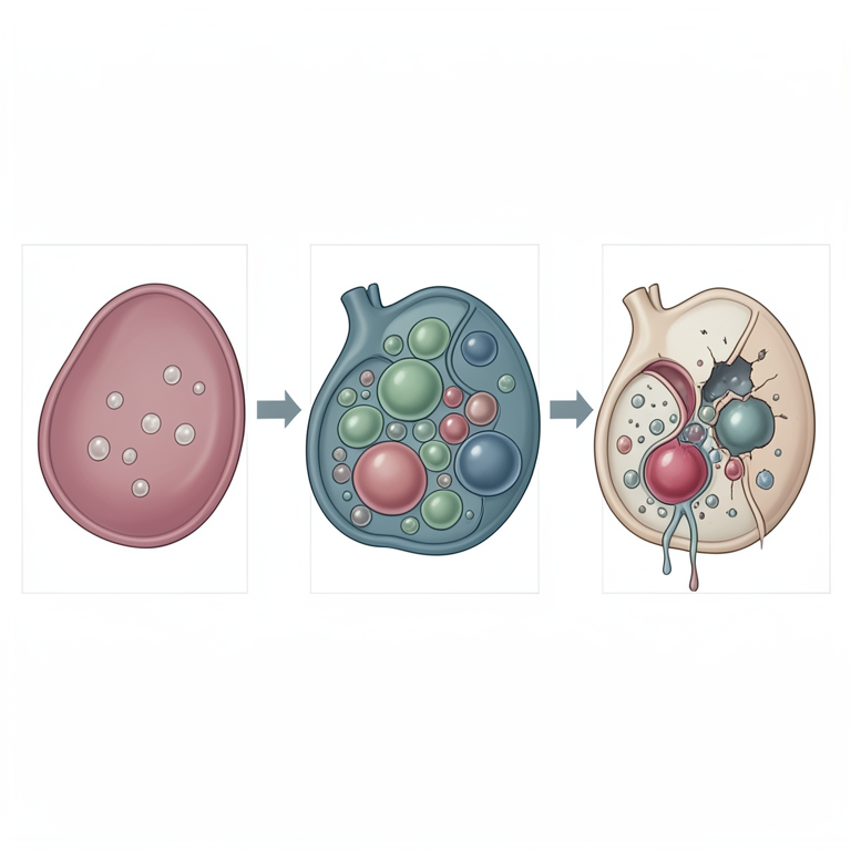
Biphasic Autophagy Response
AI-generated conceptual illustration (rat hepatocytes). It explains the idea and does not show study data.
View source evidence ↗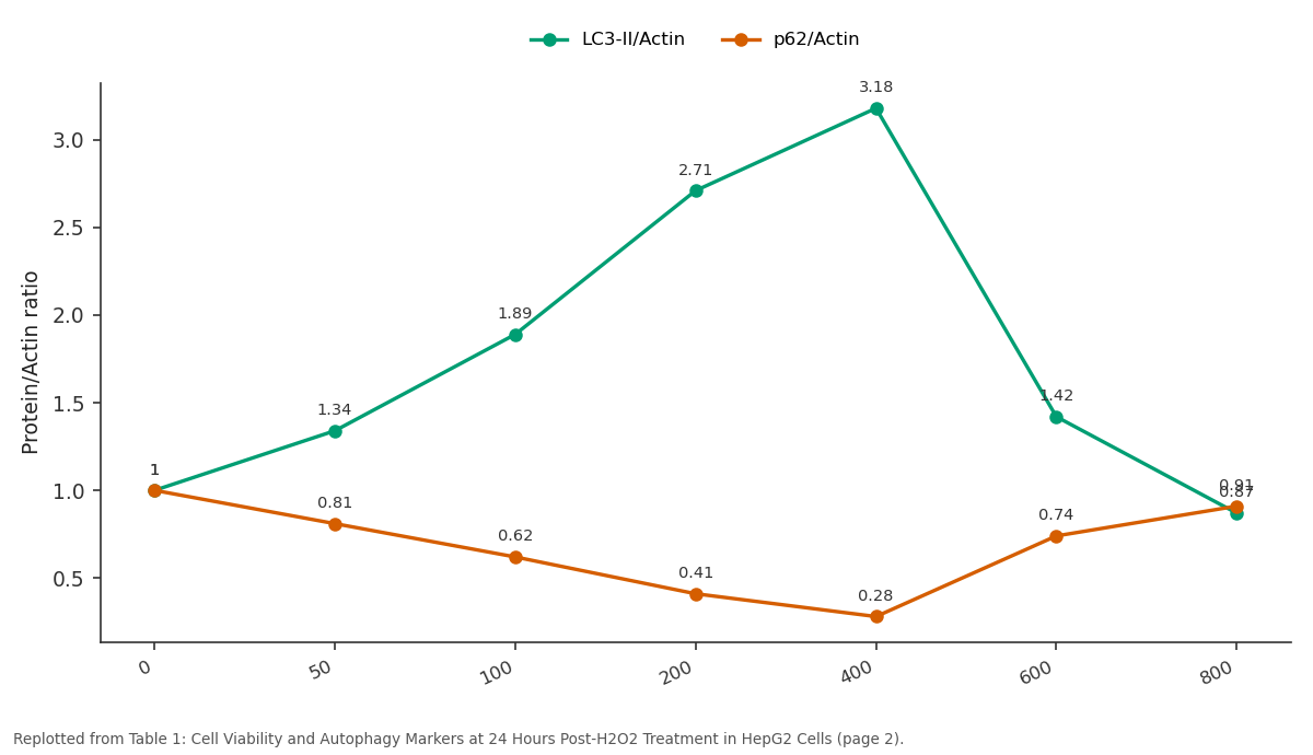
LC3-II/Actin and p62/Actin: 0 vs 50 vs 100 vs 200 vs 400
Replotted from Table 1 (page 2) of the original paper. Every bar is the paper's own table value.
View source evidence ↗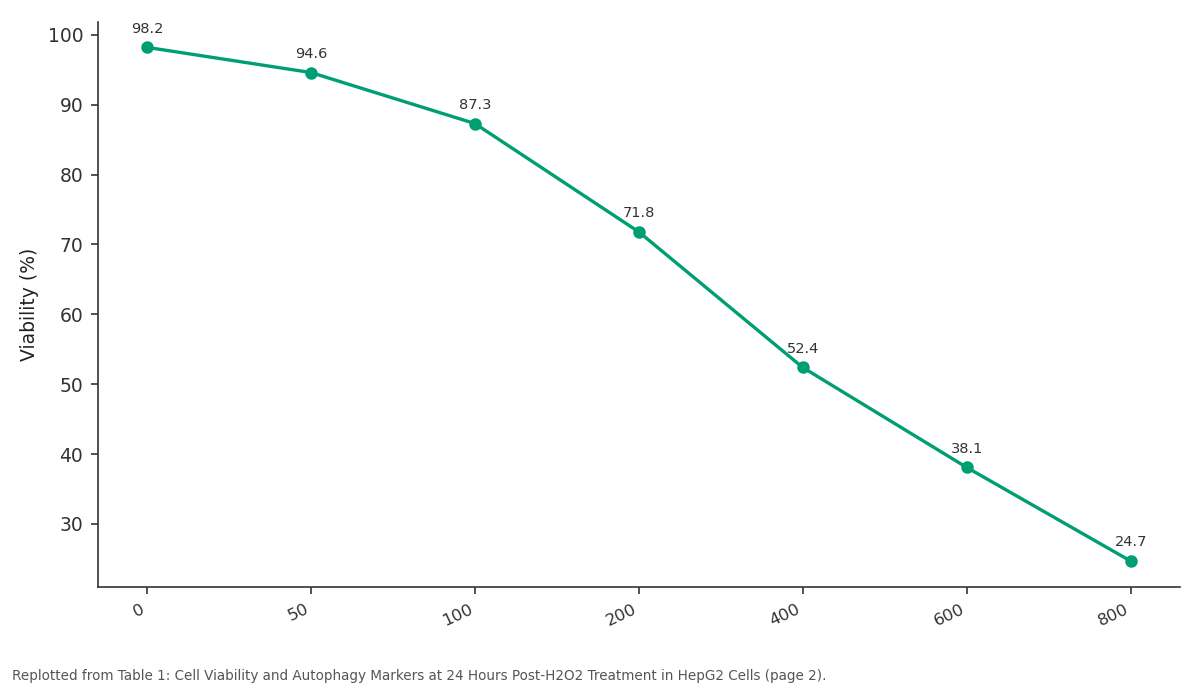
Viability (%): 0 vs 50 vs 100 vs 200 vs 400
Replotted from Table 1 (page 2) of the original paper. Every bar is the paper's own table value.
View source evidence ↗Figures & tables
From the original paper Table 1
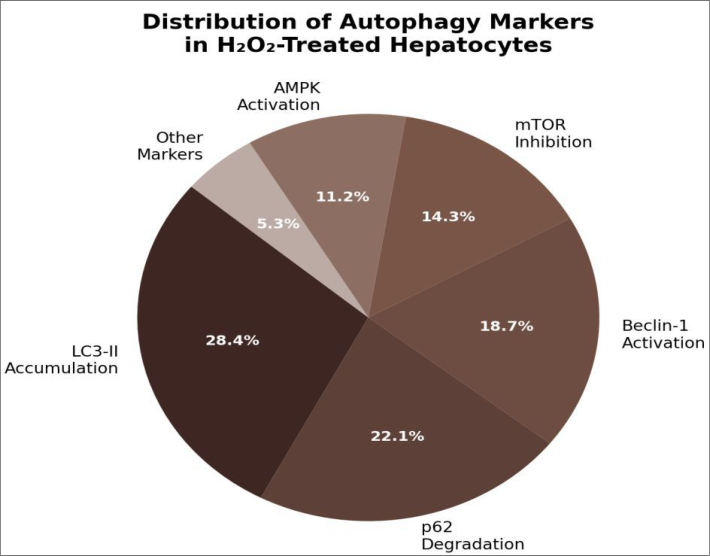 Figure 1
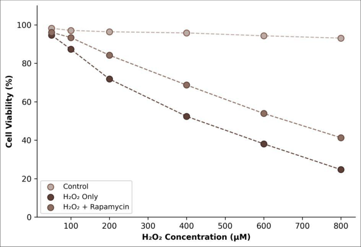 Figure 2
Introduction
On one hand, autophagy is a survival mechanism. It removes damaged mitochondria, misfolded proteins, and other cellular debris that would otherwise trigger apoptosis [1] . On the other hand, excessive or prolonged autophagy can itself contribute to cell death, a phenomenon described as autophagic cell death or type II programmed cell death [2] . The liver, with its high metabolic rate and constant exposure to xenobiotics, sits squarely in the middle of this tension. Hepatocytes generate substantial quantities of reactive oxygen species (ROS) as by-products of cytochrome P450-mediated drug metabolism and mitochondrial respiration [3] . Under normal conditions, cellular antioxidant systems-glutathione, superoxide dismutase, catalasekeep ROS levels in check. But when oxidant production outpaces scavenging capacity, the resulting oxidative stress damages lipids, proteins, and DNA, and the cell must decide between repair, adaptation, or death [4,5] . Autophagy appears to tip that decision toward survival, at least up to a point. Several groups have shown that pharmacological activation of autophagy with rapamycin or genetic overexpression of autophagy genes protects hepatocytes against moderate oxidative insults [6,7] . What hasn't been systematically explored is the precise concentration range at which this protection operates and the threshold at which it fails. In this research, we exposed murine primary hepatocytes and human HepG2 cells to a graded series of H2O2 concentrations, measured autophagy flux at each dose, and tested whether rapamycin pre-treatment could extend the protective range. The research was conducted at the Instituto de Biodiversidad Del Choco, Quibdo, Colombia.
Material and Methods
Material: Primary hepatocytes were isolated from 8-week-old male C57BL/6J mice by a two-step collagenase perfusion technique [8] . HepG2 cells (ATCC HB-8065) were cultured in Dulbecco's modified Eagle's medium supplemented with 10% foetal bovine serum and 1% penicillin-streptomycin at 37 degrees Celsius in 5% CO 2 .
In-vitro Protocol
Cells were seeded in 96-well plates (10,000 cells per well for viability) or 6-well plates (500,000 cells per well for western blotting) and allowed to attach for 24 hours. Hydrogen peroxide (Sigma-Aldrich, St Louis, MO, USA) was freshly prepared in serum-free medium at concentrations of 50, 100, 200, 400, 600, and 800 uM. For rapamycin pre-treatment groups, cells were incubated with 100 nM rapamycin (LC Laboratories, Woburn, MA, USA) for 2 hours before H2O2 challenge. Beclin-1 knockdown was achieved using small interfering RNA (siRNA; Dharmacon, Lafayette, CO, USA) transfected with Lipofectamine RNAiMAX (Invitrogen, Carlsbad, CA, USA) 48 hours before treatment.
Safety Considerations
Hydrogen peroxide at concentrations above 100 uM is a mild irritant and oxidiser. All handling was performed in a certified laminar-flow biosafety cabinet with appropriate personal protective equipment. Waste containing H2O2 was neutralised with sodium thiosulphate before disposal. Mouse procedures followed the 3R principles (replacement, reduction, refinement), and animals were housed in individually ventilated cages with environmental enrichment throughout. At the 200 uM dose-where autophagy was most active-LC3-II accumulation and p62 degradation together accounted for over half of the measured response (Figure 1). Beclin-1 activation contributed 18.7%, consistent with its role as an early autophagy initiator.
Methods
Methods
Discussion
The biphasic autophagy response we observed-activation at moderate doses, collapse at high doses-fits the concept of hormesis, where a stressor produces opposite effects depending on intensity [9,10] . Below 400 uM, H2O2 appeared to act as a signalling molecule that triggered protective autophagy through AMPK-mediated mTOR inhibition [11] . Above 600 uM, the oxidant load likely overwhelmed lysosomal function directly, since ROS can peroxidise lysosomal membranes and release cathepsins into the cytosol, thereby blocking the final degradation step of autophagy [12,13] . Rapamycin's rescue effect was greatest at the tipping point (400 uM), where a modest boost to autophagic initiation was enough to tip the balance toward survival. The near-total loss of benefit at 800 uM supports the view that the bottleneck at high oxidant loads is lysosomal integrity rather than autophagic initiation [14] .
Beclin-1 knockdown abolished the rapamycin effect, confirming dependence on canonical autophagy rather than an off-target mechanism [15,16] . Our research was limited to a 24-hour window and a single oxidant (H2O2). Chronic lowlevel oxidative stress, which better models fatty liver disease progression, might produce different autophagy dynamics [17, 18] . Also, primary hepatocytes and HepG2 cells can differ in their metabolic and autophagic capacities, so care is needed when extrapolating between the two models [19,20] .
Conclusion
This research defines a functional threshold for autophagymediated protection against oxidative stress in hepatocytes. Autophagy flux rose progressively with H2O2 concentration up to 400 uM, beyond which it collapsed alongside a sharp rise in apoptosis. Rapamycin pre-treatment extended the protective range but couldn't overcome catastrophic oxidant loads above 600 uM. Beclin-1 was confirmed as an essential mediator of this protection. These results carry implications for liver diseases in which oxidative stress is a driver, including non-alcoholic steatohepatitis and drug-induced liver injury. Therapeutic strategies aimed at boosting autophagy may be most effective when oxidative damage is moderate and lysosomal function is still intact. Identifying pharmacological agents that simultaneously stimulate autophagy and stabilise lysosomes could widen the therapeutic window.
Figures and tables from the paper
| H2O2 (uM) | Viability (%) | LC3-II/Actin | p62/Actin | Apoptosis (%) | Rapa Viability (%) | Delta |
|---|---|---|---|---|---|---|
| 0 | 98.2 | 1.00 | 1.00 | 2.3 | 97.8 | - |
| 50 | 94.6 | 1.34 | 0.81 | 4.1 | 96.1 | +1.5 |
| 100 | 87.3 | 1.89 | 0.62 | 8.7 | 93.4 | +6.1 |
| 200 | 71.8 | 2.71 | 0.41 | 17.4 | 84.2 | +12.4 |
| 400 | 52.4 | 3.18 | 0.28 | 31.6 | 68.7 | +16.3 |
| 600 | 38.1 | 1.42 | 0.74 | 48.2 | 53.9 | +15.8 |
| 800 | 24.7 | 0.87 | 0.91 | 62.3 | 41.3 | +16.6 |
Table text extracted from the original PDF; superscript letters are shown inline. The original table image is below.
View original table image
References
20 in the original paper- Mizushima N, Komatsu M. Autophagy: renovation of cells and tissues. Cell. 2011;147(4):728-741.
- Kroemer G, Levine B. Autophagic cell death: the story of a misnomer. Nature Reviews Molecular Cell Biology. 2008;9(12):1004-1010.
- Jaeschke H, McGill MR, Ramachandran A. Oxidant stress, mitochondria, and cell death mechanisms in drug-induced liver injury. Drug Metabolism Reviews. 2012;44(1):88-106.
- Schieber M, Chandel NS. ROS function in redox signaling and oxidative stress. Current Biology. 2014;24(10):R453-R462.
- Filomeni G, De Zio D, Cecconi F. Oxidative stress and autophagy: the clash between damage and metabolic needs. Cell Death and Differentiation. 2015;22(3):377388.
- Ni HM, Bockus A, Boggess N, Jaeschke H, Ding WX. Activation of autophagy protects against acetaminophen-induced hepatotoxicity. Hepatology. 2012;55(1):222-232.
- Czaja MJ, Ding WX, Donohue TM, et al. Functions of autophagy in normal and diseased liver. Autophagy. 2013;9(8):1131-1158.
- Seglen PO. Preparation of isolated rat liver cells. Methods in Cell Biology. 1976;13:29-83.
- Calabrese EJ, Baldwin LA. Hormesis: the doseresponse revolution. Annual Review of Pharmacology and Toxicology. 2003;43:175-197.
- Son Y, Cheong YK, Kim NH, Chung HT, Kang DG, Pae HO. Mitogen-activated protein kinases and reactive oxygen species. Journal of Signal Transduction. 2011;2011:792639.
- Kim J, Kundu M, Bhatt V, Guan JL. AMPK and mTOR regulate autophagy through direct phosphorylation of Ulk1. Nature Cell Biology. 2011;13(2):132-141.
- Boya P, Kroemer G. Lysosomal membrane permeabilization in cell death. Oncogene. 2008;27(50):6434-6451.
- Settembre C, Fraldi A, Medina DL, Ballabio A. Signals from the lysosome: a control centre for cellular clearance and energy metabolism. Nature Reviews Molecular Cell Biology. 2013;14(5):283-296.
- Komatsu M, Waguri S, Ueno T, et al. Impairment of starvation-induced and constitutive autophagy in Atg7deficient mice. Journal of Cell Biology. 2005;169(3):425-434.
- Pattingre S, Tassa A, Qu X, et al. Bcl-2 antiapoptotic proteins inhibit Beclin 1-dependent autophagy. Cell. 2005;122(6):927-939.
- Liang XH, Jackson S, Seaman M, et al. Induction of autophagy and inhibition of tumorigenesis by beclin 1. Nature. 1999;402(6762):672-676.
- Singh R, Kaushik S, Wang Y, et al. Autophagy regulates lipid metabolism. Nature. 2009;458(7242):1131-1135.
- Allaire M, Rautou PE, Codogno P, Lotersztajn S. Autophagy in liver diseases: time for translation? Journal of Hepatology. 2019;70(5):985-998.
- Donato MT, Tolosa L, Gomez-Lechon MJ. Culture and functional characterization of human hepatoma HepG2 cells. Protocols in In Vitro Hepatocyte Research. 2015;1250:77-93.
- Klionsky DJ, Abdelmohsen K, Abe A, et al. Guidelines for the use and interpretation of assays for monitoring autophagy (3rd edition). Autophagy. 2016;12(1):1-222.