
Research article · 2026 · Full text
International Journal of Biology SciencesExtraction and characterization of collagen from marine organisms: A comparative study of Mugil cephalus and jellyfish
Volume 8(4) · Pages 86–93 · DOI: 10.33545/26649926.2026.v8.i4a.625 ↗
Research areasCollagen: Extraction and CharacterizationSilk-based biomaterials and applicationsDyeing and Modifying Textile Fibers
Abstract
Author-writtenCollagen is an essential structural protein widely used in biomedical, pharmaceutical, cosmetic, and food industries due to its biocompatibility, biodegradability, and low antigenicity. Traditionally, commercial collagen has been derived from terrestrial animals such as bovine and porcine sources; however, concerns related to zoonotic diseases, religious restrictions, and safety issues have increased interest in alternative sources. Marine organisms have emerged as promising candidates because they provide abundant and sustainable sources of collagen with favorable biochemical properties. The present study aimed to extract and characterize collagen protein from two marine organisms, the grey mullet Mugil cephalus and jellyfish, and to compare their biochemical and structural characteristics. Collagen was extracted from fish skin and jellyfish mesoglea tissues using acid-based extraction followed by salt precipitation, dialysis, and freeze-drying. The extracted collagen samples were analyzed for yield, protein concentration, spectroscopic characteristics, and molecular weight distribution using ultraviolet (UV-visible) spectroscopy, Fourier Transform Infrared (FTIR) spectroscopy, and sodium dodecyl sulfate polyacrylamide gel electrophoresis (SDS-PAGE). The extraction yield of collagen from jellyfish (6.36%) was higher than that obtained from Mugil cephalus skin (4.70%). Protein concentration analysis also revealed a slightly greater protein content in jellyfish-derived collagen. UV-visible spectroscopic analysis showed characteristic absorption peaks near 228-229 nm, confirming the presence of peptide bonds typical of collagen proteins. FTIR spectra displayed prominent amide bands (amide A, amide I, amide II, and amide III), indicating the preservation of collagen’s triple-helical structure. SDS-PAGE electrophoresis further confirmed the presence of α1 and α2 chains with molecular weights ranging from approximately 110 to 120 kDa, along with β-dimer components typical of type I collagen. The findings demonstrate that both Mugil cephalus and jellyfish are viable marine sources of collagen with comparable structural characteristics. The higher extraction yield observed in jellyfish suggests its potential as an efficient alternative source of collagen. These results contribute to the growing field of marine biotechnology and highlight the potential utilization of marine organisms for the sustainable production of collagen for biomedical and industrial applications.
Summary
Source-checkedPlain-language reading of the full paper, prepared with AI and checked against the text. Each point links to the authors' own words.
Why it matters
- Collagen is a critical structural protein used across the food, cosmetic, pharmaceutical, and biomedical industries because it is biodegradable, biocompatible, and has low antigenicity. View evidence ↗
- Safety concerns regarding mammalian collagen have risen due to zoonotic diseases like foot-and-mouth disease and bovine spongiform encephalopathy. View evidence ↗
- Marine-derived collagen is a promising alternative to mammalian sources due to its high availability, favorable physicochemical properties, and lower risk of transmitting diseases. View evidence ↗
The question
- The study aimed to extract and characterize collagen from jellyfish and the grey mullet (Mugil cephalus) to compare their structural and biochemical properties. View evidence ↗
What they did
- The researchers collected and processed between 500 and 1000 grams of fresh tissue for each species. View evidence ↗
- Collagen was isolated from jellyfish mesoglea and fish skin using a process of acid-based extraction, dialysis, salt precipitation, and freeze-drying. View evidence ↗
- The study used SDS-PAGE, FTIR spectroscopy, and UV-visible spectroscopy to analyze the molecular weight distribution, spectroscopic characteristics, protein concentration, and yield of the extracted collagen. View evidence ↗
What they found
- The extraction yield was higher for jellyfish mesoglea (6.36%) than for Mugil cephalus skin (4.70%). View evidence ↗
- Jellyfish-derived collagen had a higher protein concentration (3.21 mg/mL) than that of Mugil cephalus (2.84 mg/mL). View evidence ↗
- Both collagen samples showed strong absorption peaks near 228-229 nm in UV-visible spectroscopic analysis, confirming the presence of peptide bonds. View evidence ↗
- FTIR analysis identified the amide I band at approximately 1652 cm⁻¹ for Mugil cephalus and 1655 cm⁻¹ for jellyfish, indicating a triple-helical configuration. View evidence ↗
- SDS-PAGE analysis showed that both sources contained α1 and α2 chains with molecular weights between approximately 112 and 120 kDa. View evidence ↗
- Both samples exhibited β-dimer bands at approximately 205-210 kDa, which are characteristic of type I collagen. View evidence ↗
What it means
- The authors conclude that both jellyfish and Mugil cephalus are viable sources of collagen with similar structural characteristics. View evidence ↗
- Due to its higher extraction yield, jellyfish is suggested as a particularly efficient alternative source of collagen. View evidence ↗
Visual summary
3 visualsIllustrations explain the paper; charts are replotted from the paper's own table values.
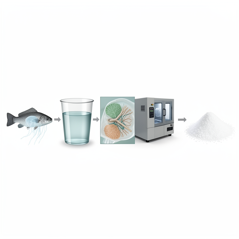
Collagen Extraction Workflow
AI-generated conceptual illustration (Grey mullet (Mugil cephalus) skin and jellyfish mesoglea). It explains the idea and does not show study data.
View source evidence ↗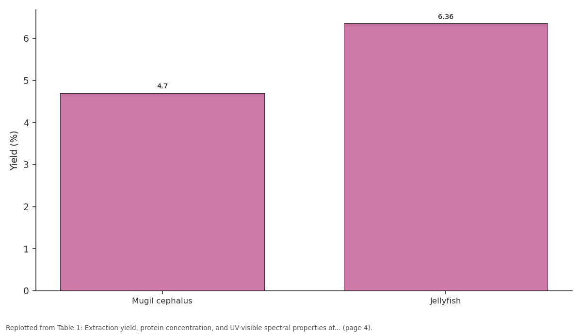
Yield (%): Mugil cephalus vs Jellyfish
Replotted from Table 1 (page 4) of the original paper. Every bar is the paper's own table value.
View source evidence ↗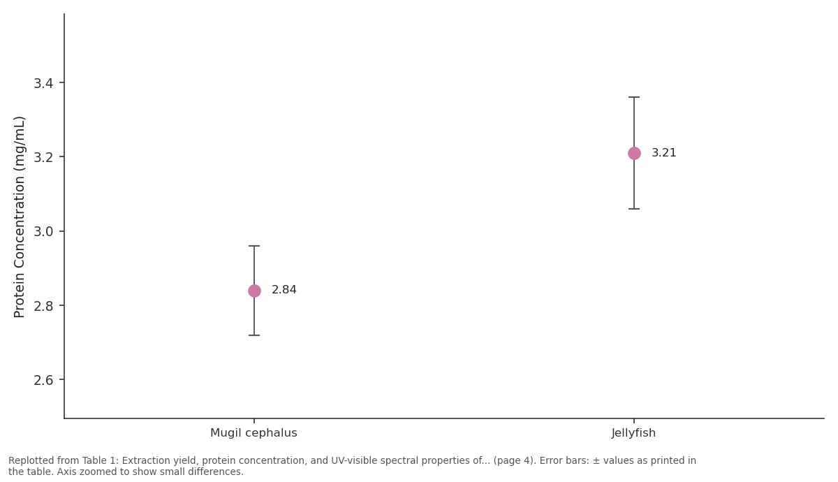
Protein Concentration (mg/mL): Mugil cephalus vs Jellyfish
Replotted from Table 1 (page 4) of the original paper. Every bar is the paper's own table value.
View source evidence ↗Figures & tables
From the original paper Table 1
 Table 2
 Table 3
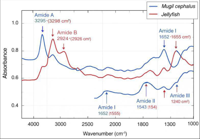 Figure 1
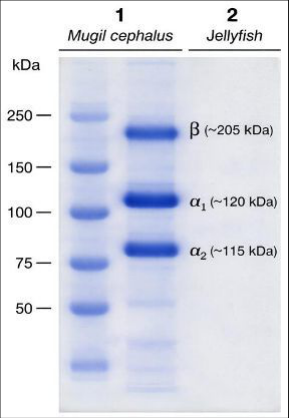 Figure 2
Introduction
Marine environments represent a vast and largely untapped reservoir of biologically active biomolecules with diverse applications in biotechnology, medicine, pharmaceuticals, and the food industry. Among these biomolecules, collagen has received considerable scientific attention due to its structural importance and wide range of industrial uses. Collagen is the most abundant structural protein in animals, constituting approximately 25-30% of the total protein content in vertebrates and forming the major component of connective tissues such as skin, tendons, bones, and cartilage (Shoulders & Raines, 2009) [9] . Owing to its biocompatibility, biodegradability, and low antigenicity, collagen has been widely utilized in biomedical materials, wound dressings, tissue engineering scaffolds, and cosmetic formulations (Silvipriya et al., 2015) [11] . Historically, collagen used in commercial and biomedical applications has been obtained predominantly from terrestrial animals, especially bovine and porcine sources (Gómez-However, the emergence of health concerns associated with zoonotic diseases such as bovine spongiform encephalopathy and foot-and-mouth disease has raised safety issues regarding mammalian collagen. In addition, religious restrictions and ethical considerations in several communities limit the acceptability of bovine and porcinederived products (Jongjareonrak et al., 2005) [6] . These concerns have stimulated growing interest in alternative collagen sources, particularly those derived from marine organisms. Marine-derived collagen has emerged as a promising substitute because of its high availability, minimal risk of transmissible diseases, and favorable physicochemical properties (Gómez-Guillén et al., 2011) [4] . Several studies indicate that collagen extracted from marine organisms often exhibits lower molecular weight and better solubility compared with mammalian collagen, characteristics that enhance its absorption and functional performance in biomedical applications (Silva et al., 2014) [10] . Furthermore, the processing of marine fish generates substantial quantities of by-products such as skin, scales, bones, and fins, which are rich in collagen and can be effectively utilized for the production of value-added biomaterials (Arvanitoyannis & Kassaveti, 2008) [2] . The utilization of such resources not only supports sustainable bioproduct development but also reduces environmental burdens associated with fishery waste. Among various marine species, the grey mullet, Mugil cephalus, has attracted attention as a potential collagen source because of its wide distribution in marine and estuarine environments and its commercial importance in fisheries (FAO, 2020) [3] . The skin and connective tissues of Mugil cephalus are particularly rich in collagen fibers, making them suitable for extraction and biochemical analysis. Previous investigations have demonstrated that fish-derived collagen possesses unique thermal and structural properties that differ from those of terrestrial animals due to variations in amino acid composition and environmental adaptation (Nagai & Suzuki, 2000) [7] . These differences can influence the stability, solubility, and potential industrial applications of marine collagen. Another promising marine source of collagen is jellyfish, members of the phylum Cnidaria. Jellyfish tissues contain large amounts of collagen-like proteins that play a structural role in maintaining the integrity of the mesoglea (Addad et al., 2011) [1] . Several studies have reported that jellyfish collagen exhibits remarkable biocompatibility and structural similarity to vertebrate collagen, making it an attractive candidate for biomedical applications, including tissue engineering and regenerative medicine (Hoyer et al., 2014) [5] . Moreover, jellyfish populations have increased significantly in many marine ecosystems, creating opportunities for their utilization as sustainable biomaterial resources (Purcell et al., 2007) [8] . Despite significant progress in marine collagen research, existing studies have largely concentrated on a limited number of commercially important fish species. Comparative investigations involving multiple marine organisms, particularly fish and jellyfish, remain relatively limited in the literature. Furthermore, the physicochemical characteristics and extraction efficiency of collagen can vary considerably among different marine species due to differences in habitat temperature, biochemical composition, and structural organization of connective tissues (Nagai & Suzuki, 2000) [7] . Consequently, systematic comparative studies are required to better understand the structural and biochemical properties of collagen derived from diverse marine organisms. The present research addresses this gap by examining collagen extracted from two marine sources: the grey mullet Mugil cephalus and jellyfish. Although individual studies have explored collagen extraction from fish or jellyfish separately, comprehensive investigations that compare collagen obtained from these organisms using standardized extraction and characterization methods remain scarce. Understanding these differences is important for identifying suitable marine sources for biomedical and industrial applications. Therefore, the primary research problem underlying this study is the limited comparative understanding of collagen characteristics derived from different marine organisms and the need to identify efficient alternative sources of highquality collagen. Addressing this issue requires the extraction, purification, and detailed characterization of collagen from selected marine species. The main aim of the present study is to extract and characterize collagen protein from marine organisms, specifically Mugil cephalus and jellyfish. In order to accomplish this aim, the study pursues several specific objectives: 1) To isolate collagen from selected marine sources using appropriate biochemical extraction techniques, 2) To determine the yield and purity of the extracted protein, 3) To investigate its structural and physicochemical characteristics using analytical methods, and 4) To compare the properties of collagen obtained from fish and jellyfish to evaluate their suitability for potential applications.
This research is expected to contribute to the expanding field of marine biotechnology by identifying alternative collagen resources that are both sustainable and economically valuable. The findings may provide useful insights into the structural characteristics of marine collagen and support its potential use in biomedical engineering, pharmaceutical formulations, cosmetic products, and food technology. Moreover, the valorization of marine organisms and fishery by-products for collagen extraction may promote environmentally responsible utilization of marine resources.
The remainder of this article is organized as follows. The materials and methods section describes the procedures used for collagen extraction and characterization. The results and discussion section presents the experimental findings and interprets the biochemical and structural properties of the isolated collagen. Finally, the conclusion summarizes the major outcomes of the study and outlines possible directions for future investigations.
Materials and Methods
The present investigation adopted a quantitative experimental research approach to extract and characterize collagen protein from selected marine organisms. A quantitative approach was considered appropriate because the study involved measurable biochemical parameters such as extraction yield, protein concentration, molecular characteristics, and spectroscopic properties. Quantitative experimental analysis allows objective measurement and statistical comparison of biochemical properties derived from different marine sources (Gómez-Guillén et al., 2011) [4] . This approach is widely used in biomolecular research where physicochemical properties of proteins are examined using analytical instruments and standardized laboratory protocols (Shoulders & Raines, 2009) [9] . Therefore, a quantitative framework provided a reliable basis for evaluating the structural and biochemical characteristics of collagen isolated from fish and jellyfish tissues.
Research Design
The study followed a laboratory-based experimental design focused on the extraction, purification, and physicochemical characterization of collagen. In this design, collagen was isolated from two marine organisms-grey mullet (Mugil cephalus) and jellyfish-using standardized biochemical procedures. The extracted protein samples were then subjected to a series of analytical techniques, including yield determination, spectroscopic analysis, and molecular characterization. This experimental design enabled systematic comparison of collagen properties derived from different biological sources while maintaining controlled laboratory conditions.
Biological Materials and Target Samples
The biological materials used in this study consisted of marine organisms known to contain significant amounts of collagen.
Fish samples
Fresh grey mullet (Mugil cephalus) specimens were obtained from a local fish market or coastal fish landing center. Skin tissues were selected for collagen extraction because fish skin contains high concentrations of collagen fibers (Nagai & Suzuki, 2000) [7] .
Jellyfish samples
Fresh jellyfish specimens were collected from marine coastal regions or obtained from local seafood suppliers. The gelatinous mesoglea tissue was used as the primary source of collagen-like proteins (Addad et al., 2011) [1] . Immediately after collection, the samples were transported to the laboratory in ice boxes to prevent protein degradation. The tissues were thoroughly washed with cold distilled water to remove impurities such as salts, blood residues, and debris.
Sampling Strategy and Sample Size
Grey mullet (Mugil cephalus) and jellyfish were chosen based on their documented collagen content and availability in marine environments (Hoyer et al., 2014) [5] . Multiple biological replicates were used to ensure reliability and reproducibility of the results.
For each species, approximately 500-1000 g of fresh tissue was collected and processed for collagen extraction. Experimental procedures were performed in triplicate to minimize analytical error and improve the statistical reliability of the results.
Chemicals and Reagents
All chemicals and reagents used in the study were of analytical grade. The primary reagents included: Acetic acid (0.5 M) for collagen extraction Sodium hydroxide (NaOH) for removal of noncollagenous proteins Sodium chloride (NaCl) for collagen precipitation Pepsin enzyme (for pepsin-soluble collagen extraction, if required) Ethanol and distilled water for washing and purification All solutions were prepared using deionized or distilled water to maintain experimental accuracy.
Collagen Extraction Procedure
The extraction of collagen was carried out using modified protocols commonly applied in marine collagen studies (Nagai & Suzuki, 2000; Gómez-Guillén et al., 2011) [7,4] .
Preparation of tissue samples
Fish skin and jellyfish tissues were cut into small pieces and washed repeatedly with cold distilled water. The samples were then treated with 0.1 M NaOH solution for approximately 24 hours at 4°C to remove non-collagenous proteins. The solution was replaced periodically during treatment to ensure effective purification.
Acid extraction
After alkaline treatment, the tissues were rinsed with distilled water until neutral pH was achieved. The cleaned tissues were then immersed in 0.5 M acetic acid solution and stirred continuously at 4°C for 48 hours. This step facilitated the solubilization of collagen fibers present in the connective tissues.
Filtration and centrifugation
The extraction mixture was filtered through cheesecloth to remove insoluble residues and subsequently centrifuged at high speed (approximately 10,000 rpm for 20 minutes). The supernatant containing dissolved collagen was carefully collected.
Salt precipitation
Collagen precipitation was achieved by adding sodium chloride to the supernatant until a final concentration of approximately 2.5 M NaCl was reached. The solution was maintained at 4°C to facilitate protein precipitation.
Dialysis
The precipitated collagen was collected by centrifugation and dissolved again in a small volume of acetic acid solution. The solution was then dialyzed against distilled water using dialysis tubing to remove excess salts and impurities.
Freeze-drying
The purified collagen solution was freeze-dried to obtain collagen in powder form. The dried collagen samples were stored at -20°C until further analysis.
Determination of Collagen Yield
The extraction yield was calculated based on the dry weight of collagen obtained relative to the initial weight of the raw tissue material. The percentage yield was determined using the following formula: Yield (%) = (Weight of extracted collagen / Weight of raw material) × 100
Characterization of Extracted Collagen
Several analytical techniques were used to evaluate the structural and biochemical characteristics of the extracted collagen.
UV-Visible Spectroscopy
UV-visible spectrophotometric analysis was performed to identify characteristic collagen absorption peaks. Protein solutions were scanned in the wavelength range of 200-400 nm, and the absorption maxima were recorded.
Fourier Transform Infrared Spectroscopy (FTIR)
FTIR analysis was conducted to determine the functional groups and secondary structure of the collagen molecules. The spectra were recorded in the range of 400-4000 cm⁻¹, allowing identification of characteristic amide bands (amide I, II, and III), which indicate the presence of collagen triplehelix structure (Silva et al., 2014) [10] .
SDS-PAGE Analysis
Sodium dodecyl sulfate polyacrylamide gel electrophoresis (SDS-PAGE) was used to determine the molecular weight distribution and subunit composition of the extracted collagen. This technique allows identification of α-chains, βcomponents, and other structural fragments associated with collagen proteins.
Protein Content Determination
The total protein content of the extracted samples was measured using standard biochemical methods Bradford assay, which are commonly used for protein quantification.
Data Analysis
Quantitative data obtained from the experimental procedures were analyzed using descriptive statistical methods, including mean values and standard deviations. Comparative analysis was performed to evaluate differences in collagen yield and physicochemical characteristics between Mugil cephalus and jellyfish samples. Graphical representations and statistical analyses were conducted using standard scientific software. Several measures were implemented to ensure the reliability and validity of the experimental results. All experiments were performed in triplicate, and standardized laboratory protocols were followed throughout the extraction and analysis procedures. Calibration of analytical instruments was conducted prior to measurements to ensure accuracy. Replication of experiments helped reduce experimental variability and increased the reproducibility of the findings.
Ethical Considerations
Although the study involved marine organisms rather than human participants, ethical guidelines related to responsible scientific research were strictly followed. Marine specimens were obtained from legally permitted sources such as local fisheries or seafood markets. No endangered or protected species were used in the study. Laboratory waste was handled and disposed of according to institutional environmental safety regulations. Proper documentation and transparent reporting of experimental procedures were maintained to ensure research integrity and reproducibility.
Results
This section presents the experimental outcomes obtained from the extraction and biochemical characterization of collagen derived from two marine organisms: the grey mullet Mugil cephalus and jellyfish. The results include quantitative measurements of collagen extraction yield, protein concentration, ultraviolet (UV-visible) spectral characteristics, Fourier Transform Infrared (FTIR) spectral profiles, and molecular weight distribution determined through SDS-PAGE analysis. These parameters were assessed to evaluate the biochemical properties of collagen extracted from both marine sources and to document observable differences in yield, purity, and structural characteristics. All experimental measurements were performed in triplicate, and the reported values represent the mean values obtained from repeated analyses.
Collagen Extraction Yield, Protein Concentration, and UV-Visible Spectral Properties
The extraction protocol resulted in the successful isolation of collagen from both Mugil cephalus skin tissue and jellyfish mesoglea tissue. Prior to extraction, approximately 500 g of fresh tissue from each organism was processed through alkaline treatment, acid extraction, salt precipitation, dialysis, and freeze-drying. The final collagen samples were obtained in dry powdered form and subsequently analyzed for protein concentration and spectral characteristics. A comparative summary of the extraction yield, protein concentration, and UV-visible absorption properties of the extracted collagen samples is presented in Table 1. The data show that the collagen extraction yield from jellyfish mesoglea tissue (6.36%) was higher than that obtained from the skin of Mugil cephalus (4.70%). The difference in extraction yield may reflect variations in tissue composition and collagen distribution within the organisms. Jellyfish mesoglea consists largely of extracellular matrix components rich in collagen-like proteins, whereas fish skin contains collagen interwoven with other structural proteins. Protein concentration analysis further indicated that the collagen solution derived from jellyfish exhibited a higher protein concentration ( The amide I band, typically considered the most significant indicator of protein secondary structure, appeared at approximately 1652 cm⁻¹ in Mugil cephalus collagen and 1655 cm⁻¹ in jellyfish collagen. This band arises from C=O stretching vibrations within the peptide bond and reflects the triple-helical configuration characteristic of collagen molecules (Fig. 1). Similarly, the amide II band detected near 1543-1546 cm⁻¹ corresponds to N-H bending vibrations combined with C-N stretching. The amide III band observed near 1238-1240 cm⁻¹ is associated with complex vibrations involving C-N stretching and N-H deformation. The presence of these bands in both samples indicates that the extracted proteins retain structural features typical of collagen.
| Source | Tissue Used | Initial Tissue Weight (g) | Collagen Yield (g) | Yield (%) | Protein Concentration (mg/mL) | Absorption Maximum (nm) | Absorbance |
|---|---|---|---|---|---|---|---|
| Mugil cephalus | Skin | 500 | 23.5 | 4.70 | 2.84 ± 0.12 | 228 | 0.86 |
| Jellyfish | Mesoglea | 500 | 31.8 | 6.36 | 3.21 ± 0.15 | 229 | 0.91 |
Table text extracted from the original PDF; superscript letters are shown inline. The original table image is below.
View original table image
SDS-PAGE Molecular Weight Profile
The molecular weight distribution and subunit composition of the extracted collagen samples were examined using SDS-PAGE electrophoresis. The electrophoretic analysis revealed distinct protein bands corresponding to collagen αchains and higher molecular weight components. The molecular weight bands detected in the SDS-PAGE analysis are summarized in Table 3. Both collagen samples exhibited prominent α1 and α2
| Source | α1 Chain (kDa) | α2 Chain (kDa) | β Components (kDa) |
|---|---|---|---|
| Mugil cephalus | ~120 | ~115 | ~210 |
| Jellyfish | ~118 | ~112 | ~205 |
Table text extracted from the original PDF; superscript letters are shown inline. The original table image is below.
View original table image
chains with molecular weights ranging from approximately 112 to 120 kDa. These bands correspond to the primary structural components of type I collagen molecules. In addition to the α-chains, β-dimer bands were observed at approximately 205-210 kDa in both samples. These βcomponents represent cross-linked dimers formed through intermolecular interactions between collagen chains (Fig. 2). The electrophoretic patterns of the two samples were largely similar, although the band intensity of the α-chains appeared slightly stronger in jellyfish-derived collagen. This observation is consistent with the higher protein concentration measured in jellyfish collagen extracts.
Discussion
The present investigation examined the extraction and biochemical characterization of collagen derived from two marine organisms, namely the grey mullet Mugil cephalus and jellyfish. The experimental procedures successfully yielded collagen from both sources using acid extraction followed by purification and analytical characterization. Quantitative analysis revealed that jellyfish tissue produced a higher extraction yield and protein concentration compared with collagen obtained from Mugil cephalus. Spectroscopic examination using UV-visible analysis showed characteristic absorption peaks near 228-229 nm, indicating the presence of peptide bonds typical of collagen proteins. Structural analysis through FTIR spectroscopy confirmed the presence of major collagen-associated functional groups, including amide A, amide I, amide II, and amide III bands. In addition, SDS-PAGE electrophoresis demonstrated the presence of α1 and α2 chains as well as βdimer components, which are commonly associated with type I collagen. Together, these findings confirm that both marine organisms represent viable sources of structurally intact collagen proteins.
Relationship of Findings to the Research Objectives
The primary objective of this study was to extract and characterize collagen proteins from selected marine organisms and to compare their biochemical properties. The results clearly demonstrate that collagen can be efficiently isolated from both Mugil cephalus and jellyfish tissues using acid-based extraction methods. The analytical techniques employed in the study provided complementary evidence regarding the structural integrity and purity of the isolated proteins. Spectroscopic data and electrophoretic profiles confirmed the presence of characteristic collagen features in both samples, thereby addressing the central research objective of identifying potential marine-derived collagen sources. The comparative results further highlight measurable differences between the two organisms in terms of extraction efficiency and protein concentration, which contributes to understanding species-specific variations in collagen composition.
Comparison with Previous Studies
The collagen yield obtained from Mugil cephalus skin in this study (4.70%) is comparable to values reported for other marine fish species. For example, Nagai and Suzuki (2000) [7] documented collagen yields ranging from 3-6% in several fish skins and connective tissues. Similarly, Jongjareonrak et al. (2005) [6] reported extraction yields between 4% and 7% for collagen isolated from marine fish skin using acidbased extraction methods. These findings suggest that the collagen content observed in Mugil cephalus is consistent with previously studied marine fish species. The higher extraction yield observed in jellyfish tissues aligns with earlier investigations indicating that jellyfish mesoglea contains large quantities of collagen-like structural proteins. Addad et al. (2011) [1] demonstrated that jellyfish collagen can be extracted efficiently due to the high proportion of extracellular matrix components present in the gelatinous tissue. Furthermore, Hoyer et al. (2014) [5] reported that collagen derived from jellyfish exhibits favorable extraction efficiency and structural stability, making it a promising biomaterial candidate. The results obtained in the present study support these earlier observations and reinforce the potential of jellyfish as an alternative marine collagen source. The UV-visible spectral peak observed near 228-229 nm corresponds to the characteristic absorbance region associated with peptide bonds in collagen molecules. Similar absorption maxima have been reported for collagen isolated from various marine organisms, including fish and invertebrates (Silva et al., 2014) [10] . The absence of significant absorption peaks in the higher wavelength region suggests minimal contamination from aromatic amino acidrich proteins, indicating effective removal of noncollagenous components during the purification process. FTIR spectral analysis further confirmed the presence of collagen-specific structural features. The amide I band detected near 1652-1655 cm⁻¹ is widely recognized as an indicator of the triple-helical structure of collagen (Shoulders & Raines, 2009) [9] . Comparable FTIR spectral patterns have been reported in marine collagen studies, including those involving fish skin and jellyfish tissues (Gómez-Guillén et al., 2011) [4] . The presence of amide II and amide III bands also supports the structural integrity of the extracted proteins. [6] . The detection of β-dimer components further supports the presence of intact collagen molecules with intermolecular cross-linking. The findings of this study largely align with the existing body of research on marine-derived collagen. The observed collagen yields, spectroscopic characteristics, and molecular weight profiles correspond closely with values reported in previous studies involving fish and jellyfish tissues. However, the slightly higher protein concentration detected in jellyfish collagen extracts compared with fish collagen may reflect differences in tissue composition and collagen organization within the organisms. Jellyfish mesoglea is primarily composed of extracellular matrix components, which may contribute to greater collagen availability and extraction efficiency. In contrast, fish skin contains collagen embedded within a more complex matrix of structural proteins, lipids, and pigments, which may reduce extraction efficiency. Similar observations have been reported in earlier investigations examining collagen extraction from marine fish tissues (Gómez-Guillén et al., 2011) [4] . Despite these differences, the overall structural characteristics of collagen from both sources remain comparable, indicating that marine organisms can provide functionally similar collagen molecules. The results contribute to the broader theoretical understanding of marine-derived collagen as a biomaterial resource. Marine collagen differs from mammalian collagen in several biochemical aspects, including amino acid composition, thermal stability, and solubility characteristics (Silva et al., 2014) [10] . The identification of collagen from both fish and jellyfish supports the concept that diverse marine organisms share common structural protein frameworks within their extracellular matrices. These findings reinforce the idea that marine biodiversity represents a valuable reservoir of biomolecules with unique biochemical properties. Additionally, the study highlights the importance of speciesspecific differences in collagen composition and extraction efficiency. Understanding these variations is essential for developing optimized extraction protocols and selecting appropriate marine organisms for specific industrial applications.
Industrial Implications
From an applied perspective, the results demonstrate the feasibility of utilizing marine organisms as alternative sources of collagen for biomedical and industrial purposes. Marine collagen has gained increasing attention in fields such as tissue engineering, wound healing, drug delivery, and cosmetic formulations due to its favorable biocompatibility and biodegradability (Silvipriya et al., 2015) [11] . The identification of collagen in both Mugil cephalus and jellyfish suggests that these organisms could serve as sustainable raw materials for collagen-based products. Furthermore, the use of fishery by-products such as fish skin for collagen extraction may contribute to waste reduction and value addition in the seafood processing industry. Arvanitoyannis and Kassaveti (2008) [2] emphasized that fish processing generates significant quantities of biological waste, which can be converted into valuable biomaterials through appropriate extraction techniques. The present study supports this concept by demonstrating the successful recovery of collagen from marine tissues. Jellyfish utilization also presents potential economic and environmental benefits. In several coastal regions, jellyfish populations have increased substantially, sometimes disrupting fisheries and marine ecosystems (Purcell et al., 2007) [8] . Converting jellyfish biomass into useful biomaterials such as collagen could provide a productive use for these abundant marine resources. The study contributes to the expanding literature on marinederived biomolecules by providing comparative data on collagen extracted from Mugil cephalus and jellyfish. While numerous investigations have examined collagen from individual marine species, fewer studies have directly compared collagen characteristics across different marine organisms using similar analytical techniques. The results therefore add valuable information regarding extraction efficiency, structural properties, and molecular composition of collagen derived from these two sources.
Figures and tables from the paper
| Functional Band | Mugil cephalus (cm⁻¹) | Jellyfish (cm⁻¹) | Structural Significance |
|---|---|---|---|
| Amide A | 3295 | 3298 | N-H stretching vibration |
| Amide B | 2924 | 2926 | CH₂ asymmetric stretching |
| Amide I | 1652 | 1655 | C=O stretching of peptide bond |
| Amide II | 1543 | 1546 | N-H bending vibration |
| Amide III | 1238 | 1240 | C-N stretching and N-H deformation |
Table text extracted from the original PDF; superscript letters are shown inline. The original table image is below.
View original table image
References
12 in the original paper- Addad S, Exposito JY, Faye C, Ricard-Blum S, Lethias C. Isolation, characterization and biological evaluation of jellyfish collagen for use in biomedical applications. Marine Drugs. 2011;9(6):967-983.
- Arvanitoyannis IS, Kassaveti A. Fish industry waste: treatments, environmental impacts, current and potential uses. International Journal of Food Science & Technology. 2008;43(4):726-745.
- Food and Agriculture Organization. The state of world fisheries and aquaculture 2020. Rome: FAO; 2020.
- Gómez-Guillén MC, Giménez B, López-Caballero ME, Montero P. Functional and bioactive properties of collagen and gelatin from alternative sources: a review. Food Hydrocolloids. 2011;25(8):1813-1827.
- Hoyer B, Bernhardt A, Heinemann S, Stachel I, Meyer M, Gelinsky M. Biomimetically mineralized jellyfish collagen scaffolds for bone tissue engineering. Acta Biomaterialia. 2014;10(2):883-892.
- Jongjareonrak A, Benjakul S, Visessanguan W, Nagai T, Tanaka M. Isolation and characterization of acid and pepsin-solubilised collagens from the skin of brownstripe red snapper (Lutjanus vitta). Food Chemistry. 2005;93(3):475-484.
- Nagai T, Suzuki N. Isolation of collagen from fish waste material-skin, bone and fins. Food Chemistry. 2000;68(3):277-281.
- Purcell JE, Uye S, Lo WT. Anthropogenic causes of jellyfish blooms and their direct consequences for humans. Marine Ecology Progress Series. 2007;350:153-174.
- Shoulders MD, Raines RT. Collagen structure and stability. Annual Review of Biochemistry. 2009;78:929-958.
- Silva TH, Moreira-Silva J, Marques ALP, Domingues A, Bayon Y, Reis RL. Marine origin collagens and their potential applications. Marine Drugs. 2014;12(12):5881-5901.
- Silvipriya KS, Krishna Kumar K, Bhat AR, Kumar BD, John A, Lakshmanan P. Collagen: animal sources and biomedical applications. Journal of Applied Pharmaceutical Science. 2015;5(3):123-127.
- Zhang Y, Liu W, Li G, Shi B, Miao Y, Wu X. Isolation and partial characterization of pepsin-soluble collagen from the skin of grass carp (Ctenopharyngodon idella). Food Chemistry. 2007;103(3):906-912.