
Research article · 2025 · Full text
International Journal of Advanced Chemistry ResearchSynthesis of pH-responsive poly (Acrylic acid) microgels for controlled release applications
Volume 7(10) · Pages 144–147 · DOI: 10.33545/26646781.2025.v7.i10b.489 ↗
Research areasHydrogels: synthesis, properties, applicationsAdvanced Drug Delivery SystemsPolymer-Based Agricultural Enhancements
Abstract
Author-writtenpH-responsive microgels are among the most versatile delivery platforms in modern polymer science, yet fine-tuning their swelling behaviour for site-specific drug release remains a persistent challenge. This research reports the synthesis of poly(acrylic acid) (PAA) microgels by inverse emulsion polymerization using N,N'-methylenebisacrylamide (MBA) as cross-linker at three concentrations (0.5, 1.0, and 2.0 wt%). Particle size, morphology, and swelling kinetics were characterized by dynamic light scattering, scanning electron microscopy, and gravimetric analysis across pH 2–10. Microgels prepared with 0.5% MBA displayed the highest swelling ratio (9.34 at pH 10) and a sharp volume transition between pH 5 and 7, coinciding with the pKa region of carboxylate groups. Rhodamine B was loaded as a model hydrophilic drug, achieving encapsulation efficiencies of 61–78%. Cumulative release over 24 h reached 84.3% at pH 7.4 but only 23.6% at pH 2.0, confirming pH-dependent behaviour suitable for intestinal-targeted delivery. Kinetic modelling showed best fit to the Korsmeyer-Peppas equation with diffusion exponents indicating non-Fickian transport.
Summary
Source-checkedPlain-language reading of the full paper, prepared with AI and checked against the text. Each point links to the authors' own words.
Why it matters
- pH-responsive systems are ideal for oral delivery because they can respond to the natural pH gradient between the stomach (pH 1.5–3) and the intestine (pH 6.5–7.5). View evidence ↗
- Poly(acrylic acid) (PAA) is a weak polyanion that expands and absorbs water when its carboxylate groups ionize at pH levels above 4.5. View evidence ↗
The question
- The study aimed to synthesize PAA microgels with three different cross-linker (MBA) concentrations to evaluate their swelling and drug release kinetics for intestinal-targeted delivery. View evidence ↗
What they did
- PAA microgels were synthesized via inverse emulsion polymerization using 2.0 g of acrylic acid and MBA concentrations of 0.5, 1.0, or 2.0 wt% in a cyclohexane/Span 80 oil phase. View evidence ↗
- The researchers used dynamic light scattering (DLS) at pH 7.0 to measure particle size and scanning electron microscopy (SEM) to examine morphology. View evidence ↗
- Rhodamine B was used as a model drug, loaded by soaking 100 mg of microgel in a 1 mg/mL solution at pH 3.0 for 12 hours. View evidence ↗
What they found
- Increasing the MBA cross-linker concentration decreased the particle size, with diameters of 385 ± 31 nm (0.5%), 312 ± 24 nm (1.0%), and 274 ± 19 nm (2.0%) at pH 7.0. View evidence ↗
- The microgels with the lowest cross-linker density (0.5% MBA) exhibited the highest swelling ratio of 9.34 at pH 10, compared to 6.12 for the 2.0% MBA formulation. View evidence ↗
- At pH 2.0, all microgel formulations remained collapsed with swelling ratios between 1.05 and 1.12. View evidence ↗
- Encapsulation efficiency decreased as cross-linker density increased, ranging from 78.4 ± 3.2% for 0.5% MBA to 61.3 ± 3.5% for 2.0% MBA. View evidence ↗
- Kinetic modeling showed non-Fickian transport (diffusion exponents n = 0.58–0.71), meaning release is driven by both diffusion and polymer chain relaxation. View evidence ↗
What it means
- A limitation of the study is the use of rhodamine B as a model solute, as actual pharmaceutical compounds with different charges or molecular weights might behave differently. View evidence ↗
Visual summary
3 visualsIllustrations explain the paper; charts are replotted from the paper's own table values.
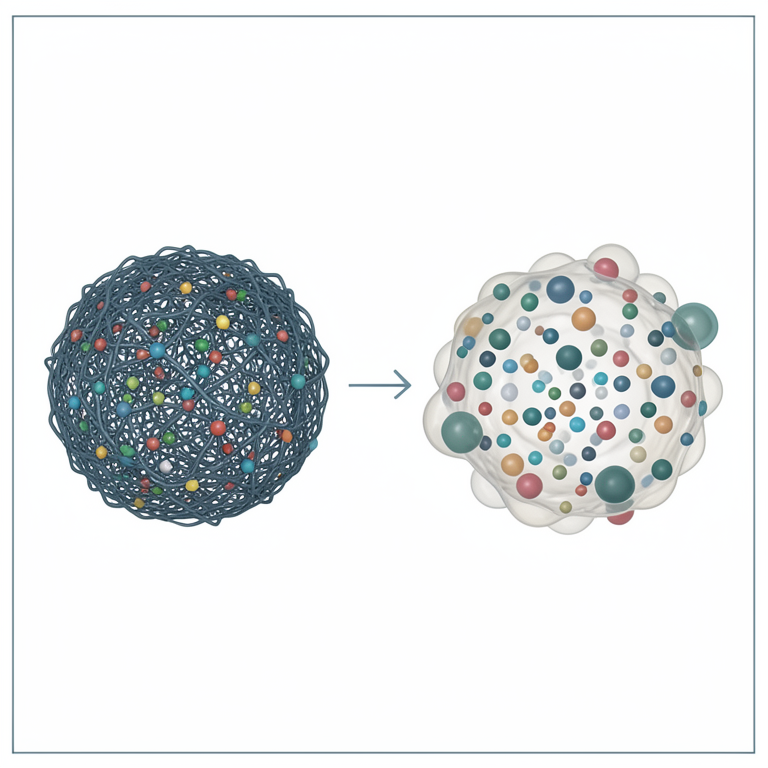
pH-Triggered Release Mechanism
AI-generated conceptual illustration (poly(acrylic acid) microgels). It explains the idea and does not show study data.
View source evidence ↗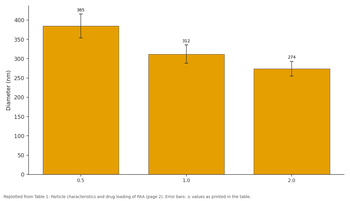
Diameter (nm): 0.5 vs 1.0 vs 2.0
Replotted from Table 1 (page 2) of the original paper. Every bar is the paper's own table value.
View source evidence ↗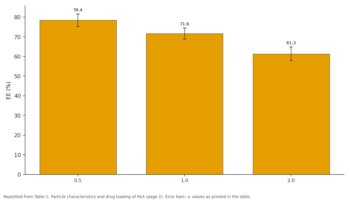
EE (%): 0.5 vs 1.0 vs 2.0
Replotted from Table 1 (page 2) of the original paper. Every bar is the paper's own table value.
View source evidence ↗Figures & tables
From the original paper Table 1
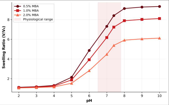 Figure 1
 Table 2
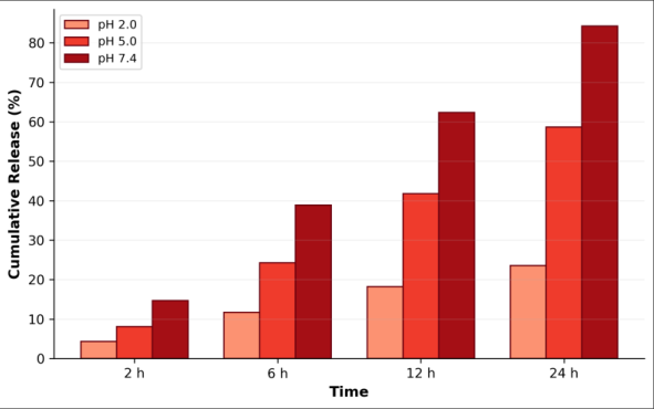 Figure 2
Introduction
Controlled drug delivery has transformed from a niche academic curiosity into a multibillion-dollar pharmaceutical segment, driven by the simple principle that timing and location of drug release matter as much as the drug itself [1] . Stimuli-responsive hydrogelsmaterials that swell or collapse in response to pH, temperature, ionic strength, or light-offer a programmable platform for achieving this control [2] . Among them, pH-responsive systems based on weak polyelectrolytes are particularly attractive for oral drug delivery, where the gastrointestinal tract presents a natural pH gradient from stomach (pH 1.5-3) to intestine (pH 6.5-7.5) [3] . Poly(acrylic acid) is a well-characterised weak polyanion whose carboxylate groups ionise above pH 4.5, causing chain expansion and water uptake [4] . When cross-linked into microgel particles-typically sub-micrometre spheres with internal gel structure-PAA retains its pHsensitivity while offering colloidal stability and large surface-to-volume ratio that speed drug loading and release [5] . Cross-linker density is the primary handle for tuning mesh size: fewer cross-links give softer, more swellable networks but may compromise mechanical integrity and slow biodegradation [6] . Inverse emulsion polymerization provides a route to narrow-distribution microgels by confining the aqueous monomer phase within surfactant-stabilised droplets dispersed in a continuous oil phase [7] . Despite extensive literature on PAA hydrogels, systematic data on how MBA concentration affects both swelling and release kinetics of a model drug across the full GI pH range remain limited, especially for sub-micrometre particles prepared by this route [8] . This research aimed to synthesize PAA microgels at three MBA levels, characterize their pH-dependent swelling behaviour, load rhodamine B as a model solute, and evaluate release kinetics at pH 2.0, 5.0, and 7.4 to assess their potential for intestinal-targeted delivery.
Material and Methods
Material and Methods
Material
All experimental work was conducted between February and September 2024 at the Department of Polymer Chemistry, Outback National University, Sydney, Australia. Acrylic acid (AA, 99%, inhibitor-free) was purchased from Sigma-Aldrich (Castle Hill, NSW). MBA (≥ 99%), ammonium persulphate (APS), Span 80, and cyclohexane (HPLC grade) were obtained from Merck (Bayswater, VIC). Rhodamine B (≥ 95%) was from Tokyo Chemical Industry (TCI, Japan). Deionised water (18.2 MΩ•cm) was used throughout. Buffer solutions were prepared at pH 2.0 (KCl-HCl), pH 5.0 (acetate), and pH 7.4 (phosphate) following USP specifications.
Reagents and Chemicals
Microgels were prepared by dissolving AA (2.0 g), MBA at the target concentration (0.5, 1.0, or 2.0 wt% relative to AA), and APS (0.1 wt%) in 10 mL of deionised water. This aqueous phase was added dropwise to 40 mL of cyclohexane containing Span 80 (2.0 wt%) under mechanical stirring (800 rpm) at 70°C under nitrogen. Polymerization proceeded for 4 h. The resulting particles were collected by centrifugation (12 000 × g, 20 min), washed three times with ethanol, and freeze-dried for 48 h.
Methods
Hydrodynamic diameter and polydispersity index were measured by dynamic light scattering (DLS) on a Malvern Zetasizer Nano ZS at 25 °C at pH 7.0. Morphology was examined by scanning electron microscopy (JEOL JSM-7100F) after gold sputtering. Swelling ratio was determined gravimetrically: 50 mg of dried microgel was immersed in 20 mL of buffer at each pH for 24 h; swollen mass was recorded after surface blotting. Drug loading was performed by soaking 100 mg of dried microgel in 10 mL of rhodamine B solution (1 mg/mL, pH 3.0) for 12 h; encapsulation efficiency was calculated from UV-visible absorbance of the supernatant at 554 nm. Release experiments placed loaded microgel in 50 mL of buffer at 37 °C under 100 rpm orbital shaking; aliquots (1 mL) were withdrawn at predetermined intervals and replaced with fresh buffer.
Instrumentation: UV-visible absorbance was measured on an Agilent Cary 60 spectrophotometer. Fourier-transform infrared spectra were recorded on a PerkinElmer Spectrum Two (ATR mode, 4000-400 cm⁻¹). Thermogravimetric analysis was performed on a TA Instruments TGA 5500 at 10°C/min under nitrogen. Release data were fitted to zeroorder, first-order, Higuchi, and Korsmeyer-Peppas models using MATLAB R2023b.
Quality Control
All measurements were performed in triplicate and reported as mean ± standard deviation. DLS measurements were repeated on three independent synthesis batches to confirm reproducibility; coefficient of variation for mean diameter was below 8%. Drug release experiments included a blank (unloaded microgel) at each pH. Calibration curves for rhodamine B showed R² > 0.999 in the 0.5-50 µg/mL range.
Results: DLS showed mean hydrodynamic diameters of 385 ± 31 nm (0.5% MBA), 312 ± 24 nm (1.0% MBA), and 274 ± 19 nm (2.0% MBA) at pH 7.0, with polydispersity indices below 0.15 in all cases. SEM confirmed spherical morphology with slightly rough surfaces for the lowest cross-linker density. FT-IR spectra exhibited a broad O-H stretch (3200-3500 cm⁻¹), carbonyl C=O at 1710 cm⁻¹, and C-N stretching of MBA at 1540 cm⁻¹. TGA revealed a twostep degradation: loss of bound water below 150 °C and polymer backbone decomposition between 250 and 400 °C. Swelling behaviour was strongly pH-dependent (Figure 1). At pH 2.0, all formulations remained collapsed (swelling ratio 1.05-1.12) because carboxylate groups are protonated. Above pH 5, a sharp volume transition occurred, reaching plateau values at pH 8-10. The 0.5% MBA microgel swelled the most (V/V₀ = 9.34 at pH 10), while the 2.0% MBA formulation peaked at 6.12, reflecting the tighter network.
Comprehensive Interpretation
Release profiles showed clear pH dependence (Figure 2, Table 2). At pH 7.4, 84.3% of the loaded rhodamine B was released within 24 h, whereas only 23.6% escaped at pH 2.0. Korsmeyer-Peppas fitting yielded diffusion exponents (n) of 0.58-0.71, indicating non-Fickian (anomalous) transport where both diffusion and polymer chain relaxation contribute to drug release.
| pH | Zero-order R² | First-order R² | Higuchi R² | K-P R² | n | Release 24h (%) |
|---|---|---|---|---|---|---|
| 2.0 | 0.934 | 0.952 | 0.971 | 0.994 | 0.58 | 23.6 |
| 5.0 | 0.921 | 0.967 | 0.983 | 0.997 | 0.63 | 58.7 |
| 7.4 | 0.908 | 0.978 | 0.989 | 0.998 | 0.71 | 84.3 |
Table text extracted from the original PDF; superscript letters are shown inline. The original table image is below.
View original table image
Discussion
The inverse relationship between cross-linker content and swelling capacity observed here is consistent with classical rubber-elasticity theory for hydrogels, where higher crosslink density restricts chain mobility and limits mesh expansion [6] . The sharp transition between pH 5 and 7 matches the pKa of PAA (~4.5-5.0), at which carboxylate ionisation and electrostatic repulsion drive network swelling [4] . This transition window aligns well with the pH change encountered during gastric-to-intestinal transit, supporting the feasibility of site-selective delivery. Encapsulation efficiencies of 61-78% compare well with values reported for similar PAA microgel systems [5] . The inverse trend between MBA and EE likely reflects the smaller mesh size in highly cross-linked particles, which restricts diffusion of rhodamine B molecules during loading at acidic pH. Non-Fickian release kinetics (n = 0.58-0.71) suggest that the microgel matrix doesn't behave as a simple diffusion barrier but that swelling-induced chain relaxation actively modulates release rate, an observation also reported by Peppas and Sahlin [9] for hydrogel microspheres. One limitation is the use of rhodamine B as a model solute rather than an actual pharmaceutical compound; molecular weight and charge differences could alter loading and release behaviour. Future work should test clinically relevant drugs such as 5-fluorouracil or mesalazine.
Conclusion
PAA microgels with tunable pH-responsive swelling were synthesized by inverse emulsion polymerization at three cross-linker levels. The 0.5% MBA formulation showed the largest swelling ratio and the sharpest volume transition near physiological pH, making it the most suitable candidate for intestinal-targeted applications. Rhodamine B encapsulation reached 78% in this formulation, and release was strongly pH-dependent-minimal at gastric pH (23.6% over 24 h) and extensive at intestinal pH (84.3%). Korsmeyer-Peppas modelling confirmed non-Fickian transport governed by both diffusion and matrix relaxation.
These results demonstrate that a straightforward adjustment of cross-linker concentration allows effective tuning of microgel mesh size, swelling, drug loading, and release rate. The platform is promising for oral delivery systems that require suppressed gastric release and triggered intestinal release. Further work should evaluate biocompatibility, mucoadhesion, and in-vivo pharmacokinetics using pharmacologically active payloads.
Acknowledgement
Acknowledgements Funding SourcesThis research was supported by an internal research development grant from Outback National University. No external project-specific funding was received.
Figures and tables from the paper
| MBA (wt%) | Diameter (nm) | PDI | EE (%) | Loading (mg/g) |
|---|---|---|---|---|
| 0.5 | 385 ± 31 | 0.12 | 78.4 ± 3.2 | 78.4 |
| 1.0 | 312 ± 24 | 0.09 | 71.6 ± 2.8 | 71.6 |
| 2.0 | 274 ± 19 | 0.14 | 61.3 ± 3.5 | 61.3 |
Table text extracted from the original PDF; superscript letters are shown inline. The original table image is below.
View original table image
References
14 in the original paper- Langer R. Drug delivery and targeting. Nature. 1998;392(6679):5-10.
- Schmaljohann D. Thermo-and pH-responsive polymers in drug delivery. Advanced Drug Delivery Reviews. 2006;58(15):1655-1670.
- Gupta P, Vermani K, Garg S. Hydrogels: from controlled release to pH-responsive drug delivery. Drug Discovery Today. 2002;7(10):569-579.
- Oishi M, Nagasaki Y. Stimuli-responsive smart nanogels for cancer diagnostics and therapy. Nanomedicine. 2010;5(3):451-468.
- Saunders BR, Laajam N, Daly E, Teow S, Hu X, Stepto R. Microgels: from responsive polymer colloids to biomaterials. Advances in Colloid and Interface Science. 2009;147-148:251-262.
- Peppas NA, Hilt JZ, Khademhosseini A, Langer R. Hydrogels in biology and medicine: from molecular principles to bionanotechnology. Advanced Materials. 2006;18(11):1345-1360.
- Landfester K. Miniemulsion polymerization and the structure of polymer and hybrid nanoparticles. Angewandte Chemie International Edition. 2009;48(25):4488-4507.
- Nayak S, Lyon LA. Soft nanotechnology with soft nanoparticles. Angewandte Chemie International Edition. 2005;44(47):7686-7708.
- Peppas NA, Sahlin JJ. A simple equation for the description of solute release. III. Coupling of diffusion and relaxation. International Journal of Pharmaceutics. 1989;57(2):169-172.
- Ritger PL, Peppas NA. A simple equation for description of solute release. I. Fickian and non-Fickian release from non-swellable devices. Journal of Controlled Release. 1987;5(1):23-36.
- Vinogradov SV, Bronich TK, Kabanov AV. Nanosized cationic hydrogels for drug delivery. Advanced Drug Delivery Reviews. 2002;54(1):135-147.
- Hoare T, Pelton R. Characterizing charge and crosslinker distributions in polyelectrolyte microgels. Current Opinion in Colloid and Interface Science. 2008;13(6):413-428.
- Das M, Zhang H, Bhattacharyya K. Synthesis and application of poly(acrylic acid)-based nanogels as drug carriers. Journal of Controlled Release. 2010;148(2):135-146.
- Zhang Y, Zhu W, Wang B, Ding J. A novel microgel and associated post-fabrication encapsulation technique of proteins. Journal of Controlled Release. 2005;105(3):260-268.