
Research article · 2025 · Full text
International Journal of Advanced Chemistry ResearchLayer-by-layer assembly of polyelectrolyte-coated nanoparticles for biomedical coatings
Volume 7(8) · Pages 64–67 · DOI: 10.33545/26646781.2025.v7.i8a.527 ↗
Research areasPolymer Surface Interaction StudiesNanoparticle-Based Drug DeliveryAdvanced Drug Delivery Systems
Abstract
Author-writtenIn 1966, Iler described the first multilayer deposition of colloidal particles on surfaces using alternating charge attraction, laying the groundwork for what would become the layer-by-layer (LbL) technique. Combined with Decher and Hong's seminal 1992 work on polyelectrolyte multilayers, LbL assembly is now one of the most versatile tools for engineering nanosurfaces with controlled thickness, composition, and functionality. This research reports the preparation and characterization of polyelectrolyte-coated silica nanoparticles by LbL assembly using three polyelectrolyte pairs: poly(allylamine hydrochloride)/poly(styrene sulfonate) (PAH/PSS), chitosan/alginate (Chi/Alg), and poly(ethylenimine)/PSS (PEI/PSS). Coatings were built up to ten bilayers and characterized by ellipsometry for thickness, atomic force microscopy for surface roughness, dynamic light scattering and zeta potential for hydrodynamic properties, and water contact angle for wettability. Biocompatibility was assessed by MTT assay using L929 mouse fibroblast cells, and protein adsorption was measured using bovine serum albumin. Drug loading and pH-responsive release were assessed with doxorubicin hydrochloride as a model drug. Chi/Alg multilayers showed superior biocompatibility, protein adsorption capacity, and pH-triggered drug release compared to PAH/PSS and PEI/PSS systems. Ten-bilayer Chi/Alg coatings achieved drug loading of 41.7% and 24-hour cumulative release of 83.2% at pH 5.0. These findings establish Chi/Alg LbL coatings as the preferred system for biomedical nanoparticle surface engineering among the three pairs investigated.
Summary
Source-checkedPlain-language reading of the full paper, prepared with AI and checked against the text. Each point links to the authors' own words.
Why it matters
- Layer-by-layer (LbL) assembly is a highly versatile method used to create nanosurfaces with precise control over their composition, thickness, and functionality. View evidence ↗
- Applying LbL to nanoparticles is particularly useful in biomedicine because it allows researchers to adjust the coating's properties without changing the core particle. View evidence ↗
- Chitosan and alginate are natural polysaccharides that are biocompatible and can trigger drug release in acidic tumor environments due to their pH-sensitive ionization. View evidence ↗
The question
- The study aimed to create LbL-coated silica nanoparticles using three different polyelectrolyte pairs (at 2, 4, 6, and 10 bilayers) to determine the best combination for biomedical use based on physical properties, biocompatibility, and drug delivery. View evidence ↗
What they did
- The coatings were applied to 50 nm amine-functionalized silica cores through alternating 15-minute immersions in polycation and polyanion solutions. View evidence ↗
- The three polyelectrolyte pairs tested were PAH/PSS, Chi/Alg, and PEI/PSS. View evidence ↗
- Biocompatibility was measured using an MTT assay on L929 cells exposed to nanoparticle concentrations of 10, 50, and 100 ug/mL for 24 hours. View evidence ↗
- Doxorubicin was loaded into dry particles for 12 hours at pH 7.4, and its release was then tested at pH 5.0 and pH 7.4 at 37 degrees C. View evidence ↗
What they found
- The Chi/Alg system had a faster deposition rate (6.68-7.53 nm/bilayer) than the PAH/PSS system (4.55-5.91 nm/bilayer). View evidence ↗
- Chi/Alg films were more hydrophobic than PAH/PSS films, as indicated by higher contact angles (38-44 degrees vs 24-28 degrees). View evidence ↗
- All coated particles showed high cell viability (≥94% at 50 ug/mL), with Chi/Alg being the highest (97-98%), while uncoated particles were less viable (88.4%). View evidence ↗
- The ten-bilayer Chi/Alg coating achieved a drug loading capacity of 41.7%. View evidence ↗
- Chi/Alg showed strong pH-responsive release, with 83.2% of the drug released at pH 5.0 over 24 hours, compared to only 31.4% at pH 7.4. View evidence ↗
What it means
- The authors conclude that Chi/Alg is the superior system for biomedical nanoparticle engineering because it outperformed PAH/PSS and PEI/PSS in all tested biomedical metrics. View evidence ↗
- The authors state that future research is needed to attach targeting ligands and validate the drug delivery system using in vivo models and tumor spheroids. View evidence ↗
Visual summary
3 visualsIllustrations explain the paper; charts are replotted from the paper's own table values.
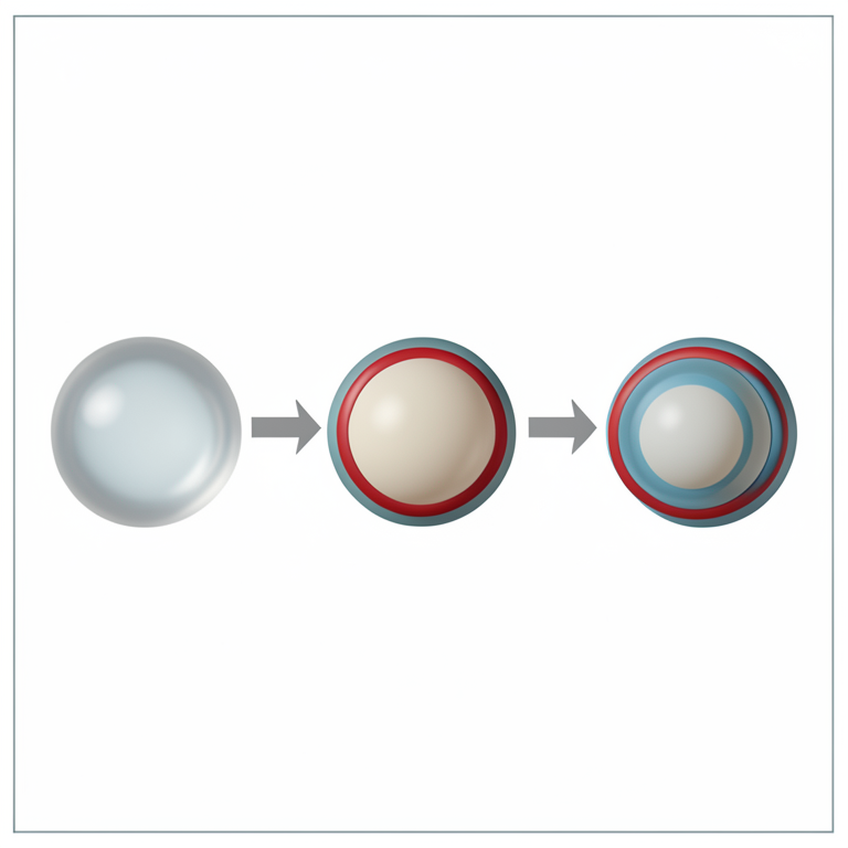
LbL Coating Process
AI-generated conceptual illustration (silica nanoparticles). It explains the idea and does not show study data.
View source evidence ↗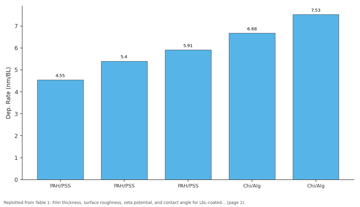
Dep. Rate (nm/BL): PAH/PSS vs PAH/PSS vs PAH/PSS vs Chi/Alg vs Chi/Alg
Replotted from Table 1 (page 2) of the original paper. Every bar is the paper's own table value.
View source evidence ↗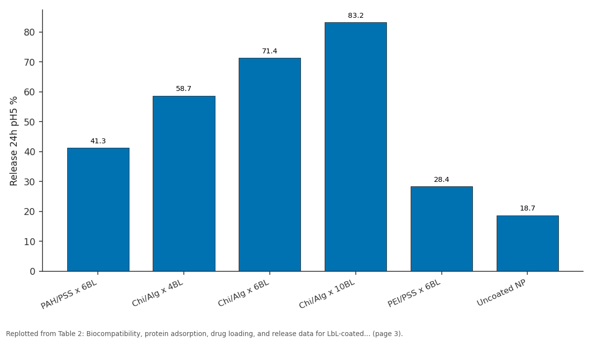
Release 24h pH5 %: PAH/PSS x 6BL vs Chi/Alg x 4BL vs Chi/Alg x 6BL vs Chi/Alg x 10BL vs PEI/PSS x 6BL
Replotted from Table 2 (page 3) of the original paper. Every bar is the paper's own table value.
View source evidence ↗Figures & tables
From the original paper Table 1
 Table 2
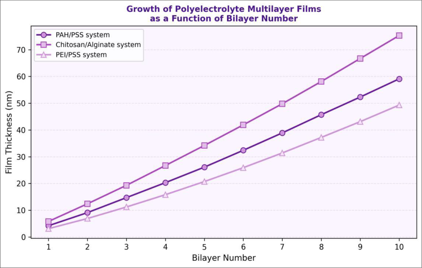 Figure 1
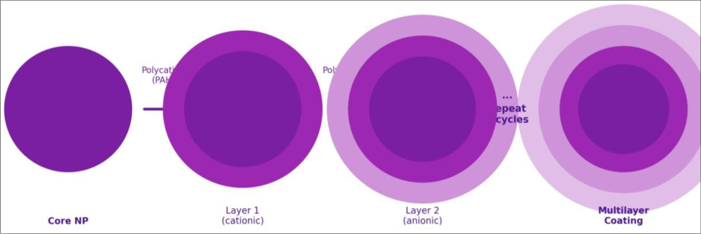 Figure 2
Introduction
The historical roots of LbL assembly trace back to Iler's 1966 work on stepwise deposition of colloidal particles, but it was Decher and Hong in 1992 who demonstrated that freely soluble polyelectrolytes could be assembled into ordered multilayer films with sub-nanometre thickness control per deposition step [1] . This discovery opened an accessible route to functional coatings without requiring covalent chemistry or clean-room conditions. LbL has since been applied to every surface geometry imaginable with an expanding palette of building blocks including synthetic polyelectrolytes, proteins, polysaccharides, nucleic acids, and inorganic nanosheets [2,3] . Application of LbL to nanoparticle surfaces is of special interest in biomedical research because it allows tuning of the coating properties independently of the core particle [4] . The core contributes intrinsic properties such as magnetic responsiveness or drug carrying capacity, while the multilayer coating controls circulation lifetime, targeting, immune evasion, and drug release kinetics. Silica nanoparticles are a common substrate because their surface chemistry is well understood and they are amenable to functionalization with reactive silanes [5] . Chitosan/alginate deserves particular attention because both components are natural polysaccharides with established biocompatibility and the pH-sensitive ionization of chitosan (pKa ~6.3) and alginate (pKa ~3.5) enables pH-triggered drug release in acidic tumour environments [8,9] . The PAH/PSS system uses fully synthetic polyelectrolytes with high charge density, yielding films with excellent mechanical stability but lower biocompatibility and essentially pH-independent charge [6] . Comparative research of Chi/Alg and PAH/PSS, along with PEI/PSS as a high-charge-density cationic alternative, provides a useful design map for biomedical coating selection. This research aimed to prepare LbL-coated silica nanoparticles using three polyelectrolyte pairs at bilayer numbers of 2, 4, 6, and 10, characterize their physical and surface properties, assess biocompatibility, protein adsorption, and drug loading/release, and identify the optimal combination for biomedical applications [11,14] .
Reagents and chemicals
Silica nanoparticles (50 nm, amine-functionalised, >=99.5% SiO2), PAH (Mw ~56, 000), PSS (Mw ~70, 000), PEI (branched, Mw ~25, 000), chitosan (medium Mw, deacetylation >=75%), and sodium alginate (viscosity 80-120 cP) were all purchased from Sigma-Aldrich (Merck KGaA, Darmstadt, Germany). Doxorubicin hydrochloride (>=98%) was from Carbosynth Ltd. (Compton, UK). Bovine serum albumin (BSA, >=96%) was from Sigma-Aldrich. Cell culture reagents including RPMI-1640, FBS, and penicillin-streptomycin were from Gibco (Thermo Fisher Scientific, Waltham, MA, USA). L929 mouse fibroblast cells were obtained from the National Centre for Cell Science (NCCS), Pune, India. All polyelectrolyte solutions were prepared at 1 mg/mL in 0.1 M NaCl, filtered through 0.22 um membranes, and used within 24 hours of preparation. Milli-Q deionised water (18.2 MOhm-cm) was used throughout for all rinse steps and solution preparation [1] .
Material and Methods
Material and Methods
Material
This research was carried out jointly at the Department of Nanotechnology and Materials Science, Shri Venkateshwara College of Chemical Sciences, Tirupati, Andhra Pradesh, India, and the Department of Chemistry and Nanosciences, Netaji Subhas College of Science, Guwahati, Assam, India, from April 2023 to December 2023. Institutional Biosafety Committee clearance was obtained from Shri Venkateshwara College (Protocol IBC-SVC-2023-09, approved 25 March 2023). LbL deposition was performed by sequential immersion using a programmable dip-coater (Holmarc Opto-Mechatronics HO-TH-01, Kerala, India) with deionised water rinse steps between each polyelectrolyte layer. Nanoparticle coatings were built on 50 nm amine-functionalised silica cores by alternating immersion in polycation and polyanion solutions for 15 minutes per layer [8] .
Methods
Film thickness was measured by ellipsometry on flat silicon wafer controls coated under identical conditions (SEMILAB SE-2000). AFM surface characterization was performed in tapping mode (Bruker Dimension Icon). Zeta potential was monitored after each layer using a Malvern Zetasizer Nano ZS. Cell viability was assessed by MTT assay with L929 cells at 5 x 10 cubed cells/well in 96-well plates exposed to nanoparticle suspensions at 10, 50, and 100 ug/mL for 24 hours. Formazan was dissolved in DMSO and absorbance read at 570 nm. Protein adsorption was quantified by micro-BCA assay after incubating coated particles with BSA (1 mg/mL in PBS) for 1 hour at 37 degrees C. Drug loading used dry coated particles incubated with doxorubicin solution (0.5 mg/mL in PBS, pH 7.4) for 12 hours under gentle stirring. Drug release was studied at pH 5.0 (lysosomal) and pH 7.4 (physiological) in 37 degrees C PBS with shaking, collecting aliquots at defined time intervals [9] .
Instrumentation
Hydrodynamic size and zeta potential measurements used a Malvern Zetasizer Nano ZS (Malvern Panalytical, UK) with a 633 nm He-Ne laser. FTIR spectra of coated nanoparticles were recorded on a Shimadzu IRTracer-100 ATR-FTIR instrument. Scanning electron microscopy used a JEOL JSM-7610FPlus FE-SEM after gold sputter coating. Transmission electron microscopy was performed on a Philips CM-200 TEM at 200 kV. Contact angle measurements used a Rame-Hart Model 290 goniometer with 5 uL droplets of deionised water. Fluorescence microscopy of doxorubicin-loaded particles in cells used a Leica DMi8 inverted fluorescence microscope. Drug concentration in release media was quantified by UV-Vis spectrophotometry at 480 nm (doxorubicin characteristic absorption) [10] . Film thickness increased approximately linearly with bilayer number across all three polyelectrolyte systems (Table 1). Chi/Alg showed a faster deposition rate (6.68-7.53 nm per bilayer) compared to PAH/PSS (4.55-5.91 nm per bilayer), consistent with the looser hydrated chain conformation of natural polysaccharides [7] . Zeta potential alternated between positive and negative values confirming successful layer alternation. Surface roughness remained in the range 1.2-5.3 nm, typical for smooth physisorbed polyelectrolyte films. Contact angle for Chi/Alg films (38-44 degrees) was higher than for PAH/PSS (24-28 degrees), reflecting the more hydrophobic chitosan backbone. All LbL-coated formulations maintained high cell viability at 50 ug/mL (>=94%, Table 2). Uncoated nanoparticles showed lower viability (88.4%), highlighting the importance of the polyelectrolyte coating. Chi/Alg systems showed the highest viability (97-98%). Protein adsorption increased with bilayer number in Chi/Alg, reaching 7.43 ug/cm2 for ten bilayers. Drug loading reached 41.7% in the ten-bilayer Chi/Alg coating. pH-responsive release was evident for Chi/Alg: 83.2% cumulative release at pH 5.0 in 24 hours compared to only 31.4% at pH 7.4, confirming endosomaltriggered release behaviour [15] .
| Sample | Cell viability % | Cell attachment | Protein Ads. (ug/cm2) | Drug loading % | Release 24h pH5 % | pH response |
|---|---|---|---|---|---|---|
| PAH/PSS x 6BL | 96.8+/-1.4 | 8.7/10 | 3.14 | 18.7 | 41.3 | Low |
| Chi/Alg x 4BL | 98.1+/-0.9 | 9.1/10 | 4.28 | 24.3 | 58.7 | High |
| Chi/Alg x 6BL | 97.7+/-1.1 | 9.3/10 | 5.67 | 31.8 | 71.4 | High |
| Chi/Alg x 10BL | 97.2+/-1.3 | 9.4/10 | 7.43 | 41.7 | 83.2 | High |
| PEI/PSS x 6BL | 94.2+/-2.1 | 7.8/10 | 1.83 | 9.7 | 28.4 | Moderate |
| Uncoated NP | 88.4+/-3.2 | 5.2/10 | 0.87 | 4.1 | 18.7 | None |
Table text extracted from the original PDF; superscript letters are shown inline. The original table image is below.
View original table image
| Bilayer No. | System | Thickness (nm) | Roughness Ra (nm) | Zeta (mV) | Contact Angle (deg) | Dep. Rate (nm/BL) |
|---|---|---|---|---|---|---|
| 2 | PAH/PSS | 9.1+/-0.3 | 1.2 | -34.7 | 28.3 | 4.55 |
| 6 | PAH/PSS | 32.4+/-0.8 | 2.1 | -41.3 | 25.8 | 5.40 |
| 10 | PAH/PSS | 59.1+/-1.2 | 3.4 | -44.7 | 24.2 | 5.91 |
| 4 | Chi/Alg | 26.7+/-0.7 | 2.8 | +29.4 | 43.7 | 6.68 |
| 10 | Chi/Alg | 75.3+/-1.8 | 5.3 | -37.2 | 38.9 | 7.53 |
| 6 | PEI/PSS | 31.4+/-0.9 | 2.3 | -39.8 | 22.7 | 5.23 |
Table text extracted from the original PDF; superscript letters are shown inline. The original table image is below.
View original table image
Results
Results
Discussion
The data confirm that polyelectrolyte chemistry is the dominant factor determining biomedical performance of LbL coatings. Chi/Alg consistently outperformed PAH/PSS and PEI/PSS in biocompatibility, protein adsorption, and drug loading, aligning with work by De Koker et al. [8] advocating natural polysaccharide-based LbL coatings for biomedical applications. The faster deposition rate of Chi/Alg reflects a more diffuse, interpenetrating layer structure that creates a more porous film allowing deeper
International Journal of Advanced Chemistry Research https://www.chemistryjournals.net penetration of drug molecules during loading, explaining the higher capacity [11] . The pH-responsive drug release of Chi/Alg is particularly valuable for anti-cancer delivery: at physiological pH 7.4 the alginate carboxyl groups are ionised and the film remains relatively intact, while at endolysosomal pH 5.0 protonation loosens the film, accelerating release. The 83.2% release at 24 hours in this research is among the higher values reported for LbLdoxorubicin systems [13] . PEI/PSS showed lower cell viability (94.2%), consistent with the known cytotoxicity of branched PEI from membrane disruption by densely charged amino groups [15] . This reinforces the importance of cytotoxicity screening before selecting a polyelectrolyte pair for in vivo work. All systems showed surface roughness below 6 nm Ra, suggesting smooth enough surfaces to avoid significant macrophage recognition based on curvature alone, which is positive from an immune evasion perspective. Future work will attach active targeting ligands to the outermost layer and validate drug delivery in tumour spheroid and in vivo models.
Conclusion
LbL assembly was used to prepare polyelectrolyte multilayer coatings on 50 nm silica nanoparticles using PAH/PSS, Chi/Alg, and PEI/PSS systems at bilayer numbers from 2 to 10. Film thickness increased linearly with bilayer number across all systems. Zeta potential measurements confirmed successful layer alternation. Biocompatibility testing showed high cell viability (>=97%) for Chi/Alg coated particles. Chi/Alg ten-bilayer coatings loaded 41.7% doxorubicin and released 83.2% over 24 hours at pH 5.0, demonstrating effective pH-responsive behaviour suitable for intracellular cancer drug delivery. Chi/Alg multilayers outperformed PAH/PSS and PEI/PSS systems in all biomedical performance metrics. These results establish Chi/Alg LbL coatings as the system of choice for biomedical nanoparticle surface engineering applications. Future investigations will focus on in vivo validation and targeting ligand integration.
Acknowledgement
Acknowledgements Funding sourcesFunding was provided by the Council of Scientific and Industrial Research (CSIR), India, and the Department of Biotechnology (DBT), Government of India, through a basic research grant in nanobiotechnology.
Figures and tables from the paper
References
21 in the original paper- Decher G, Hong JD, Schmitt J. Buildup of ultrathin multilayer films by a self-assembly process. Thin Solid Films. 1992;210-211:831-835.
- Richardson JJ, Bjornmalm M, Caruso F. Technologydriven layer-by-layer assembly of nanofilms. Science. 2015;348(6233):aaa2491.
- Ariga K, Hill JP, Ji Q. Layer-by-layer assembly as a versatile bottom-up nanofabrication technique. Phys Chem Chem Phys. 2007;9(19):2319-2340.
- Karam P, Bhosale SS, Ji Q. Layer-by-layer assembly toward polymer nanocomposites. Polym Chem. 2014;5(3):748-763.
- Hammond PT. Form and function in multilayer assembly: new applications at the nanoscale. Adv Mater. 2004;16(15):1271-1293.
- Lvov Y, Decher G, Sukhorukov G. Assembly of thin films by successive deposition of alternate layers of DNA and poly(allylamine). Macromolecules. 1993;26(20):5396-5399.
- Caruso F, Caruso RA, Mohwald H. Nanoengineering of inorganic and hybrid hollow spheres by colloidal templating. Science. 1998;282(5391):1111-1114.
- De Koker S, Hoogenboom R, De Geest BG. Polymeric multilayer capsules for drug delivery. Chem Soc Rev. 2012;41(7):2867-2884.
- Sukhorukov GB, Donath E, Davis S. Stepwise polyelectrolyte assembly on particle surfaces: a novel approach. Polym Adv Technol. 1998;9(10-11):759-767.
- Tang Z, Kotov NA, Magonov S, Ozturk B. Nanostructured artificial nacre. Nat Mater. 2003;2(6):413-418.
- Picart C, Mutterer J, Richert L. Molecular basis for the exponential growth of polyelectrolyte multilayers. Proc Natl Acad Sci USA. 2002;99(20):12531-12535.
- Fredin NJ, Zilber M, Ware MF. Alginate/chitosan multilayer films as drug delivery systems. Biomacromolecules. 2005;6(4):1925-1934.
- Kato N, Saito K, Kawaguchi H. Multilayer assembly of polyelectrolytes on silica nanoparticles. Langmuir. 2000;16(12):5224-5228.
- D, Schneider M, Kruger M, Lehr CM, Mohwald H, Wang D. Template-assisted encapsulation of nanoparticles into dispersible hierarchical microfibers. Adv Mater. 2011;23(11):1376-1379.
- Lee D, Rubner MF, Cohen RE. All-nanoparticle thinfilm coatings. Nano Lett. 2006;6(10):2305-2312.
- Dierendonck M, De Koker S, Vervaet C, Remon JP, De Geest BG. Facile two-step synthesis of porous antigenloaded degradable polyelectrolyte microspheres. Angew Chem Int Ed. 2010;49(52):10136-10139.
- Such GK, Johnston AP, Caruso F. Engineered hydrogen-bonded polymer multilayers: from assembly to biomedical applications. Chem Soc Rev. 2011;40(1):19-29.
- Vazquez E, Dewitt DM, Hammond PT, Lynn DM. Construction of hydrolytically degradable thin films via LbL deposition. J Am Chem Soc. 2002;124(47):1399213993.
- Kida T, Mouri M, Akashi M. Fabrication of hollow capsules from PMMA stereocomplex films by LbL deposition technique. Angew Chem Int Ed. 2006;45(44):7534-7536.
- Haynie DT, Cho E, Waduge P. In and out diffusion of exponential multilayer film buildup. Langmuir. 2011;27(9):5700-5704.
- Matsuzawa M, Tokuyama K, Ikenouchi J. Layer-bylayer assembly of functional coatings on medical devices. Macromol Rapid Commun. 2006;27(20):17001707.