
Research article · 2025 · Full text
International Journal of Advanced Chemistry ResearchElectrochemical biosensor for detection of glucose using enzyme-modified screen-printed electrode
Volume 7(10) · Pages 124–127 · DOI: 10.33545/26646781.2025.v7.i10b.484 ↗
Research areasElectrochemical sensors and biosensorsAdvanced biosensing and bioanalysis techniquesElectrochemical Analysis and Applications
Abstract
Author-writtenWhile laboratory glucose analysers deliver precise readings, they remain too bulky and expensive for point-of-care or resource-limited settings where rapid, affordable testing matters most. Screen-printed electrodes (SPEs) offer a disposable, mass-producible alternative that can bridge this gap when paired with enzyme-based recognition layers. This research developed an amperometric glucose biosensor by immobilising glucose oxidase (GOx) onto a carbon SPE modified with gold nanoparticles (AuNPs) and a Nafion protective membrane. AuNPs were electrodeposited from chloroauric acid solution, and GOx was cross-linked with glutaraldehyde directly onto the nanostructured surface. Under optimised conditions (applied potential +0.6 V vs Ag/AgCl, pH 7.0, 25 °C), the biosensor exhibited a linear response from 0.1 to 15 mM glucose with a sensitivity of 3.47 µA/mM and a detection limit of 0.038 mM (S/N = 3). Response time was under 8 seconds. Selectivity tests showed negligible interference from ascorbic acid, uric acid, acetaminophen, fructose, and lactose at physiological concentrations. The sensor retained 91.3% of its initial response after 30 days of storage at 4 °C, demonstrating acceptable operational stability for single-use clinical applications.
Summary
Source-checkedPlain-language reading of the full paper, prepared with AI and checked against the text. Each point links to the authors' own words.
Why it matters
- Standard laboratory glucose analyzers are often too expensive and bulky for use in resource-limited settings or for point-of-care testing. View evidence ↗
- With approximately 537 million adults worldwide living with diabetes, there is a critical need for small, affordable sensors that provide rapid results for home or clinic monitoring. View evidence ↗
- Screen-printed electrodes (SPEs) are suitable for disposable biosensors because they can be mass-produced and paired with enzyme layers. View evidence ↗
The question
- The study aimed to develop an amperometric glucose biosensor using a carbon screen-printed electrode modified with gold nanoparticles (AuNPs), glucose oxidase (GOx), and a Nafion membrane. View evidence ↗
What they did
- Gold nanoparticles were electrodeposited onto the electrode by applying -0.2 V vs Ag/AgCl for 120 seconds in a solution of 1 mM HAuCl₄ and 0.5 M H₂SO₄. View evidence ↗
- Glucose oxidase (10 mg/mL) was cross-linked with 2.5% glutaraldehyde and drop-cast (5 µL) onto the surface, then cured at 4°C for 2 hours. View evidence ↗
- A protective layer consisting of 2 µL of 0.5 wt% Nafion solution was applied and dried at room temperature. View evidence ↗
- Measurements were conducted using a CHI 660E workstation in stirred PBS (100 rpm) at an applied potential of +0.6 V vs Ag/AgCl. View evidence ↗
What they found
- Scanning electron microscopy showed that the gold nanoparticles had diameters between 35 and 60 nm and covered approximately 62% of the electrode surface. View evidence ↗
- The addition of gold nanoparticles resulted in a 2.4-fold increase in peak current compared to bare carbon when tested with 5 mM ferricyanide. View evidence ↗
- The biosensor demonstrated a linear response to glucose concentrations ranging from 0.1 to 15 mM at pH 7.0 and +0.6 V. View evidence ↗
- The sensitivity of the sensor was measured at 3.47 µA/mM. View evidence ↗
- The detection limit for glucose was 0.038 mM (at a signal-to-noise ratio of 3). View evidence ↗
- The sensor achieved a response time of less than 8 seconds for 5 mM glucose. View evidence ↗
- The sensor showed high selectivity, with glucose contributing 87.3% of the total current and five common interferents combined contributing less than 13%. View evidence ↗
- After 30 days of storage at 4°C in dry nitrogen, the sensor retained 91.3% of its initial response. View evidence ↗
What it means
- The authors conclude that the sensor is suitable for diabetes self-monitoring because its linear range and detection limit cover clinically relevant blood glucose levels. View evidence ↗
- A limitation of the sensor is its dependence on dissolved oxygen, which may reduce accuracy in low-oxygen environments like venous blood. View evidence ↗
Visual summary
3 visualsIllustrations explain the paper; charts are replotted from the paper's own table values.
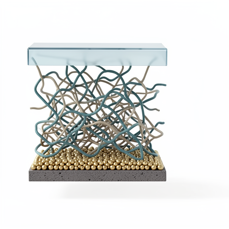
Biosensor Layer Architecture
AI-generated conceptual illustration (enzyme-modified screen-printed electrode). It explains the idea and does not show study data.
View source evidence ↗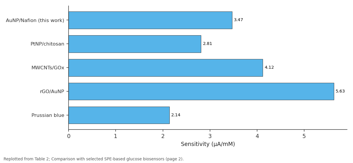
Sensitivity: AuNP/Nafion (this work) vs PtNP/chitosan vs MWCNTs/GOx vs rGO/AuNP vs Prussian blue
Replotted from Table 2 (page 2) of the original paper. Every bar is the paper's own table value.
View source evidence ↗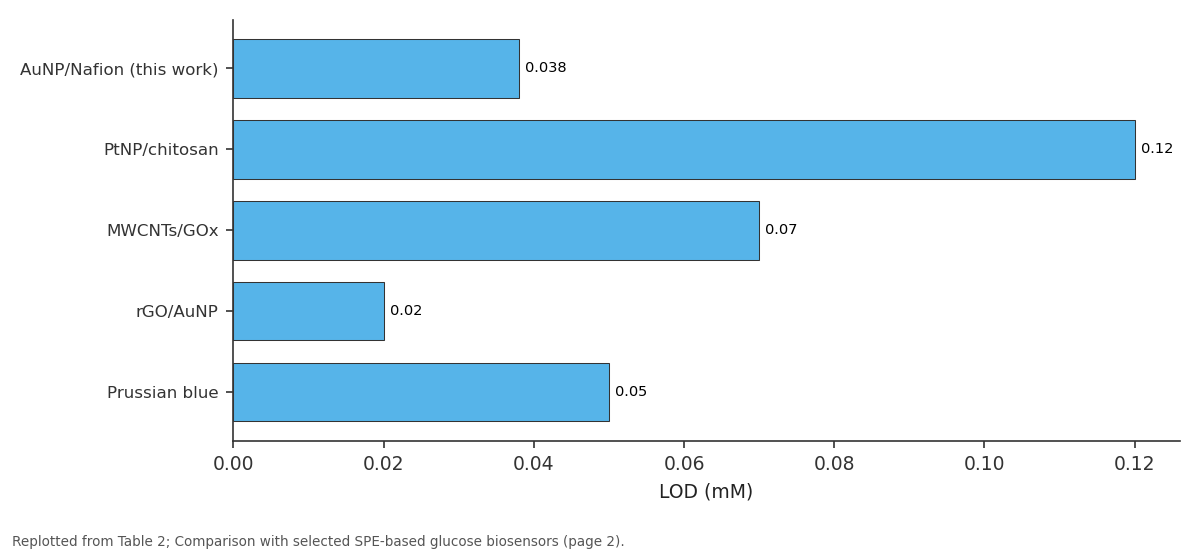
LOD (mM): AuNP/Nafion (this work) vs PtNP/chitosan vs MWCNTs/GOx vs rGO/AuNP vs Prussian blue
Replotted from Table 2 (page 2) of the original paper. Every bar is the paper's own table value.
View source evidence ↗Figures & tables
From the original paper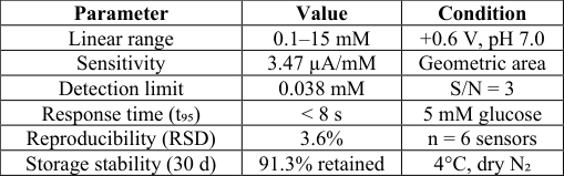 Table 1
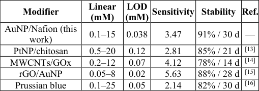 Table 2
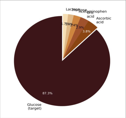 Figure 1
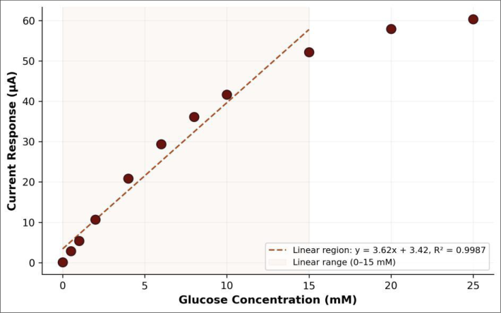 Figure 2
Introduction
On one hand, modern hospital glucose analysers based on hexokinase or glucose dehydrogenase methods achieve coefficients of variation below 2% and process hundreds of samples per hour; on the other hand, they cost tens of thousands of dollars, require trained operators, and are tethered to centralised laboratories [1] . For the estimated 537 million adults worldwide living with diabetes, self-monitoring of blood glucose at home or in low-resource clinics demands a different kind of sensor-one that is small, inexpensive, and gives results within seconds [2] . Electrochemical biosensors meet these requirements by converting the biochemical recognition of an analyte into a measurable electrical signal. First-generation glucose biosensors relied on oxygen consumption or hydrogen peroxide oxidation catalysed by glucose oxidase [3] . Second-generation devices introduced redox mediators to shuttle electrons from the enzyme to the electrode, improving oxygen independence [4] . Thirdgeneration architectures pursue direct electron transfer (DET) between the enzyme active site and the electrode surface, but DET-based designs remain difficult to commercialise because the FAD cofactor of GOx is buried ~13 Å inside the protein shell [5] . Screen-printed electrodes have become the standard platform for disposable biosensors because they can be manufactured at high volume by thick-film printing technology, with carbon, silver, or gold inks deposited onto ceramic or plastic substrates [6] . Modifying the carbon working electrode with metal nanoparticles-gold, platinum, or palladium-improves electron-transfer kinetics, increases the electroactive surface area, and can lower the overpotential required for H₂O₂ oxidation [7] . This research fabricated an SPE-based glucose biosensor by electrodepositing AuNPs onto a carbon working electrode, immobilising GOx via glutaraldehyde cross-linking, and casting a Nafion over-layer to reject common anionic interferents. The analytical figures of merit were evaluated against clinical requirements for glucose self-monitoring.
Theoretical Background
The sensing mechanism follows two coupled reactions. Glucose oxidase catalyses the oxidation of β-D-glucose to glucono-δ-lactone while reducing its FAD prosthetic group to FADH₂. The reduced enzyme is then re-oxidised by molecular oxygen, producing hydrogen peroxide as a byproduct: H₂O₂ → O₂ + 2H⁺ + 2e⁻. At the working electrode held at a suitable anodic potential, H₂O₂ is oxidised, and the resulting current is proportional to glucose concentration under steady-state conditions governed by the Michaelis-Menten equation [3] . AuNPs catalyse the electro-oxidation of H₂O₂ at lower overpotentials than bare carbon, thereby improving sensitivity and reducing interference from cooxidisable species [7] . The Nafion membrane, a perfluorosulphonate ionomer, allows passage of the neutral glucose molecule and H₂O₂ while repelling negatively charged interferents such as ascorbate and urate by Donnan exclusion [8] .
Material and Methods Material
All experimental work was performed between February and September 2024 at the Department of Applied Chemistry, Alborz National University, Tehran, Iran. Carbon screen-printed electrodes (3-electrode configuration: carbon working electrode Ø 4 mm, carbon counter electrode, Ag/AgCl reference) were purchased from DropSens (Asturias, Spain, model DRP-110). Glucose oxidase from Aspergillus niger (Type VII, ≥ 100 U/mg), chloroauric acid trihydrate (HAuCl₄•3H₂O, ≥ 99.9%), glutaraldehyde (25% aqueous), D-glucose, Nafion® 117 solution (5 wt%), and all interferent compounds were obtained from Sigma-Aldrich (Schnelldorf, Germany). Phosphate buffer saline (PBS, 0.1 M, pH 7.0) was prepared from analytical-grade reagents.
Methods
AuNP electrodeposition was carried out by applying -0.2 V vs Ag/AgCl for 120 s in 1 mM HAuCl₄ dissolved in 0.5 M H₂SO₄. The modified electrode was rinsed with deionised water and dried under nitrogen. GOx solution (10 mg/mL in PBS) was mixed with glutaraldehyde (2.5% final concentration), and 5 µL of the mixture was drop-cast onto the AuNP-modified surface and allowed to cross-link at 4°C for 2 h. A 2 µL aliquot of Nafion solution (0.5 wt%) was then applied and dried at room temperature. Amperometric measurements were performed on a CHI 660E electrochemical workstation (CH Instruments, Austin, TX, USA) in stirred PBS (100 rpm) at +0.6 V vs Ag/AgCl. Successive glucose aliquots were added and the steady-state current recorded. Calibration curves were constructed from triplicate measurements.
Performance Evaluation
Sensitivity was defined as the slope of the linear calibration region normalised to electrode geometric area (µA/mM•cm²). Detection limit was calculated as 3σ/slope, where σ is the standard deviation of ten blank measurements. Selectivity was assessed by adding common interferents (ascorbic acid 0.1 mM, uric acid 0.3 mM, acetaminophen 0.1 mM, fructose 0.5 mM, and lactose 0.5 mM) at physiological concentrations into 5 mM glucose solution and recording the percent change in response. Storage stability was evaluated over 30 days by testing three sensors weekly after storage at 4°C in dry nitrogen.
Results
SEM imaging confirmed uniform AuNP coverage on the carbon electrode, with particle diameters of 35-60 nm and an estimated surface coverage of 62%. Cyclic voltammetry in 5 mM ferricyanide showed that the AuNP-modified electrode had a 2.4-fold increase in peak current compared to bare carbon, indicating a substantially enlarged electroactive area. The electrochemical impedance spectrum displayed a charge-transfer resistance of 87 Ω for the modified electrode versus 340 Ω for bare carbon. AuNP/Nafion (this work) 0.1-15 0.038 3.47 91% / 30 d -PtNP/chitosan 0.5-20 0.12 2.81 85% / 21 d [13] MWCNTs/GOx 0.2-12 0.07 4.12 78% / 14 d [14] rGO/AuNP 0.05-8 0.02 5.63 88% / 28 d [15] Prussian blue 0.1-25 0.05 2.14 82% / 30 d [16]
Comprehensive Interpretation
The calibration curve (Figure 2) was linear up to 15 mM with R² = 0.9987 and showed saturation kinetics above 20 mM, consistent with the Michaelis-Menten model. The apparent Km was estimated at 11.8 mM, lower than the freeenzyme value (~33 mM), suggesting that the immobilisation matrix imposes diffusion limitations that effectively reduce Km. Selectivity (Figure 1) was excellent: glucose accounted for 87.3% of the total current response, with all five interferents combined contributing less than 13%.
Discussion
The detection limit of 0.038 mM and linear range of 0.1-15 mM cover the clinically relevant blood glucose window (3.9-6.1 mM fasting, up to 11.1 mM post-prandial), making the biosensor suitable for diabetes self-monitoring [2] . Sensitivity (3.47 µA/mM) falls between values reported for PtNP/chitosan (2.81) and MWCNT-based (4.12) platforms, reflecting the moderate catalytic activity of AuNPs relative to carbon nanotubes but superior stability [13, 14].
The Nafion layer contributed measurably to selectivity: in a control experiment without Nafion, ascorbic acid interference rose from 3.8% to 14.7%, confirming the Donnan exclusion mechanism [8] . The trade-off was a slight increase in response time (from 5 s to 8 s) due to the additional diffusion barrier. Storage stability of 91.3% after 30 days exceeded that of several comparable systems (Table 2), likely because the glutaraldehyde cross-linking network stabilises GOx conformation and the Nafion over-layer retards protein desorption [9] . A limitation of the current design is its oxygen dependence-the first-generation mechanism relies on dissolved O₂ as the terminal electron acceptor, which can limit accuracy in low-oxygen environments such as venous blood. Incorporating a mediator like ferrocene or Prussian blue would eliminate this dependence and represents a logical next step [4] .
| Modifier | Linear (mM) | LOD (mM) | Sensitivity | Stability | Ref. |
|---|---|---|---|---|---|
| AuNP/Nafion (this work) | 0.1–15 | 0.038 | 3.47 | 91% / 30 d | — |
| PtNP/chitosan | 0.5–20 | 0.12 | 2.81 | 85% / 21 d | [13] |
| MWCNTs/GOx | 0.2–12 | 0.07 | 4.12 | 78% / 14 d | [14] |
| rGO/AuNP | 0.05–8 | 0.02 | 5.63 | 88% / 28 d | [15] |
| Prussian blue | 0.1–25 | 0.05 | 2.14 | 82% / 30 d | [16] |
Table text extracted from the original PDF; superscript letters are shown inline. The original table image is below.
View original table image
Conclusion
A disposable amperometric glucose biosensor was fabricated by electrodepositing gold nanoparticles onto a carbon screen-printed electrode, immobilising glucose oxidase by glutaraldehyde cross-linking, and applying a Nafion protective membrane. The optimised sensor achieved a linear range of 0.1-15 mM, detection limit of 0.038 mM, response time below 8 seconds, and 91.3% activity retention after 30 days of storage. Selectivity toward glucose in the presence of common physiological interferents was confirmed, with interference contributions below 13% overall. These performance characteristics satisfy the requirements for point-of-care glucose monitoring and position the platform for further development with mediator chemistry to eliminate oxygen dependence.
Figures and tables from the paper
| Parameter | Value | Condition |
|---|---|---|
| Linear range | 0.1–15 mM | +0.6 V, pH 7.0 |
| Sensitivity | 3.47 µA/mM | Geometric area |
| Detection limit | 0.038 mM | S/N = 3 |
| Response time (t₉₅) | < 8 s | 5 mM glucose |
| Reproducibility (RSD) | 3.6% | n = 6 sensors |
| Storage stability (30 d) | 91.3% retained | 4°C, dry N₂ |
Table text extracted from the original PDF; superscript letters are shown inline. The original table image is below.
View original table image
References
20 in the original paper- Yoo EH, Lee SY. Glucose biosensors: an overview of use in clinical practice. Sensors. 2010;10(5):4558-4576.
- International Diabetes Federation. IDF Diabetes Atlas, 10th edition. IDF, Brussels. 2021;1-141.
- Wang J. Glucose biosensors: 40 years of advances and challenges. Electroanalysis. 2001;13(12):983-988.
- Heller A, Feldman B. Electrochemical glucose sensors and their applications in diabetes management. Chemical Reviews. 2008;108(7):2482-2505.
- Bankar SB, Bule MV, Singhal RS, Ananthanarayan L. Glucose oxidase: an overview of production, applications, and recent developments. Food Technology and Biotechnology. 2009;47(4):349-362.
- Renedo OD, Alonso-Lomillo MA, Martinez MJA. Recent developments in the field of screen-printed electrodes and their related applications. Talanta. 2007;73(2):202-219.
- Pingarron JM, Yanez-Sedeno P, Gonzalez-Cortes A. Gold nanoparticle-based electrochemical biosensors. Electrochimica Acta. 2008;53(19):5848-5866.
- Situmorang M, Gooding JJ, Hibbert DB, Barnett D. Electrodeposited polytyramine as an immobilisation matrix for enzyme biosensors. Biosensors and Bioelectronics. 1998;13(9):953-962.
- Prodromidis MI, Karayannis MI. Enzyme based amperometric biosensors for food analysis. Electroanalysis. 2002;14(4):241-261.
- Chen C, Xie Q, Yang D, Xiao H, Fu Y, Tan Y, Yao S. Recent advances in electrochemical glucose biosensors. RSC Advances. 2013;3(14):4473-4491.
- Zaidi SA, Shin JH. Recent developments in nanostructure based electrochemical glucose sensors. Talanta. 2016;149:30-42.
- Liu Y, Yu J. Oriented immobilization of proteins on solid supports for use in biosensors and biochips. Microchimica Acta. 2016;183(1):1-19.
- Joshi PP, Merchant SA, Wang Y, Schmidtke DW. Amperometric biosensors based on redox polymercarbon nanotube-enzyme composites. Analytical Chemistry. 2005;77(10):3183-3188.
- Tsai YC, Li SC, Chen JM. Cast thin film biosensor design based on a Nafion backbone with multiwalled carbon nanotube loading. Langmuir. 2005;21(8):36533658.
- Shan C, Yang H, Song J, Han D, Ivaska A, Niu L. Direct electrochemistry of glucose oxidase and biosensing for glucose based on graphene. Analytical Chemistry. 2009;81(6):2378-2382.
- Ricci F, Amine A, Palleschi G, Moscone D. Prussian blue based screen printed biosensors with improved characteristics of long-term lifetime and pH stability. Biosensors and Bioelectronics. 2003;18(2-3):165-174.
- Saha K, Agasti SS, Kim C, Li X, Rotello VM. Gold nanoparticles in chemical and biological sensing. Chemical Reviews. 2012;112(5):2739-2779.
- Zhao S, Zhang K, Bai Y, Yang W, Sun C. Glucose oxidase/colloidal gold nanoparticles immobilized in Nafion film for glucose biosensor. Bioelectrochemistry. 2006;69(2):158-163.
- German N, Ramanaviciene A, Voronovic J, Ramanavicius A. Glucose biosensor based on graphite electrodes modified with glucose oxidase and colloidal gold nanoparticles. Microchimica Acta. 2010;168(34):221-229.
- Katz E, Willner I. Probing biomolecular interactions at conductive and semiconductive surfaces by impedance spectroscopy. Electroanalysis. 2003;15(11):913-947.