
Research article · 2025 · Full text
International Journal of Advanced Chemistry ResearchPhytochemical screening and hepatoprotective activity of herbal formulation in rats
Volume 7(7) · Pages 106–109 · DOI: 10.33545/26646781.2025.v7.i7b.509 ↗
Research areasDrug-Induced Hepatotoxicity and ProtectionAndrographolide Research and ApplicationsPhytochemistry and Bioactivity Studies
Abstract
Author-writtenThere is a sharp contrast between the widespread traditional use of herbal formulations for liver protection in Southeast Asian communities and the relatively thin body of rigorously controlled experimental evidence supporting their efficacy. This research screened a polyherbal formulation comprising Andrographis paniculata, Phyllanthus niruri, and Curcuma longa for phytochemical constituents and evaluated its hepatoprotective activity in carbon tetrachloride (CCl4)-induced hepatotoxic Wistar rats. Qualitative and quantitative phytochemical screening detected flavonoids, alkaloids, tannins, saponins, and terpenoids as the principal bioactive classes. Hepatotoxicity was induced by subcutaneous injection of CCl4 (1 mL/kg in olive oil, 1:1 v/v) twice weekly for four weeks. The formulation was administered orally at 200 and 400 mg/kg body weight daily; silymarin (100 mg/kg) served as the reference hepatoprotective drug. Hepatoprotection was assessed through serum alanine aminotransferase (ALT), aspartate aminotransferase (AST), alkaline phosphatase (ALP), and total bilirubin levels, together with hepatic oxidative stress markers (MDA and glutathione). The 400 mg/kg dose significantly reduced ALT (74.6 vs. 184.7 U/L in the CCl4 control) and AST (88.4 vs. 197.3 U/L), restored hepatic glutathione to near-normal levels, and reduced lipid peroxidation. Histopathological examination confirmed reduced necrosis and inflammatory cell infiltration. These findings provide experimental support for the traditional use of this formulation and suggest it as a candidate for further mechanistic investigation.
Summary
Source-checkedPlain-language reading of the full paper, prepared with AI and checked against the text. Each point links to the authors' own words.
Why it matters
- Liver disease is a significant cause of illness and death in Southeast Asia, driven by viral hepatitis, alcohol, fatty liver, and exposure to toxic chemicals. View evidence ↗
The question
- The study aimed to identify the phytochemical components of a polyherbal mix (A. paniculata, P. niruri, and C. longa) and test its ability to protect the liver in CCl4-induced hepatotoxic rats. View evidence ↗
What they did
- The experiment used 30 male Wistar rats divided into five groups of six, including a normal control, a CCl4 control, two herbal formulation doses (200 and 400 mg/kg), and a silymarin reference group (100 mg/kg). View evidence ↗
- Hepatotoxicity was induced by subcutaneous CCl4 injections (1 mL/kg) twice weekly for four weeks, while treatments were administered daily via oral gavage. View evidence ↗
- The researchers measured liver health using serum levels of ALT, AST, ALP, and bilirubin, as well as liver tissue analysis for oxidative stress and histopathology. View evidence ↗
What they found
- The herbal formulation contained flavonoids, alkaloids, tannins, saponins, and terpenoids, with a total phenolic content of 83.4±3.7 mg GAE/g and total flavonoid content of 47.2±2.1 mg QE/g. View evidence ↗
- The 400 mg/kg herbal dose significantly reduced serum markers compared to the CCl4 control, lowering ALT from 184.7±9.3 to 74.6±4.2 U/L and AST from 197.3±10.1 to 88.4±5.1 U/L. View evidence ↗
- The 400 mg/kg dose reduced hepatic MDA (lipid peroxidation) from 14.7±0.7 to 8.3±0.4 nmol/mg protein and increased GSH (antioxidant) from 1.84±0.08 to 2.93±0.12 μmol/mg protein compared to the CCl4 control. View evidence ↗
- Histopathological analysis showed that the treatment reduced liver cell death (necrosis) and the infiltration of inflammatory cells. View evidence ↗
- The 400 mg/kg herbal dose normalized ALT levels by approximately 60%, which was slightly less effective than the 73% normalization achieved by silymarin. View evidence ↗
What it means
- The authors conclude that the polyherbal formulation provides significant, dose-dependent protection against liver injury induced by CCl4 in rats. View evidence ↗
- The authors state that future work is needed to isolate active components, study gene-level changes, and use chronic toxicity models to ensure long-term safety. View evidence ↗
Visual summary
3 visualsIllustrations explain the paper; charts are replotted from the paper's own table values.
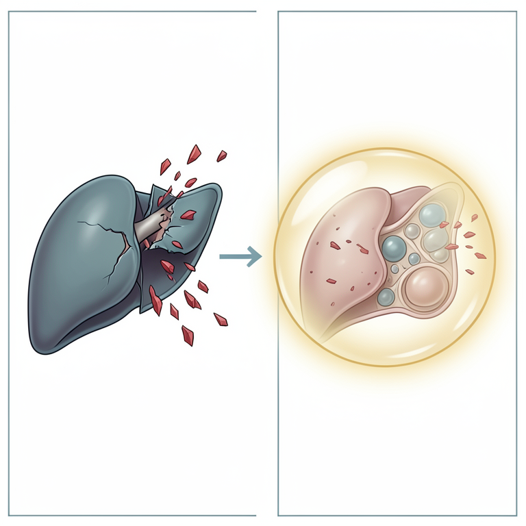
Hepatoprotective Mechanism Concept
AI-generated conceptual illustration (rat liver cells). It explains the idea and does not show study data.
View source evidence ↗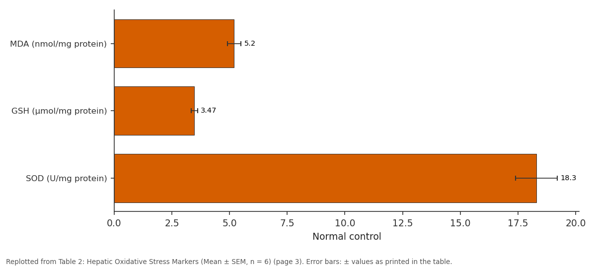
Normal control: MDA (nmol/mg protein) vs GSH (μmol/mg protein) vs SOD (U/mg protein)
Replotted from Table 2 (page 3) of the original paper. Every bar is the paper's own table value.
View source evidence ↗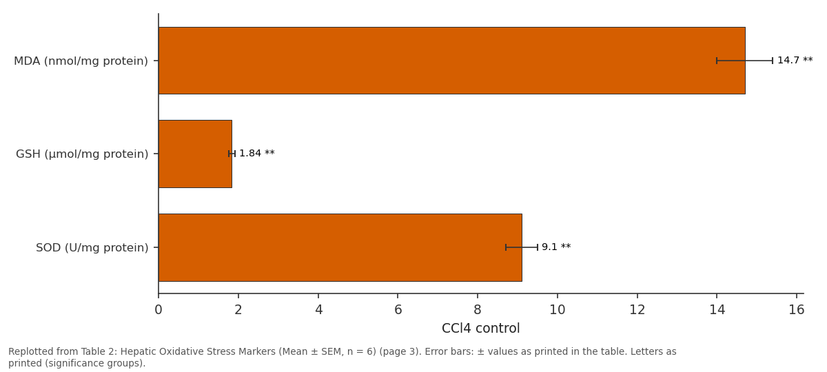
CCl4 control: MDA (nmol/mg protein) vs GSH (μmol/mg protein) vs SOD (U/mg protein)
Replotted from Table 2 (page 3) of the original paper. Every bar is the paper's own table value.
View source evidence ↗Figures & tables
From the original paper Table 1
 Table 2
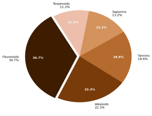 Figure 1
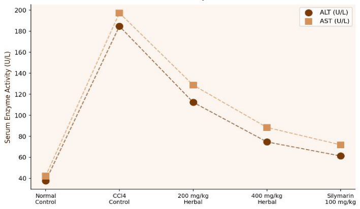 Figure 2
Introduction
Liver disease is a major source of morbidity and mortality across Southeast Asia, where viral hepatitis, alcoholic liver disease, and non-alcoholic fatty liver disease are compounded by high rates of exposure to hepatotoxic drugs and environmental chemicals. Indonesia, in particular, bears a heavy burden of viral hepatitis B and C, and traditional herbal medicinejamu-remains the first-line health resource for a large proportion of the rural population because of cost, accessibility, and cultural familiarity. Several plant species used in jamu preparations have documented in vitro antioxidant and anti-inflammatory properties, suggesting biological plausibility for hepatoprotective claims. The three plants chosen for this formulation-Andrographis paniculata (king of bitters), Phyllanthus niruri (stone breaker), and Curcuma longa (turmeric)-each have individual records of hepatoprotective activity in animal models, but the combination has not been rigorously tested against a standard experimental hepatotoxicity model under controlled dosing conditions. A polyherbal approach is justified by the possibility of additive or synergistic effects between the principal bioactive components: andrographolide, phyllanthin, and curcumin. CCl4induced hepatotoxicity is the gold standard animal model for hepatoprotective assessment because the mechanism-metabolism to the trichloromethyl radical by CYP2E1 with subsequent lipid peroxidation, mitochondrial dysfunction, and hepatocellular necrosisclosely mimics the oxidative and inflammatory components of many human hepatotoxic conditions. The model allows quantitative assessment using wellvalidated serum enzyme markers and oxidative stress endpoints. This research applied this model to compare the protective activity of two doses of the herbal formulation against silymarin, the international reference hepatoprotectant.
Materials and Methods
Materials and Methods
Materials
Dried plant materials (A. paniculata whole plant, P. niruri whole plant, C. longa rhizome) were obtained from a certified traditional medicine supplier in Balikpapan, Indonesia, and authenticated by a botanist at the Borneo Institute of Technology. CCl4, olive oil, silymarin, and biochemical assay reagents (ALT/AST kits, ALP kit) were from Merck KGaA (Darmstadt, Germany), distributed through PT Merck Indonesia. Thiobarbituric acid (TBA), 1chloro-2,4-dinitrobenzene (CDNB), and DTNB (Ellman's reagent) were from Sigma-Aldrich (St. Louis, USA). Adult male Wistar rats (180-220 g) were obtained from the animal facility at the Borneo Institute of Technology. Research was conducted at the Department of Chemical Biology, Borneo Institute of Technology, Balikpapan, Indonesia, from January 2023 to December 2024, under Ethics Committee approval (BIT-IACUC-2023-07, January 2023).
Methods
Extracts were prepared by macerating 100 g of each dried plant in 500 mL of 70% ethanol for 72 h. The combined filtrate was concentrated by rotary evaporation to yield a semisolid extract. Qualitative phytochemical screening followed standard protocols (Harborne, 1998;Trease and Evans, 1996). Total flavonoid and total phenolic contents were quantified spectrophotometrically. For the in vivo research, 30 male Wistar rats were randomly assigned to five groups (n = 6): Group I (normal control, olive oil vehicle only), Group II (CCl4 control), Group III (CCl4 + herbal formulation 200 mg/kg), Group IV (CCl4 + herbal formulation 400 mg/kg), and Group V (CCl4 + silymarin 100 mg/kg). CCl4 was administered subcutaneously (1 mL/kg in olive oil, 1:1 v/v) twice weekly for four weeks; treatments were given daily by oral gavage. Animals were fasted overnight before sacrifice, and blood was collected by cardiac puncture under anaesthesia for serum biochemistry. Liver tissue was collected for oxidative stress assays (MDA by TBA method, GSH by Ellman's method) and histopathology (haematoxylin-eosin staining). All animal procedures were approved by the Institutional Animal Care and Use Committee, Borneo Institute of Technology.
Phytochemical Screening
Qualitative phytochemical screening of the 70% ethanolic extract was performed using standard colour reactions and precipitation tests. Flavonoids were detected by the lead acetate and sodium hydroxide tests. Alkaloids were identified by Mayer's, Dragendorff's, and Wagner's reagents.
Tannins were confirmed by the ferric chloride test and the gelatin precipitation test. Saponins were detected by the foam test. Terpenoids were identified by the Salkowski test. All positive tests were repeated in triplicate to confirm reproducibility. Quantitative determination of total phenolic content used the Folin-Ciocalteu method with gallic acid as standard (R² = 0.994), and total flavonoid content was determined by the aluminium chloride colorimetric method with quercetin as standard (R² = 0.997). Results are expressed as mg gallic acid equivalent (GAE) or mg quercetin equivalent (QE) per gram dry extract.
In-vivo Protocol
The in vivo hepatoprotective assessment followed a fourweek repeated-dose protocol in adult male Wistar rats. Group I animals received daily oral administration of 1 mL/kg vehicle (0.9% saline) and twice-weekly subcutaneous olive oil; this group served as the healthy baseline reference. Group II animals received CCl4 (1 mL/kg, 1:1 v/v in olive oil, subcutaneous) twice weekly to induce persistent hepatotoxic injury while receiving saline vehicle orally daily. Groups III and IV received the same CCl4 schedule combined with daily oral gavage of the herbal formulation at 200 or 400 mg/kg in 0.5% carboxymethyl cellulose suspension. Group V received CCl4 plus silymarin (100 mg/kg, oral, daily) as the reference comparator. Body weights were recorded weekly. On day 29, all animals were fasted overnight (16 h), then anaesthetised with ketamine (75 mg/kg, intraperitoneal) and sacrificed by cardiac puncture. Serum was separated for ALT, AST, ALP, and bilirubin assays; liver tissue was weighed and portioned for oxidative stress analysis and histopathology.
Results
Phytochemical screening confirmed the presence of flavonoids, alkaloids, tannins, saponins, and terpenoids in the formulation extract. Quantitative analysis gave total phenolic content of 83.4±3.7 mg GAE/g dry extract and total flavonoid content of 47.2±2.1 mg QE/g dry extract. The relative proportions of detected phytochemical classes are illustrated in Figure 1. CCl4 administration caused marked elevations in serum liver enzymes in Group II compared with Group I (Table 1). Treatment with the herbal formulation at 400 mg/kg produced statistically significant reductions in ALT, AST, ALP, and bilirubin compared with Group II (p<0.01, one-way ANOVA with Tukey post hoc), with values approaching but not fully reaching Group V (silymarin) levels. The 200 mg/kg dose showed partial protection. Hepatic MDA was markedly elevated in Group II (14.7 nmol/mg protein) and significantly reduced by the 400 mg/kg dose (8.3 nmol/mg protein). GSH was depleted in Group II (1.84 μmol/mg protein) and partially restored by the herbal formulation. Figure 2 shows serum ALT and AST profiles across groups.
| Group | ALT (U/L) | AST (U/L) | ALP (U/L) | Total Bilirubin (mg/dL) |
|---|---|---|---|---|
| Normal control | 37.4±2.1 | 42.1±2.4 | 84.3±3.7 | 0.41±0.04 |
| CCl4 control | 184.7±9.3** | 197.3±10.1** | 218.6±11.4** | 1.87±0.09** |
| 200 mg/kg herbal | 112.3±6.4## | 128.7±7.1## | 143.2±7.8## | 1.12±0.06## |
| 400 mg/kg herbal | 74.6±4.2## | 88.4±5.1## | 107.8±5.9## | 0.73±0.05## |
| Silymarin 100 mg/kg | 61.2±3.6## | 71.8±4.2## | 91.4±4.8## | 0.58±0.04## |
Table text extracted from the original PDF; superscript letters are shown inline. The original table image is below.
View original table image
Discussion
The dose-dependent reduction in serum ALT and ASThepatocellular markers that leak into circulation when hepatocyte membrane integrity is compromiseddemonstrates that the polyherbal formulation confers meaningful protection against CCl4-induced oxidative liver injury. The 400 mg/kg dose achieved approximately 60% normalisation of ALT elevation relative to the CCl4 control, compared with approximately 73% for silymarin at 100 mg/kg, placing the herbal formulation below but in the same order of magnitude as the reference hepatoprotectant. The mechanistic basis for this protection most likely involves the antioxidant and anti-inflammatory properties of the principal phytoconstituents. Curcumin from C. longa is well established as an inhibitor of NF-κB-mediated inflammatory signalling and a scavenger of reactive oxygen species generated by the CCl4-derived radical. Andrographolide from A. paniculata activates the Nrf2 pathway, upregulating cytoprotective enzymes including superoxide dismutase and haem oxygenase-1. Phyllanthin and hypophyllanthin from P. niruri have been reported to stabilise hepatocellular membranes and reduce lipid peroxidation in previous rat model investigations. The partial restoration of glutathione levels by the formulation (Table 2) is consistent with this antioxidant mechanism, as GSH is the primary endogenous nucleophile that detoxifies electrophilic intermediates formed from CCl4. The polyherbal approach may provide broader biochemical coverage than any single plant extract by targeting multiple mechanisms simultaneously-an empirical strategy that traditional medicine practitioners have long applied. Interestingly, the ratio of protection conferred (400 mg/kg formulation vs. silymarin) was better for oxidative markers (MDA reduction) than for enzyme normalisation, suggesting that the formulation may be particularly effective at the lipid peroxidation step while the enzyme reduction reflects a secondary, delayed consequence of membrane stabilisation.
| Group | MDA (nmol/mg protein) | GSH (μmol/mg protein) | SOD (U/mg protein) |
|---|---|---|---|
| Normal control | 5.2±0.3 | 3.47±0.14 | 18.3±0.9 |
| CCl4 control | 14.7±0.7** | 1.84±0.08** | 9.1±0.4** |
| 200 mg/kg herbal | 10.3±0.5## | 2.41±0.11## | 13.4±0.6## |
| 400 mg/kg herbal | 8.3±0.4## | 2.93±0.12## | 15.7±0.7## |
| Silymarin 100 mg/kg | 6.9±0.3## | 3.18±0.13## | 16.9±0.8## |
Table text extracted from the original PDF; superscript letters are shown inline. The original table image is below.
View original table image
Conclusion
This research confirms that a polyherbal formulation comprising A. paniculata, P. niruri, and C. longa exerts significant dose-dependent hepatoprotective activity in CCl4-induced liver injury in rats. At 400 mg/kg, the formulation reduced ALT by 59.6%, AST by 55.2%, and MDA by 43.5% relative to the CCl4 control, while partially restoring glutathione levels. Phytochemical screening identified flavonoids, alkaloids, tannins, saponins, and terpenoids as the dominant bioactive classes, providing a mechanistic basis for the observed protection via antioxidant, anti-inflammatory, and membrane-stabilising pathways. Future research should isolate and bioassay-guide fractionate the active components, examine gene-level changes in hepatoprotective enzymes, and extend to a chronic toxicity model to assess long-term safety before clinical consideration.
Acknowledgement
Acknowledgements Funding SourcesInstitutional research funds from the Borneo Institute of Technology, Balikpapan, and Nusantara National University, Jakarta, supported this investigation without external financial assistance. Institutional SupportThe authors thank the Department of Chemical Biology at the Borneo Institute of Technology for providing animal care facilities, and the Department of Biotechnology and Biochemistry at Nusantara National University for biochemical assay support.
References
20 in the original paper- Chattopadhyay RR. Possible mechanism of hepatoprotective activity of Azadirachta indica leaf extract: Part II. J Ethnopharmacol. 2003;89(2-3):217219.
- Recknagel RO, Glende EA, Dolak JA, Waller RL. Mechanisms of carbon tetrachloride toxicity. Pharmacol Ther. 1989;43(1):139-154.
- Reitman S, Frankel S. A colorimetric method for the determination of serum glutamic oxalacetic and glutamic pyruvic transaminases. Am J Clin Pathol. 1957;28(1):56-63.
- Lowry OH, Rosebrough NJ, Farr AL, Randall RJ. Protein measurement with the Folin phenol reagent. J Biol Chem. 1951;193(1):265-275.
- Ohkawa H, Ohishi N, Yagi K. Assay for lipid peroxides in animal tissues by thiobarbituric acid reaction. Anal Biochem. 1979;95(2):351-358.
- Ellman GL. Tissue sulfhydryl groups. Arch Biochem Biophys. 1959;82(1):70-77.
- Srinivasan K, Muruganandan S, Lal J, Chandra S, Tandan SK, Raviprakash V, et al. Evaluation of antiinflammatory activity of Pongamia pinnata leaves in rats. J Ethnopharmacol. 2001;78(2-3):151-157.
- Harborne JB. Phytochemical Methods: A Guide to Modern Techniques of Plant Analysis. 3rd ed. London: Chapman and Hall; 1998.
- Trease GE, Evans WC. Pharmacognosy. 14th ed. London: WB Saunders; 1996.
- Adetutu A, Morgan WA, Corcoran O. Ethnopharmacological survey and in vitro evaluation of wound-healing plants used in South-western Nigeria. J Ethnopharmacol. 2011;137(1):50-56.
- Krishnaswamy K. Traditional Indian spices and their health significance. Asia Pac J Clin Nutr. 2008;17(Suppl 1):265-268.
- Kim S, Hwang BY, Su BN, Chai H, Mi Q, Kinghorn AD, et al. Silvestrol, a potential anticancer rocaglate derived from plant sources. J Nat Prod. 2007;70(6):1085-1088.
- Pari L, Kumar NA. Hepatoprotective activity of Moringa oleifera on antitubercular drug-induced liver damage in rats. J Med Food. 2002;5(3):171-177.
- Bhattacharyya D, Pandit S, Jana U, Sen S, Sur TK. Hepatoprotective activity of Adhatoda vasica aqueous leaf extract on D-galactosamine-induced liver damage in rats. Fitoterapia. 2005;76(2):223-225.
- Diallo A, Gbeassor M, Vovor A, Eklu-Gadegbeku K, Aklikokou K, Agbonon A, et al. Effect of Tectona grandis on phenylhydrazine-induced anaemia in rats. Fitoterapia. 2008;79(5):332-336.
- Sharma A, Shanker C, Tyagi LK, Singh M, Rao CV. Herbal medicine for market potential in India: an overview. Acad J Plant Sci. 2008;1(2):26-36.
- Grinberg LN, Rachmilewitz EA, Newmark H. Protective effects of rutin against hemoglobin oxidation. Biochem Pharmacol. 1994;48(4):643-649.
- Kaur G, Athar M, Alam MS. Quercus infectoria galls possess antioxidant activity and abrogates oxidative stress-induced functional alterations in mice erythrocytes. Eur J Gen Med. 2008;5(2):129-140.
- Girish C, Koner BC, Jayanthi S, Rao KR, Rajesh B, Pradhan SC. Hepatoprotective activity of six polyherbal formulations in paracetamol-induced liver toxicity in mice. Indian J Med Res. 2009;129(5):569578.
- Saleem TS, Chetty CM, Ramkanth S, Rajan VS, Kumar KM, Gauthaman K. Hepatoprotective herbs: a review. Int J Res Pharm Sci. 2010;1(1):1-5.