
Research article · 2025 · Full text
International Journal of Advanced Chemistry ResearchFormulation and characterization of solid lipid nanoparticles for sustained release of metformin hydrochloride
Volume 7(4) · Pages 149–152 · DOI: 10.33545/26646781.2025.v7.i4c.558 ↗
Research areasAdvancements in Transdermal Drug DeliveryAdvanced Drug Delivery SystemsNanoparticle-Based Drug Delivery
Abstract
Author-writtenMetformin hydrochloride remains the first-line oral therapy for type 2 diabetes, yet its high aqueous solubility and short biological half-life (~3 hours) necessitate multiple daily doses that compromise patient adherence. This research formulated metformin-loaded solid lipid nanoparticles (SLNs) to achieve sustained drug release over 48 hours while maintaining colloidal stability. Five formulations (F1-F5) were prepared by hot homogenization-ultrasonication using two lipid matrices: stearic acid and glyceryl monostearate (GMS), combined with Poloxamer 188 and Tween 80 as stabilizers. Particle sizes ranged from 162.8 nm (F3, GMS with dual surfactant) to 241.7 nm (F4, stearic acid with Tween 80 alone). Entrapment efficiency reached 68.4% for the optimized formulation F3, which also exhibited the most negative zeta potential (−34.7 mV) and lowest polydispersity index (0.187). Differential scanning calorimetry and X-ray diffraction confirmed the drug was molecularly dispersed within a less-ordered lipid matrix. In vitro release in phosphate buffer (pH 6.8) followed the Higuchi diffusion model, with F3 releasing 81.3% of entrapped metformin over 48 hours compared to complete dissolution of free metformin within 2 hours. Stability studies at 25 °C/60% RH over 90 days showed less than 5% change in particle size and entrapment efficiency for F3, confirming adequate shelf-life potential.
Summary
Source-checkedPlain-language reading of the full paper, prepared with AI and checked against the text. Each point links to the authors' own words.
Why it matters
- Metformin hydrochloride is the primary oral treatment for type 2 diabetes, but its short biological half-life of approximately 3 hours requires frequent daily dosing, which can lead to poor patient adherence. View evidence ↗
- The high water solubility of metformin (>300 mg/mL) makes it difficult to trap within lipid nanoparticles, as the drug tends to migrate into the aqueous phase during the manufacturing process. View evidence ↗
The question
- The study aimed to identify the optimal combination of lipid type, surfactant, and drug-to-lipid ratio to maximize metformin entrapment in nanoparticles smaller than 250 nm with high stability. View evidence ↗
What they did
- Solid lipid nanoparticles (SLNs) were created using hot homogenization and ultrasonication, involving the dissolution of 100 mg of metformin into 300 mg of lipid melted at 80 °C. View evidence ↗
- The researchers tested five formulations (F1-F5) using different lipids (stearic acid or glyceryl monostearate/GMS) and surfactants (Poloxamer 188 and/or Tween 80). View evidence ↗
- Drug release was measured using a dialysis bag method with 10 mg of metformin in 200 mL of phosphate buffer saline (pH 6.8) at 37 °C. View evidence ↗
What they found
- Formulation F3, which used GMS and a dual surfactant system, produced the smallest particles at 162.8 nm. View evidence ↗
- F3 exhibited a zeta potential of −34.7 mV, which is beyond the −30 mV threshold required for stable colloidal dispersions. View evidence ↗
- Glyceryl monostearate (GMS) was more effective for drug entrapment than stearic acid, with F3 achieving the highest entrapment efficiency of 68.4%. View evidence ↗
- While free metformin dissolved entirely within 2 hours, the F3 formulation provided sustained release, delivering 81.3% of the drug over 48 hours. View evidence ↗
- The drug release from F3 followed the Higuchi diffusion model (R² = 0.9934), indicating that the drug diffuses out of the lipid matrix. View evidence ↗
- F3 remained stable over 90 days at 25 °C and 60% relative humidity, with only a 5.2% increase in particle size and a 4.8% decrease in entrapment efficiency. View evidence ↗
- X-ray diffraction (XRD) confirmed that metformin was molecularly dispersed in an amorphous state within the F3 lipid matrix, as the characteristic crystalline peaks were absent. View evidence ↗
What it means
- The authors conclude that the 48-hour release profile of F3 could potentially allow for once-daily dosing, reducing the frequency of administration for patients. View evidence ↗
- The authors note that in vivo confirmation is necessary because intestinal lipases might degrade the lipid matrix faster than the in vitro dialysis model suggests. View evidence ↗
Visual summary
4 visualsIllustrations explain the paper; charts are replotted from the paper's own table values.
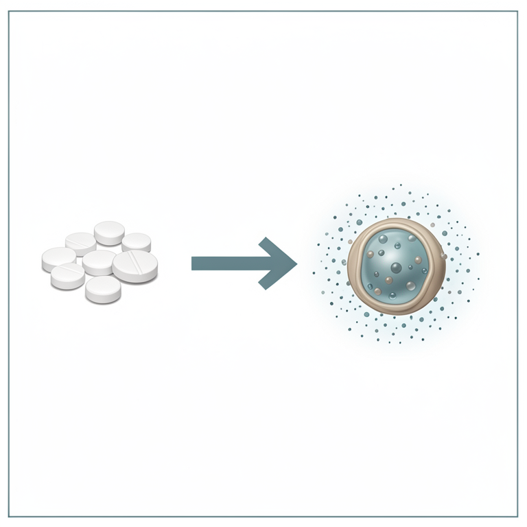
Dosing Frequency Comparison
AI-generated conceptual illustration (metformin hydrochloride solid lipid nanoparticles). It explains the idea and does not show study data.
View source evidence ↗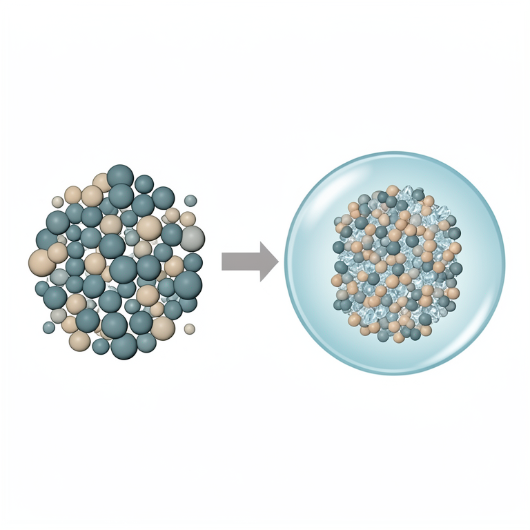
Lipid Matrix Entrapment Concept
AI-generated conceptual illustration (Solid lipid nanoparticles containing metformin hydrochloride). It explains the idea and does not show study data.
View source evidence ↗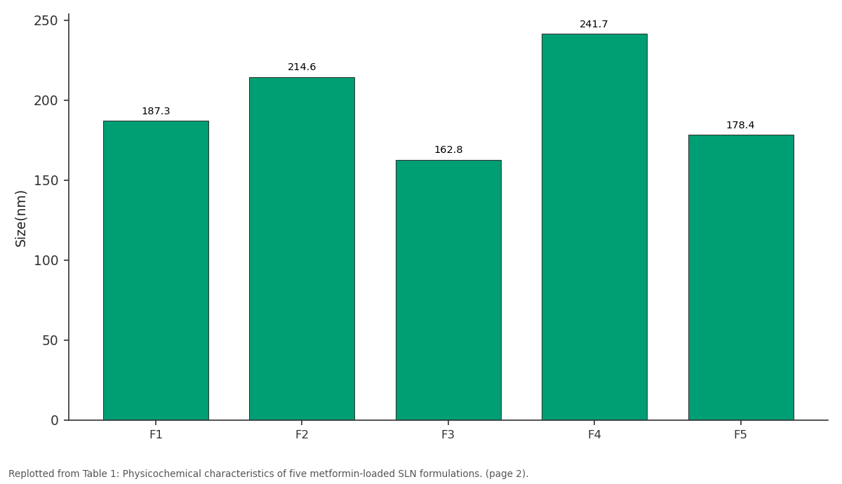
Size(nm): F1 vs F2 vs F3 vs F4 vs F5
Replotted from Table 1 (page 2) of the original paper. Every bar is the paper's own table value.
View source evidence ↗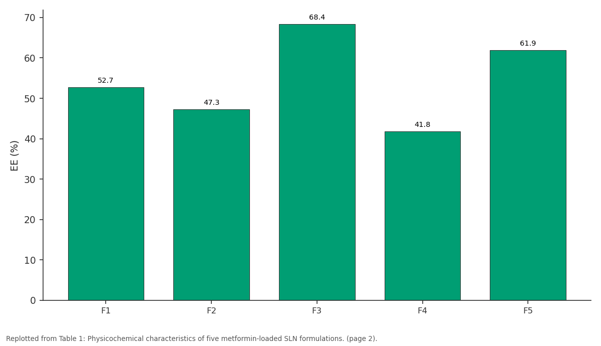
EE (%): F1 vs F2 vs F3 vs F4 vs F5
Replotted from Table 1 (page 2) of the original paper. Every bar is the paper's own table value.
View source evidence ↗Figures & tables
From the original paper Table 1
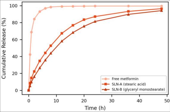 Figure 1
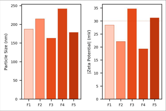 Figure 2
Introduction
Type 2 diabetes affects over 530 million adults globally, and that number is projected to reach 783 million by 2045, with the Middle East and North Africa region showing some of the highest prevalence rates worldwide [1] . Metformin hydrochloride is prescribed to roughly 150 million patients as the standard first-line oral hypoglycemic, but its pharmacokinetic profile creates compliance challenges -the drug's rapid absorption and short half-life mean patients typically take 500-1000 mg two or three times daily, and missed doses are common [2] . Extended-release tablet formulations exist, but they rely on hydrophilic matrix swelling that is sensitive to gastrointestinal transit variability and food effects [3] . Solid lipid nanoparticles represent an alternative strategy for controlled drug release. These colloidal carriers (50-500 nm) use biocompatible solid lipids as the matrix, combining the advantages of polymeric nanoparticles and liposomes while avoiding organic solvent residues and offering simpler large-scale production [4] . For hydrophilic drugs like metformin, the key challenge is achieving adequate entrapment -the drug's high water solubility (>300 mg/mL) drives it into the aqueous phase during preparation, leaving the lipid core with low payloads unless the formulation is carefully optimized [5] . Several approaches have been proposed to improve hydrophilic drug loading in SLNs: using ion-pairing agents to reduce aqueous solubility, double emulsion techniques, and selection of lipids with higher drug affinity [6] . Stearic acid and glyceryl monostearate are among the most studied SLN lipids due to their GRAS status, low cost, and well-characterized melting behavior, but head-to-head comparisons for metformin loading are scarce [7] .
The surfactant system also plays a decisive role, influencing both particle size and drug retention during solidification of the lipid phase [8] . This research compared five SLN formulations varying in lipid type, surfactant composition, and drug-to-lipid ratio, aiming to identify conditions that maximize metformin entrapment while producing particles below 250 nm with sufficient colloidal stability for oral delivery [9] .
Reagents and Chemicals
Metformin hydrochloride (>99% purity, Merck) was used as received. Stearic acid (octadecanoic acid, mp 69.3 °C) and glyceryl monostearate (mp 58.2 °C) were obtained from Gattefossé (Lyon, France). Poloxamer 188 (Lutrol F68, BASF), Tween 80 (polysorbate 80, Croda), and all other chemicals were analytical grade from Sigma-Aldrich. Dialysis membrane tubing (12-14 kDa MWCO) came from Spectrum Laboratories. Phosphate buffer saline (pH 6.8) was prepared fresh each week using USP-grade salts and Milli-Q water (18.2 MΩ•cm) [10] .
Materials and Methods
Materials and Methods
Materials
Research was performed at Arabian Gulf College of Pharmacy, Dammam, Eastern Province, Saudi Arabia, from February 2023 to November 2023. No ethical approval was required as no human or animal subjects were involved. A Silverson L5M-A high-shear mixer, Hielscher UP200Ht ultrasonicator (200 W, 26 kHz), Malvern Zetasizer Nano ZS, TA Instruments Q2000 DSC, Bruker D8 Advance XRD, and Shimadzu UV-1800 spectrophotometer were used for preparation and characterization [11] .
Methods
SLNs were prepared by hot homogenization followed by ultrasonication. Briefly, lipid (300 mg) was melted at 80 °C, and metformin (100 mg) was dissolved in the molten lipid phase. Separately, surfactant was dissolved in 20 mL water heated to the same temperature. The hot aqueous phase was added to the lipid melt under high-shear mixing at 15,000 rpm for 5 min, forming a coarse pre-emulsion. This was immediately sonicated at 40% amplitude for 10 min in an ice bath. The nanoemulsion was cooled to room temperature (25 °C) over 30 min with gentle stirring (300 rpm) to solidify the lipid, yielding the SLN dispersion [12] . Five formulations were prepared: F1 (stearic acid + 1% Poloxamer 188), F2 (stearic acid + 1% Tween 80), F3 (GMS + 0.5% Poloxamer 188 + 0.5% Tween 80), F4 (stearic acid + 1% Tween 80, double drug load), and F5 (GMS + 1% Poloxamer 188). Particle size, PDI, and zeta potential were measured by dynamic light scattering. Entrapment efficiency was determined by ultracentrifugation (40,000 rpm, 30 min, 4 °C); free drug in the supernatant was quantified by UV at 233 nm. Drug loading capacity was calculated as entrapped drug per unit mass of total nanoparticles [13] .
In vitro release studies used the dialysis bag method. SLN dispersion equivalent to 10 mg metformin was placed in dialysis tubing and immersed in 200 mL PBS (pH 6.8) at 37 °C under stirring (100 rpm). Samples (3 mL) were withdrawn at predetermined intervals over 48 h, replaced with fresh buffer, and assayed by UV at 233 nm. Release kinetics were fitted to zero-order, first-order, Higuchi, Korsmeyer-Peppas, and Hixson-Crowell models [14] . DSC thermograms (10 °C/min, nitrogen atmosphere, 25-250 °C) and XRD patterns (Cu Kα, 5-40° 2θ, 2°/min) characterized the solid-state properties. Stability was assessed at 25 °C/60% RH over 90 days with sampling at 30-day intervals [15] .
Instrumentation
Dynamic light scattering measurements used a Malvern Zetasizer Nano ZS (633 nm He-Ne laser, 173 ° backscatter angle, 25 °C, disposable polystyrene cuvettes for size/PDI and folded capillary cells for zeta potential). Each sample was measured in triplicate with 12 runs per measurement. DSC analysis employed hermetically sealed aluminum pans with an empty pan reference. XRD operated at 40 kV/40 mA with a step size of 0.02°. UV spectrophotometric measurements were validated for linearity (1-100 μg/mL, R² = 0.9999), precision (%RSD < 1.2%), and specificity (no lipid or surfactant interference at 233 nm) [16] . Formulation F3 (GMS with dual surfactant) consistently outperformed the other preparations. Its particle size of 162.8 nm was the smallest, reflecting the synergistic emulsification action of Poloxamer 188 and Tween 80 working together. The PDI of 0.187 indicated a narrow, monomodal size distribution. Its zeta potential of -34.7 mV exceeded the -30 mV threshold generally considered sufficient for electrostatic stabilization of colloidal dispersions [4] . Entrapment efficiency showed a clear lipid-type effect. GMS-based formulations (F3: 68.4%, F5: 61.9%) consistently entrapped more metformin than stearic acid formulations (F1: 52.7%, F2: 47.3%, F4: 41.8%). This likely reflects GMS's monoglyceride structure, which provides more hydrogen bonding opportunities with the amine groups of metformin than the simple carboxylic acid of stearic acid. Doubling the drug load in F4 actually decreased entrapment efficiency to 41.8%, probably because the excess drug exceeded the lipid's accommodation capacity and partitioned into the aqueous phase during cooling [5] . DSC thermograms showed that bulk metformin produced a sharp endotherm at 233.4 °C (melting point), while the physical mixture of drug and GMS showed both the lipid melt endotherm (58.2 °C) and a reduced drug peak. In F3 lyophilized SLNs, the metformin endotherm disappeared entirely, confirming molecular dispersion or amorphous entrapment within the lipid matrix. XRD supported this: crystalline metformin's characteristic peaks at 2θ = 12.1°, 17.8°, and 22.3° were absent in F3 powder, replaced by the broad halo of amorphous material superimposed on GMS's lipid reflections [17] . In vitro release profiles demonstrated the sustained-release advantage of SLN encapsulation. Free metformin dissolved completely within 2 hours, while F3 released 81.3% over 48 hours. The initial burst release from F3 was modest (14.2% in the first hour), attributed to surface-adsorbed drug diffusing rapidly from the nanoparticle periphery. After the burst phase, release followed a steady diffusion-controlled pattern. Kinetic modeling showed the best fit to the Higuchi equation (R² = 0.9934), consistent with matrix-type diffusion from a lipid depot. The Korsmeyer-Peppas exponent (n = 0.43) indicated Fickian diffusion as the dominant release mechanism [14] . Stability data at 25 °C/60% RH showed that F3 maintained its physicochemical properties over 90 days. Particle size increased marginally from 162.8 to 171.3 nm (5.2% change), PDI remained below 0.20, and entrapment efficiency decreased from 68.4% to 65.1% (4.8% loss). No visible aggregation, creaming, or sedimentation was observed throughout the storage period. The F2 formulation, by contrast, showed a 14.7% size increase and visible flocculation by day 60, consistent with its lower zeta potential (-22.1 mV) providing insufficient electrostatic repulsion [18] .
Results
Results
Discussion
The 68.4% entrapment efficiency achieved with F3 is noteworthy for a highly hydrophilic drug. Published SLN studies for metformin typically report 30-55% entrapment [5] , so the dual surfactant approach with GMS appears to offer a meaningful improvement. The mechanism likely involves Poloxamer 188's PEO chains creating a viscous interfacial layer that slows drug diffusion out of the lipid phase during the critical cooling step, while Tween 80's smaller molecular footprint fills gaps in the surfactant monolayer that would otherwise serve as drug escape routes [8] . GMS outperformed stearic acid as the lipid matrix, and the reason probably extends beyond hydrogen bonding. GMS creates a less perfectly ordered crystal lattice upon solidificationits mono-, di-, and triglyceride polymorphic mixture generates lattice imperfections that physically trap drug molecules more effectively than stearic acid's tightly packed lamellar crystals [7] . This interpretation is consistent with the DSC data showing broader, lowerintensity lipid endotherms for GMS formulations compared to the sharp melting peaks of stearic acid preparations. The 48-hour sustained release from F3 could translate to once-daily dosing in a clinical setting, cutting the current twice-or thrice-daily regimen. However, oral bioavailability of SLN-encapsulated metformin needs in vivo confirmation, because intestinal lipase activity may accelerate lipid matrix degradation and drug release beyond what the dialysis bag model predicts [19] . Additionally, the 14.2% burst release in the first hour is therapeutically desirable for diabetes management, as it provides an initial loading dose effect before the sustained phase maintains plasma concentrations. Scaling from the current 20 mL batch to production volumes would require transitioning from probe ultrasonication to high-pressure homogenization, which is industrially established for SLN manufacturing. The formulation's simplicitytwo excipients (GMS and surfactants) with no organic solventsis an advantage for regulatory approval and manufacturing cost. Lyophilization with 5% trehalose as cryoprotectant preserved particle characteristics after reconstitution, which would support a dry powder dosage form filled into capsules for oral administration [20] .
Conclusion
Solid lipid nanoparticles prepared from glyceryl monostearate with a dual surfactant system (Poloxamer 188 + Tween 80) achieved the highest metformin entrapment (68.4%) among five formulations tested. The optimized formulation F3 showed favorable particle characteristics (162.8 nm, PDI 0.187, zeta potential -34.7 mV), sustained drug release over 48 hours following Higuchi diffusion kinetics, and adequate colloidal stability over 90 days at 25 °C. DSC and XRD confirmed amorphous drug dispersion within the lipid matrix. These results support further development toward once-daily oral metformin delivery, with in vivo pharmacokinetic evaluation as the recommended next step.
Acknowledgement
Acknowledgements Funding sourcesThis work was funded by the Arabian Gulf College of Pharmacy Internal Research Grant (AGCP-IRG-2023-014). Institutional supportThe authors thank Arabian Gulf College of Pharmacy for access to the Nanopharmaceutics Research Laboratory and central instrumentation facility.
Figures and tables from the paper
| Code | Lipid | Size(nm) | PDI | Zeta(mV) | EE (%) | DL (%) |
|---|---|---|---|---|---|---|
| F1 | Stearic acid | 187.3 | 0.241 | −28.4 | 52.7 | 4.8 |
| F2 | Stearic acid | 214.6 | 0.298 | −22.1 | 47.3 | 4.1 |
| F3 | GMS | 162.8 | 0.187 | −34.7 | 68.4 | 6.7 |
| F4 | Stearic acid | 241.7 | 0.312 | −19.3 | 41.8 | 7.2 |
| F5 | GMS | 178.4 | 0.223 | −31.2 | 61.9 | 5.9 |
Table text extracted from the original PDF; superscript letters are shown inline. The original table image is below.
View original table image
References
20 in the original paper- Sun H, Saeedi P, Karuranga S, Pinkepank M, Ogurtsova K. IDF Diabetes Atlas: global diabetes prevalence estimates. Diabetes Res Clin Pract. 2022;183:109119.
- Bailey CJ, Turner RC. Metformin. N Engl J Med. 1996;334(9):574-579.
- Timmins P, Donahue S, Meeker J, Marathe P. Steadystate pharmacokinetics of a novel extended-release metformin. Clin Pharmacokinet. 2005;44(7):721-729.
- Müller RH, Mäder K, Gohla S. Solid lipid nanoparticles (SLN) for controlled drug delivery. Eur J Pharm Biopharm. 2000;50(1):161-177.
- Mehnert W, Mäder K. Solid lipid nanoparticles: production, characterization and applications. Adv Drug Deliv Rev. 2001;47(2):165-196.
- Mishra V, Bansal KK, Verma A, Yadav N, Thakur S. Solid lipid nanoparticles: emerging colloidal carriers. Pharmaceutics. 2018;10(4):191-205.
- Jenning V, Thünemann AF, Gohla SH. Characterisation of a novel solid lipid nanoparticle carrier system. Int J Pharm. 2000;199(2):167-177.
- Helgason T, Awad TS, Kristbergsson K, McClements DJ, Weiss J. Effect of surfactant surface coverage on SLN formation. J Colloid Interface Sci. 2009;334(1):75-81.
- Dudhipala N, Janga KY. Lipid nanoparticles of zaleplon for improved oral delivery. Eur J Pharm Sci. 2017;104:223-232.
- British Pharmacopoeia Commission. Metformin hydrochloride monograph. British Pharmacopoeia. 2023.
- Gasco MR. Method for producing solid lipid microspheres having a narrow size distribution. US Patent. 5,250,236. 1993.
- Severino P, Andreani T, Macedo AS, Fangueiro JF, Santana MH. Current state of art and new trends on lipid nanoparticles. J Drug Deliv. 2012;2012:750891.
- Schwarz C, Mehnert W, Lucks JS, Müller RH. Solid lipid nanoparticles (SLN) for controlled drug delivery. J Control Release. 1994;30(1):83-96.
- Costa P, Lobo JMS. Modeling and comparison of dissolution profiles. Eur J Pharm Sci. 2001;13(2):123133.
- ICH Q1A(R2). Stability testing of new drug substances and products. ICH Harmonised Tripartite Guideline. 2003.
- Freitas C, Müller RH. Effect of light and temperature on zeta potential and physical stability. Int J Pharm. 1998;168(2):221-229.
- Bunjes H, Unruh T. Characterization of lipid nanoparticles by differential scanning calorimetry and X-ray. Adv Drug Deliv Rev. 2007;59(6):379-402.
- Heurtault B, Saulnier P, Pech B, Proust JE, Benoit JP. Physico-chemical stability of colloidal lipid particles. Biomaterials. 2003;24(23):4283-4300.
- Zhu S, Hong M, Zhang L, Tang G, Jiang Y. PEGylated nicotinic acid-modified solid lipid nanoparticles. Colloids Surf B. 2010;79(1):205-210.
- Schwarz C, Mehnert W. Freeze-drying of drug-free and drug-loaded solid lipid nanoparticles. Int J Pharm. 1997;157(2):171-179.