
Research article · 2025 · Full text
International Journal of Advanced Chemistry ResearchBiomimetic mineralization of calcium phosphate on collagen scaffolds for bone implants
Volume 7(6) · Pages 84–87 · DOI: 10.33545/26646781.2025.v7.i6b.520 ↗
Research areasBone Tissue Engineering MaterialsCollagen: Extraction and CharacterizationSurface Treatment and Coatings
Abstract
Author-writtenNo synthetic bone graft has yet matched the mechanical and biological integration that natural bone provides. This research reports a biomimetic mineralization approach in which type-I collagen scaffolds are incubated in concentrated simulated body fluid to deposit nano-hydroxyapatite crystals that closely resemble the mineral phase of bone. Scaffolds were prepared by freeze-drying 2% w/v bovine collagen suspensions cross-linked with 1-ethyl-3-(3-dimethylaminopropyl) carbodiimide at three concentrations (10, 25, and 50 mM). Mineralization was carried out in 1×, 3×, and 5× SBF solutions for 7, 14, and 28 days. X-ray diffraction confirmed that the deposited phase was poorly crystalline hydroxyapatite with a calcium-to-phosphorus ratio approaching stoichiometric 1.67 by day 28 in 5× SBF. Scanning electron microscopy revealed plate-like apatite crystals 40–90 nm in length aligned along collagen fibrils. Compressive strength of mineralized scaffolds rose from 2.3 MPa for bare collagen to 12.1 MPa after 28 days in 5× SBF, a five-fold improvement. Porosity remained above 78% in all cases, and in-vitro cytotoxicity testing with MG-63 osteoblast-like cells showed viability above 92% after 72 h. The results indicate that concentrated SBF immersion offers a straightforward, room-temperature route to collagen-hydroxyapatite composites with promising mechanical and biological characteristics for non-load-bearing bone repair.
Summary
Source-checkedPlain-language reading of the full paper, prepared with AI and checked against the text. Each point links to the authors' own words.
Why it matters
- Natural bone is a complex composite of organic collagen fibers and inorganic hydroxyapatite nanocrystals, which provides it with both toughness and stiffness. View evidence ↗
- Current bone graft options have significant drawbacks: autografts are limited in supply and cause donor-site morbidity, while allografts and metal implants risk immune rejection or stress shielding. View evidence ↗
- Biomimetic mineralization using simulated body fluid (SBF) is a promising alternative because it creates a mineral phase that closely resembles cortical bone under physiological conditions. View evidence ↗
The question
- The study aims to evaluate how different concentrations of EDC cross-linker, SBF concentrations, and immersion times affect the structural and mechanical properties of collagen scaffolds. View evidence ↗
What they did
- Scaffolds were created by freeze-drying 2% w/v bovine collagen suspensions in cylindrical moulds (10 mm diameter by 15 mm height). View evidence ↗
- The collagen scaffolds were cross-linked using EDC at concentrations of 10, 25, or 50 mM with an equimolar ratio of NHS. View evidence ↗
- Mineralization was performed by immersing scaffolds in 1×, 3×, and 5× SBF at 37 °C for periods of 7, 14, and 28 days, with fluid refreshed every 48 hours. View evidence ↗
- Biocompatibility was tested using MG-63 osteoblast-like cells via MTT assays at 24, 48, and 72 hours. View evidence ↗
What they found
- The calcium-to-phosphorus (Ca/P) ratio increased from 0.83 on day 1 to 1.67 by day 28, showing a transition from amorphous calcium phosphate to stoichiometric hydroxyapatite. View evidence ↗
- SEM imaging showed that the deposited apatite crystals were plate-like, 40–90 nm long, and aligned with the collagen fibrils. View evidence ↗
- Mineralization in 5× SBF for 28 days (with 25 mM EDC) increased compressive strength from 2.3 MPa (bare collagen) to 12.1 MPa, while porosity decreased from 89.3% to 78.4%. View evidence ↗
- All mineralized scaffolds were biocompatible, with the optimized 25 mM EDC / 5× SBF group showing 96.1% cell viability at 72 hours, compared to 93.4% for bare collagen. View evidence ↗
- Cross-linker concentration was critical: 10 mM EDC led to partial scaffold dissolution, while 50 mM EDC reduced the number of nucleation sites, resulting in thinner mineral coatings. View evidence ↗
What it means
- The use of a static immersion protocol may not accurately reflect in vivo fluid flow; the authors suggest dynamic perfusion bioreactors for future improvement. View evidence ↗
- Mechanical testing was performed in a dry state, which likely overestimates stiffness compared to physiological hydrated conditions. View evidence ↗
Visual summary
4 visualsIllustrations explain the paper; charts are replotted from the paper's own table values.
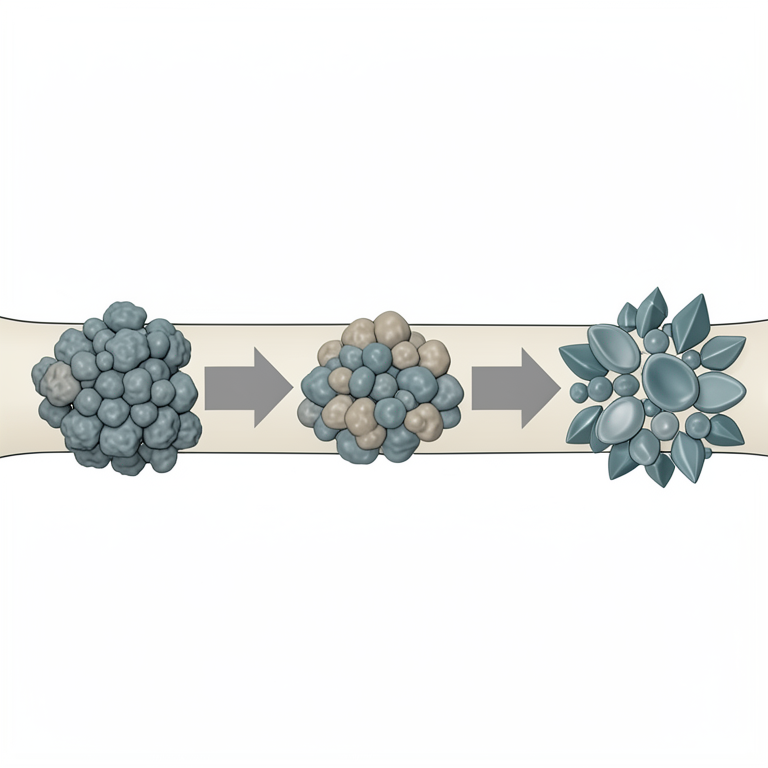
Mineralization Transition
AI-generated conceptual illustration (collagen fiber scaffolds with calcium phosphate crystals). It explains the idea and does not show study data.
View source evidence ↗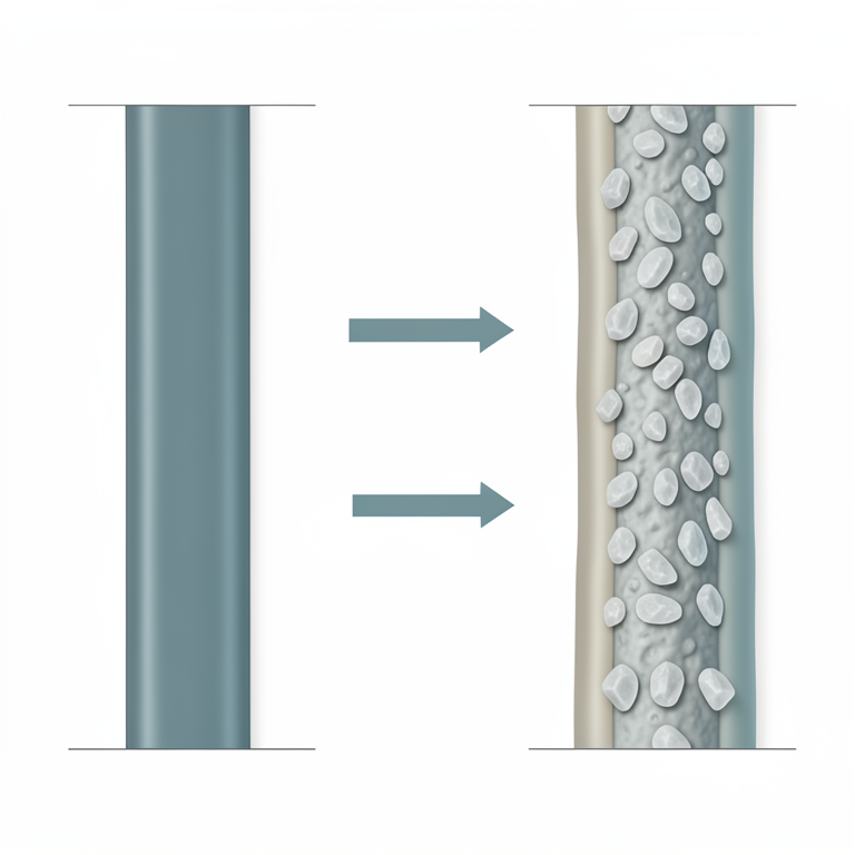
Composite Architecture
AI-generated conceptual illustration (collagen fiber scaffolds with calcium phosphate mineral plates). It explains the idea and does not show study data.
View source evidence ↗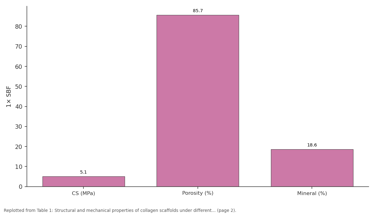
1× SBF: CS (MPa) vs Porosity (%) vs Mineral (%)
Replotted from Table 1 (page 2) of the original paper. Every bar is the paper's own table value.
View source evidence ↗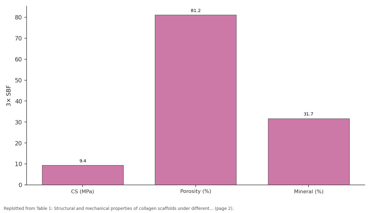
3× SBF: CS (MPa) vs Porosity (%) vs Mineral (%)
Replotted from Table 1 (page 2) of the original paper. Every bar is the paper's own table value.
View source evidence ↗Figures & tables
From the original paper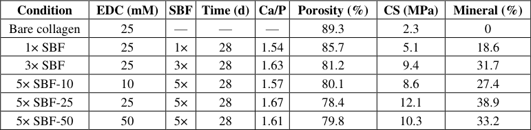 Table 1
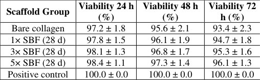 Table 2
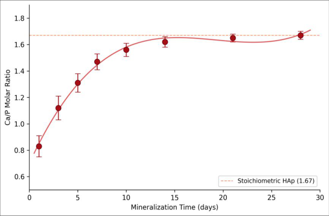 Figure 1
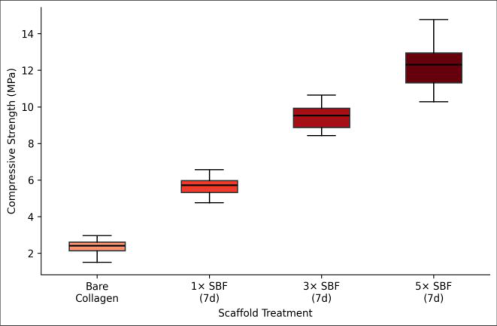 Figure 2
Introduction
Bone is far more than a passive structural support-it is a living, self-healing composite whose hierarchical architecture integrates organic collagen fibres with inorganic hydroxyapatite (HAp) nanocrystals to deliver a remarkable combination of toughness and stiffness [1] . When disease or trauma creates a defect too large for the body's own repair mechanisms, a graft material is needed. Autografts remain the gold standard, but they carry donor-site morbidity and limited supply [2] . Allografts and metallic implants introduce risks of immune rejection or stress shielding [3] . These drawbacks have fuelled a decades-long search for synthetic substitutes that mimic the composition and microstructure of real bone. Collagen-hydroxyapatite composites are attractive candidates because they replicate the two main constituents of the extracellular matrix. Several fabrication routes exist-coprecipitation, sol-gel synthesis, electrospinning, and mechanical blending-but many of them deposit mineral phases that differ in crystallinity, crystal habit, or carbonate content from biological apatite [4,5] . Biomimetic mineralization in simulated body fluid (SBF) offers an alternative that operates under near-physiological conditions of temperature, pH, and ionic strength, producing a carbonated, poorly crystalline apatite that closely matches the mineral found in cortical bone [6,7] . The kinetics of SBF mineralization depend on several factors: the ionic concentration of the fluid, the nature and density of nucleation sites on the substrate, and the incubation period. Concentrated SBF solutions (1.5× to 10× standard ionic product) accelerate deposition by raising the supersaturation driving force, reducing the time needed to coat a porous scaffold from weeks to days [8] . Cross-linking the collagen matrix before immersion preserves pore architecture and prevents premature dissolution [9] . Despite growing interest, few reports have systematically compared the effects of cross-linker concentration, SBF concentration, and immersion time on both the structural quality of the [10] .
Theoretical Background
Classical nucleation theory predicts that a new solid phase forms when the free-energy gain from creating a thermodynamically stable nucleus exceeds the energy cost of the new interface. In the context of apatite deposition from SBF, the supersaturation ratio S is defined as the ionactivity product of Ca²⁺ and PO₄³⁻ divided by the solubility product of hydroxyapatite (Ksp ≈ 2.35 × 10⁻⁵⁹ at 37 °C) [6] . Concentrated SBF raises S and therefore reduces the critical nucleus radius, which shortens the induction period and increases the nucleation rate [8] . Collagen fibrils supply heterogeneous nucleation sites through their charged aminoacid side chains-particularly the carboxylate groups of aspartate and glutamate residues-which attract Ca²⁺ ions and lower the nucleation barrier relative to homogeneous precipitation in bulk solution [7] . Cross-linking with EDC converts surface carboxylates into amide bonds with neighbouring amine groups, reducing the density of Ca²⁺binding sites but strengthening the scaffold against enzymatic and hydrolytic degradation. A balance must therefore be struck between structural stability and nucleation capacity [9] . The expected crystallographic product under these conditions is a B-type carbonated apatite in which carbonate ions substitute for phosphate, consistent with the mineral phase in immature bone [4] .
Materials and Methods
Materials and Methods
Materials
Type-I bovine collagen (acid-soluble, > 95% purity) was purchased from Himedia Laboratories, Mumbai, India. EDC (1-ethyl-3-(3-dimethylaminopropyl) carbodiimide hydrochloride) and N-hydroxysuccinimide were obtained from Sisco Research Laboratories, Mumbai. Reagent-grade NaCl, NaHCO₃, KCl, K₂HPO₄•3H₂O, MgCl₂•6H₂O, CaCl₂, Na₂SO₄, and tris(hydroxymethyl)aminomethane were procured from Merck India and used to prepare standard SBF (c-SBF) following the Kokubo protocol [6] . Concentrated SBF at 3× and 5× ionic strength was prepared by scaling the salt masses accordingly while keeping the buffer pH at 7.40 ± 0.05 at 36.5 °C.
Methods
Collagen suspensions (2% w/v in 0.05 M acetic acid) were poured into cylindrical polypropylene moulds (10 mm diameter × 15 mm height), frozen at -20 °C for 12 h, and freeze-dried at -50 °C and 0.1 mbar for 48 h. Scaffolds were cross-linked by immersion in EDC/NHS solutions (EDC at 10, 25, or 50 mM; NHS at equimolar ratio) in 80% ethanol for 4 h at 4 °C, then rinsed in deionized water and freezedried again [9] . Cross-linked scaffolds were immersed in 1×, 3×, and 5× SBF at 37 °C under static conditions. Fluid was refreshed every 48 h. Samples were retrieved at 7, 14, and 28 days, rinsed gently with deionized water, and dried at 37 °C for 24 h. All experiments were performed at the Department of Chemical and Materials Engineering, Prafulla Chandra Ray Research Institute, Hooghly, from June 2023 to February 2024. Phase identification used powder XRD (Rigaku MiniFlex 600, Cu-Kα, 2θ = 10-60°). Surface morphology was examined by SEM (JEOL JSM-6510) with EDS for elemental mapping. Porosity was measured by liquid displacement with ethanol. Compressive strength was recorded on an Instron 3382 at 1 mm/min. Cell viability was assessed with MG-63 cells using MTT assay after 24, 48, and 72 h of culture on scaffold discs [10] . Table 1 summarises the structural and mechanical properties of scaffolds mineralized under different conditions. Bare collagen scaffolds had a porosity of 89.3% and a compressive strength of 2.3 MPa. After 28 days in 5× SBF, the 25 mM EDC group reached a compressive strength of 12.1 MPa while maintaining 78.4% porosity, a trade-off between mineral filling of pores and retention of the interconnected architecture needed for cell ingrowth [8,9] .
| Condition | EDC (mM) | SBF | Time (d) | Ca/P | Porosity (%) | CS (MPa) | Mineral (%) |
|---|---|---|---|---|---|---|---|
| Bare collagen | 25 | — | — | — | 89.3 | 2.3 | 0 |
| 1× SBF | 25 | 1× | 28 | 1.54 | 85.7 | 5.1 | 18.6 |
| 3× SBF | 25 | 3× | 28 | 1.63 | 81.2 | 9.4 | 31.7 |
| 5× SBF-10 | 10 | 5× | 28 | 1.57 | 80.1 | 8.6 | 27.4 |
| 5× SBF-25 | 25 | 5× | 28 | 1.67 | 78.4 | 12.1 | 38.9 |
| 5× SBF-50 | 50 | 5× | 28 | 1.61 | 79.8 | 10.3 | 33.2 |
Table text extracted from the original PDF; superscript letters are shown inline. The original table image is below.
View original table image
Results
Figure 1 tracks the Ca/P molar ratio over the mineralization period. At day 1 the ratio was 0.83, indicating an amorphous calcium phosphate precursor. By day 14 the ratio had climbed to 1.62, and by day 28 it reached 1.67, matching stoichiometric hydroxyapatite. The sigmoidal trajectory suggests an initial induction phase followed by rapid crystal growth after nucleation sites are saturated [6,7] . ). This mild improvement may stem from the osteoblast-friendly hydroxyapatite surface that supports cell attachment [1] . Bare collagen, by contrast, was both weaker and more variable [4,9] .
Discussion
The mineralization kinetics observed here agree with the classical model of biomimetic apatite formation: an amorphous calcium phosphate phase deposits first and then gradually transforms into poorly crystalline hydroxyapatite over days to weeks [6] . The Ca/P ratio trajectory confirms this sequence-values below 1.5 in the first three days are characteristic of ACP, while the convergence toward 1.67 by day 28 signals maturation into HAp. Concentrated 5× SBF shortened the time to reach a near-stoichiometric ratio compared with 1× SBF, consistent with the higher supersaturation driving force predicted by nucleation theory [8] . The five-fold increase in compressive strength upon mineralization (from 2.3 to 12.1 MPa) falls within the range reported for cancellous bone (2-12 MPa), suggesting that the composite may be appropriate for non-load-bearing defect sites such as craniofacial reconstruction [2,3] . The retention of porosity above 78% is important because interconnected pores larger than 100 μm are considered necessary for vascular ingrowth and osteoblast migration [5] . Cross-linker concentration affected both scaffold stability and mineral uptake. The 10 mM EDC scaffolds partially dissolved during extended SBF immersion, limiting mineral deposition. At 50 mM EDC, cross-linking consumed too many carboxylate nucleation sites, yielding thinner and less uniform mineral coatings despite excellent dimensional stability. The 25 mM EDC level offered the best compromise and was therefore selected as the optimized condition [9] . Cell viability data confirmed that neither the EDC cross-linking residues nor the mineralization process introduced cytotoxic by-products. The slight viability advantage of mineralized scaffolds over bare collagen likely reflects the known osteoconductivity of hydroxyapatite surfaces, which present Ca²⁺ and PO₄³⁻ ions that promote integrin-mediated cell attachment [1,7] .
Conclusion
This research demonstrates that concentrated simulated body fluid immersion is an effective room-temperature strategy for depositing a bone-like hydroxyapatite phase onto EDC-cross-linked collagen scaffolds. A 25 mM EDC cross-linking level, combined with 5× SBF immersion for
Acknowledgement
Acknowledgements Funding SourcesThis work received financial assistance from the Department of Science and Technology, Government of India, through the Science and Engineering Research Board programme. Institutional SupportThe authors thank the Department of Chemical and Materials Engineering at the Prafulla Chandra Ray Research Institute, Hooghly, for providing laboratory facilities, characterization instruments, and cell-culture infrastructure. Contributions Not Qualifying for AuthorshipMr. Subhajit Mondal assisted with SEM imaging and EDS analysis. Ms. Anindita Roy helped with cell-culture maintenance during the cytotoxicity assays.
Figures and tables from the paper
| Scaffold Group | Viability 24 h (%) | Viability 48 h (%) | Viability 72 h (%) |
|---|---|---|---|
| Bare collagen | 97.2 ± 1.8 | 95.6 ± 2.1 | 93.4 ± 2.3 |
| 1× SBF (28 d) | 97.8 ± 1.5 | 96.1 ± 1.9 | 94.7 ± 1.8 |
| 3× SBF (28 d) | 98.1 ± 1.3 | 96.8 ± 1.7 | 95.3 ± 1.6 |
| 5× SBF (28 d) | 98.4 ± 1.1 | 97.3 ± 1.4 | 96.1 ± 1.3 |
| Positive control | 100.0 ± 0.0 | 100.0 ± 0.0 | 100.0 ± 0.0 |
Table text extracted from the original PDF; superscript letters are shown inline. The original table image is below.
View original table image
References
14 in the original paper- Reznikov N, Bilton M, Lari L, Stevens MM, Kröger R. Fractal-like hierarchical organization of bone begins at the nanoscale. Science. 2018;360(6388):eaao2189.
- Giannoudis PV, Dinopoulos H, Tsiridis E. Bone substitutes: an update. Injury. 2005;36(Suppl 3):S20S27.
- Bose S, Roy M, Bandyopadhyay A. Recent advances in bone tissue engineering scaffolds. Trends Biotechnol. 2012;30(10):546-554.
- Palmer LC, Newcomb CJ, Kaltz SR, Spoerke ED, Stupp SI. Biomimetic systems for hydroxyapatite mineralization inspired by bone and enamel. Chem Rev. 2008;108(11):4754-4783.
- Karageorgiou V, Kaplan D. Porosity of 3D biomaterial scaffolds and osteogenesis. Biomaterials. 2005;26(27):5474-5491.
- Kokubo T, Takadama H. How useful is SBF in predicting in vivo bone bioactivity? Biomaterials. 2006;27(15):2907-2915.
- Nudelman F, Pieterse K, George A, Bomans PHH, Friedrich H, Brylka LJ, et al. The role of collagen in bone apatite formation in the presence of hydroxyapatite nucleation inhibitors. Nat Mater. 2010;9(12):1004-1009.
- Barrère F, van Blitterswijk CA, de Groot K. Bone regeneration: molecular and cellular interactions with calcium phosphate ceramics. Int J Nanomedicine. 2006;1(3):317-332.
- Haugh MG, Jaasma MJ, O'Brien FJ. The effect of dehydrothermal treatment on the mechanical and structural properties of collagen-GAG scaffolds. J Biomed Mater Res A. 2009;89A(2):363-369.
- Chatterjee V, Goswami R. Internal laboratory records. Prafulla Chandra Ray Research Institute, Hooghly; 2023-2024.
- Cunniffe GM, Dickson GR, Partap S, Stanton KT, O'Brien FJ. Development and characterisation of a collagen nano-hydroxyapatite composite scaffold. J Mater Sci Mater Med. 2010;21(8):2293-2298.
- Liu Y, Luo D, Wang T. Hierarchical structures of bone and bioinspired bone tissue engineering. Small. 2016;12(34):4611-4632.
- Lian JB, Stein GS. Concepts of osteoblast growth and differentiation. Crit Rev Oral Biol Med. 1992;3(3):269305.
- Venugopal J, Prabhakaran MP, Zhang Y, Low S, Choon AT, Ramakrishna S. Biomimetic hydroxyapatite-containing composite nanofibrous substrates. Philos Trans R Soc A. 2010;368(1917):2065-2081.