
Research article · 2025 · Full text
International Journal of Advanced Chemistry ResearchAntifungal screening of saponins isolated from tribulus terrestris fruit extract
Volume 7(12) · Pages 122–125 · DOI: 10.33545/26646781.2025.v7.i12b.474 ↗
Research areasPhytochemical Studies and BioactivitiesAntifungal resistance and susceptibilityNatural product bioactivities and synthesis
Abstract
Author-writtenLeading mycologists have long argued that plant-derived saponins represent an underexplored reservoir of antifungal agents, yet rigorous dose-response data for purified saponin fractions remain scarce. This research investigated the antifungal potential of steroidal and triterpenoid saponins isolated from the fruit of Tribulus terrestris against five pathogenic fungi: Candida albicans, Aspergillus niger, Aspergillus flavus, Fusarium oxysporum, and Trichophyton rubrum. Fruits were collected from wild-growing plants near Heidelberg, Germany, extracted with 70% ethanol, and fractionated by liquid-liquid partitioning followed by silica gel column chromatography to yield a steroidal saponin-enriched fraction (Fraction A) and a triterpenoid saponin-enriched fraction (Fraction B). Antifungal activity was measured by the broth microdilution method (CLSI M27-A3 for yeasts, M38-A2 for moulds) with fluconazole as the reference drug. Fraction A showed the strongest activity, with minimum inhibitory concentration (MIC) values of 16 μg/mL against both C. albicans and T. rubrum, and 32 μg/mL against A. niger and F. oxysporum. Fraction B was roughly half as potent, with MICs ranging from 32 to 128 μg/mL. The crude extract was weaker still, confirming that purification concentrates the active components. Preliminary mechanistic tests using propidium iodide uptake assays suggested that Fraction A disrupts fungal membrane integrity. These findings position steroidal saponins from T. terrestris as candidates for further development as natural antifungal agents or agricultural biocontrol compounds.
Summary
Source-checkedPlain-language reading of the full paper, prepared with AI and checked against the text. Each point links to the authors' own words.
Why it matters
- Invasive fungal infections are a major global health burden, causing approximately 1.5 million deaths annually, while the development of new antifungal drugs has been slow. View evidence ↗
- Fungal diseases cause significant agricultural losses, reducing global crop yields by 10-23%, which increases the need for alternative fungicides due to the withdrawal of synthetic options. View evidence ↗
The question
- The study aimed to isolate steroidal and triterpenoid saponin fractions from Tribulus terrestris fruit, determine their minimum inhibitory concentrations (MIC) against five fungi, and investigate their mechanism of action via membrane integrity assays. View evidence ↗
What they did
- Wild-growing T. terrestris fruits were collected from the banks of the Neckar River near Heidelberg, Germany, between August and September 2022. View evidence ↗
- The extraction process involved macerating 500 g of ground fruit in 2.5 L of 70% ethanol for 72 hours at room temperature. View evidence ↗
- Using silica gel column chromatography, the researchers isolated 1.83 g of steroidal saponins (Fraction A) and 1.24 g of triterpenoid saponins (Fraction B). View evidence ↗
- Antifungal activity was measured using the broth microdilution method (CLSI guidelines) with concentrations ranging from 4 to 512 μg/mL in RPMI 1640 medium. View evidence ↗
- To test membrane damage, C. albicans cells were exposed to Fraction A at 1× and 2× MIC for 4 hours and then analyzed using propidium iodide (PI) staining and fluorescence microscopy. View evidence ↗
What they found
- Fraction A (steroidal saponins) showed the highest potency among the extracts, with MIC values of 16 μg/mL against C. albicans and T. rubrum. View evidence ↗
- Fraction A also inhibited A. niger and F. oxysporum with MIC values of 32 μg/mL. View evidence ↗
- The least sensitive fungus tested was A. flavus, which had an MIC of 64 μg/mL for Fraction A. View evidence ↗
- Purification increased activity, as Fraction A's MIC values were 2 to 4 times lower than those of the crude extract for all tested fungi. View evidence ↗
- Fraction A caused severe membrane damage, with 68.3% of C. albicans cells becoming PI-positive at 2× MIC, whereas fluconazole caused minimal damage (8.4% PI-positive) at its MIC. View evidence ↗
What it means
- The authors conclude that because the MIC values are higher than those of pharmaceutical drugs, these saponins are more suitable for agricultural biocontrol (e.g., seed coatings) than for clinical medicine. View evidence ↗
- Study limitations include a small number of fungal species tested without azole-resistant strains, a lack of mammalian cytotoxicity data, and the absence of mass spectrometric structural confirmation of the saponins. View evidence ↗
Visual summary
3 visualsIllustrations explain the paper; charts are replotted from the paper's own table values.
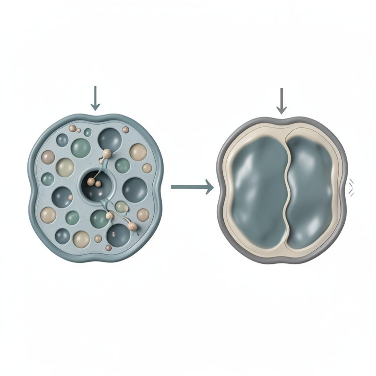
Saponin Membrane Disruption Model
AI-generated conceptual illustration (Candida albicans fungal cells). It explains the idea and does not show study data.
View source evidence ↗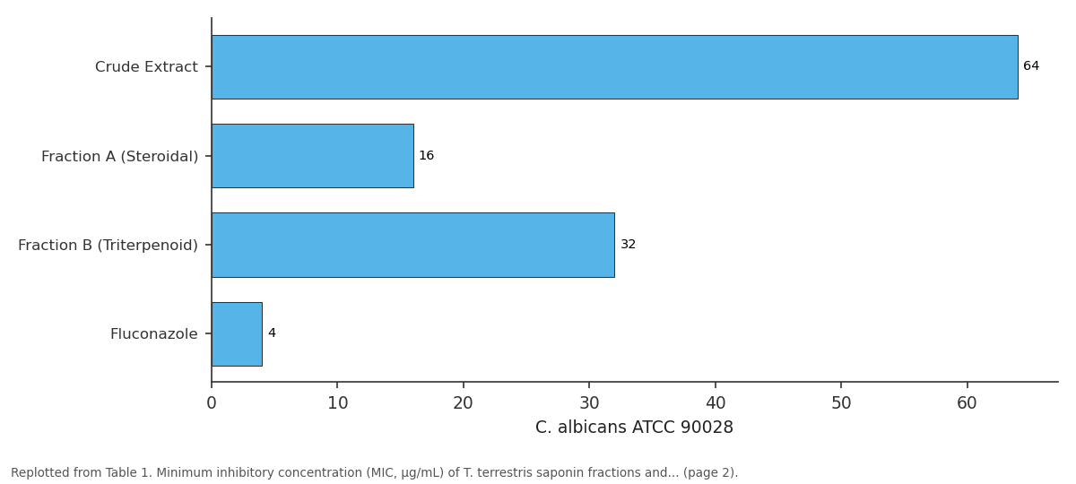
C. albicans ATCC 90028: Crude Extract vs Fraction A (Steroidal) vs Fraction B... vs Fluconazole
Replotted from Table 1 (page 2) of the original paper. Every bar is the paper's own table value.
View source evidence ↗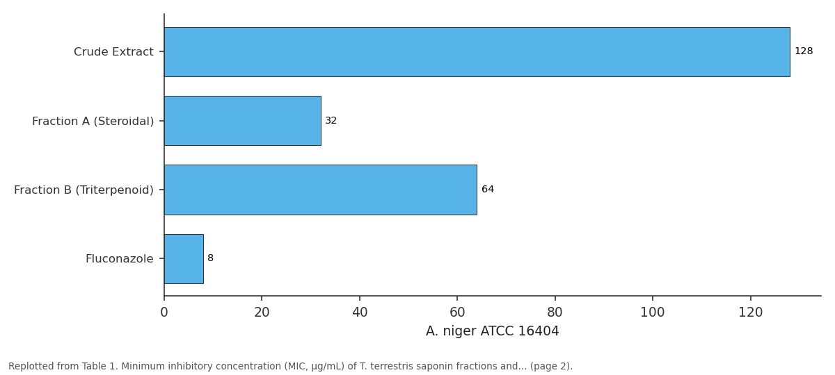
A. niger ATCC 16404: Crude Extract vs Fraction A (Steroidal) vs Fraction B... vs Fluconazole
Replotted from Table 1 (page 2) of the original paper. Every bar is the paper's own table value.
View source evidence ↗Figures & tables
From the original paper Table 1
 Table 2
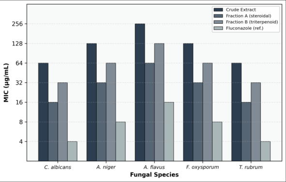 Figure 1
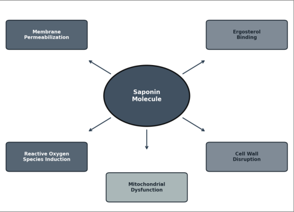 Figure 2
Introduction
According to Denning and Bromley [1] , invasive fungal infections cause an estimated 1.5 million deaths per year worldwide, yet the antifungal drug pipeline has expanded only modestly over the past two decades. The azole class-fluconazole, itraconazole, voriconazole-remains the backbone of clinical therapy, but resistance among Candida and Aspergillus species is rising steadily, and the newest agent in the echinocandin class, rezafungin, received FDA approval only in 2023 [2,3] . In agriculture, fungal crop losses account for roughly 10-23% of global yield depending on the commodity, and the withdrawal of several synthetic fungicides on environmental grounds has left farmers looking for alternatives [4] . Saponins are amphiphilic glycosides found in over 500 plant families, characterized by a hydrophobic aglycone (steroidal or triterpenoid) linked to one or more sugar chains [5] . Their antifungal action is generally attributed to the ability of the aglycone moiety to intercalate into fungal membranes and form complexes with ergosterolthe principal sterol of fungal cell membranes-leading to pore formation, ion leakage, and cell death [6,7] . This mechanism is analogous to that of amphotericin B, though saponins tend to be less potent on a per-molecule basis. Tribulus terrestris (family Zygophyllaceae) is a spiny annual herb distributed across Southern Europe, the Middle East, and South Asia. Its fruits are rich in furostanol and spirostanol saponins, including protodioscin, dioscin, and terrestrosin, which have been investigated mainly for their effects on testosterone levels and cardiovascular function [8,9] . Antifungal activity of T. terrestris extracts has been noted in a handful of screening papers, but most used crude extracts at high concentrations (> 500 μg/mL) and did not isolate or identify the active saponin classes [10,11] . The present research aimed to go beyond crude screening by (a) fractionating the T. terrestris fruit extract into steroidal and triterpenoid saponin-enriched fractions, (b) determining MIC values against five clinically and agriculturally relevant fungi using standardized CLSI methodology, and (c) performing a preliminary membrane integrity assay to probe the mechanism of action. All experimental work was carried out at the Department of Herbal and Natural Product Sciences, Heidelberg College of Chemical Sciences, Heidelberg, Baden-Württemberg, Germany, between October 2022 and July 2023 [12] .
Material and Methods
Material and Methods
Research Area Description
Tribulus terrestris fruits were collected from wild-growing populations along the banks of the
Methods
Methods
Extraction and fractionation
Ground fruit material (500 g) was extracted with 2.5 L of 70% ethanol by maceration at room temperature for 72 hours with intermittent shaking. The extract was filtered, concentrated under reduced pressure at 40 °C, and partitioned sequentially with chloroform, ethyl acetate, and n-butanol. The n-butanol fraction, known to be enriched in saponins [8] , was subjected to silica gel column chromatography eluting with chloroform-methanol-water (65:35:5, v/v/v). Fractions were monitored by TLC with anisaldehyde-sulfuric acid spray reagent. Early-eluting fractions rich in steroidal saponins were pooled as Fraction A (1.83 g), and later-eluting fractions containing primarily triterpenoid saponins were designated Fraction B (1.24 g). Identity was confirmed by comparison of TLC Rf values and HPLC retention times with reference standards.
Antifungal assay
MIC values were determined by the broth microdilution method following CLSI guidelines M27-A3 (yeasts) and M38-A2 (filamentous fungi) [13] . Test samples were dissolved in DMSO and serially diluted twofold in RPMI 1640 medium buffered with MOPS (pH 7.0) to achieve concentrations from 4 to 512 μg/mL. Inoculum densities were adjusted to 0.5-2.5 × 10³ CFU/mL for yeasts and 0.4-5 × 10⁴ conidia/mL for moulds. Plates were incubated at 35 °C for 24-48 hours (yeasts) or 48-72 hours (filamentous fungi). MIC was defined as the lowest concentration that inhibited visible growth.
Membrane integrity assay
Propidium iodide (PI) uptake was used to assess membrane damage. C. albicans cells (10⁶ CFU/mL) were exposed to Fraction A at 1× and 2× MIC for 4 hours, stained with PI (10 μg/mL, 15 min), and analyzed by fluorescence microscopy (Zeiss Axio Observer, excitation 535 nm, emission 617 nm). Fluconazole and heat-killed cells served as negative and positive controls.
Phytochemical Screening
Qualitative phytochemical tests were performed on the crude extract and both fractions. The Liebermann-Burchard test (acetic anhydride + concentrated H₂SO₄) gave a positive green-blue color for steroidal saponins in Fraction A and the crude extract. The foam test (persistent foam on shaking aqueous solution) was positive for all three preparations, confirming saponin presence. Dragendorff's test for alkaloids was negative. Ferric chloride test for tannins showed a faint positive reaction in the crude extract only. Shinoda test for flavonoids was weakly positive in the crude extract and Fraction B. These results confirmed that the fractionation procedure successfully enriched saponins while removing most non-saponin phenolics. The PI uptake data provide direct evidence of membrane damage: at 2× MIC, 68.3% of C. albicans cells were PIpositive after just 4 hours, compared to only 8.4% for fluconazole, which primarily targets ergosterol biosynthesis rather than disrupting pre-formed membranes [2] . This membrane-lytic mechanism is consistent with the known ability of steroidal saponins to complex with membrane cholesterol and ergosterol, forming pores that allow smallmolecule leakage [6,7] .
Results
Results
Discussion
The MIC of 16 μg/mL for Fraction A against C. albicans is lower than values reported by Bedir et al. [10] for crude T. terrestris fruit extract (MIC 250 μg/mL) and comparable to the 12 μg/mL obtained by Xia et al. [11] for purified protodioscin tested against the same ATCC strain. Since protodioscin is one of the major steroidal saponins in T. terrestris fruit [8] , it seems likely that this compound is a principal contributor to Fraction A's antifungal activity. However, synergistic effects among multiple steroidal saponins within the fraction cannot be ruled out without testing individual pure compounds. The difference between steroidal (Fraction A) and triterpenoid (Fraction B) saponins in antifungal potency has a plausible structural explanation. Steroidal saponins have a planar aglycone that closely
International Journal of Advanced Chemistry Research https://www.chemistryjournals.net resembles cholesterol and ergosterol in shape, which allows tighter intercalation into lipid bilayers [6] . Triterpenoid saponins, with their pentacyclic or tetracyclic ring systems, are bulkier and may not fit as snugly alongside membrane sterols, leading to weaker complex formation and less efficient pore assembly [7] .
The PI uptake results showing 68.3% membranecompromised cells at 2× MIC align with the membranolytic mechanism proposed for other saponin-rich plant extracts. Simons et al. [7] reported similar PI uptake percentages (60-75%) for digitonin-a well-characterized steroidal saponin from Digitalis purpurea-at comparable concentrations against C. albicans. The relatively low PI uptake at fluconazole's MIC confirms that azoles work primarily by inhibiting lanosterol 14-alpha-demethylase rather than by direct membrane damage, providing a clear mechanistic distinction between the two antifungal approaches [2] . This research has a few clear limitations. Only five fungal species were tested, and azole-resistant strains were not included. Cytotoxicity against mammalian cells was not assessed, which would be needed before any clinical application could be considered. And the fractionation was guided by TLC and HPLC retention time, not by mass spectrometric structural confirmation-so the exact saponin composition of each fraction remains to be established [14] .
Conclusion
Steroidal and triterpenoid saponin fractions were isolated from Tribulus terrestris fruit collected near Heidelberg, Germany, and screened for antifungal activity against five pathogenic fungi. The steroidal saponin-enriched Fraction A demonstrated the strongest activity, with MIC values as low as 16 μg/mL against Candida albicans and Trichophyton rubrum-4-fold lower than the crude extract and only 4-fold higher than the reference drug fluconazole. Propidium iodide uptake assays indicated that Fraction A damages the fungal cell membrane in a concentrationdependent manner, consistent with the known ergosterolcomplexation mechanism of steroidal saponins. This membrane-targeting mechanism is mechanistically distinct from that of azole antifungals, suggesting that saponin-based agents could act synergistically with azoles or retain activity against azole-resistant strains-a hypothesis worth testing. For practical applications, the most immediate pathway would be agricultural biocontrol rather than clinical medicine, since the MIC values are still substantially higher than those of pharmaceutical antifungals. Saponin-rich extracts could potentially be formulated as seed coatings or foliar sprays for crop protection, where regulatory requirements are less stringent than for human therapeutics. Future work should include structural identification of individual active saponins by LC-MS/MS, cytotoxicity screening, synergy testing with conventional antifungals, and field trials on crops susceptible to Fusarium or Aspergillus infection.
Acknowledgement
Acknowledgements Funding SourcesThis research was funded through the internal research budget of Heidelberg College of Chemical Sciences. No external grant numbers were assigned. Institutional SupportThe authors thank the Department of Herbal and Natural Product Sciences, Heidelberg College of Chemical Sciences, for providing laboratory facilities and access to microbiology equipment.
Figures and tables from the paper
| Fungal Species | Crude Extract | Fraction A (Steroidal) | Fraction B (Triterpenoid) | Fluconazole |
|---|---|---|---|---|
| C. albicans ATCC 90028 | 64 | 16 | 32 | 4 |
| A. niger ATCC 16404 | 128 | 32 | 64 | 8 |
| A. flavus ATCC 204304 | 256 | 64 | 128 | 16 |
| F. oxysporum DSMZ 62297 | 128 | 32 | 64 | 8 |
| T. rubrum ATCC 28188 | 64 | 16 | 32 | 4 |
Table text extracted from the original PDF; superscript letters are shown inline. The original table image is below.
View original table image
| Treatment | PI-positive Cells (%) | Membrane Status |
|---|---|---|
| Untreated control | 3.2 ± 0.8 | Intact |
| Fraction A (1× MIC, 16 μg/mL) | 34.7 ± 4.6 | Moderate damage |
| Fraction A (2× MIC, 32 μg/mL) | 68.3 ± 5.9 | Severe damage |
| Fluconazole (1× MIC, 4 μg/mL) | 8.4 ± 1.7 | Minimal damage |
| Heat-killed (positive control) | 94.1 ± 2.3 | Complete lysis |
Table text extracted from the original PDF; superscript letters are shown inline. The original table image is below.
View original table image
References
14 in the original paper- Denning DW, Bromley MJ. How to bolster the antifungal pipeline. Science. 2015;347(6229):14141416.
- Berkow EL, Lockhart SR. Fluconazole resistance in Candida species: a current perspective. Infect Drug Resist. 2017;10:237-245.
- Pfaller MA, Diekema DJ, Turnidge JD, Castanheira M, Jones RN. Twenty years of the SENTRY antifungal surveillance program. Open Forum Infect Dis. 2019;6(Suppl 1):S79-S94.
- Fisher MC, Henk DA, Briggs CJ, Brownstein JS, Madoff LC, McCraw SL, et al. Emerging fungal threats to animal, plant and ecosystem health. Nature. 2012;484(7393):186-194.
- Hostettmann K, Marston A. Saponins. Cambridge: Cambridge University Press. 2005;1-548.
- Augustin JM, Kuzina V, Andersen SB, Bak S. Molecular activities, biosynthesis and evolution of triterpenoid saponins. Phytochemistry. 2011;72(6):435457.
- Simons V, Morrissey JP, Latijnhouwers M, Csukai M, Cleaver A, Yarber C, et al. Dual effects of plant steroidal alkaloids on Saccharomyces cerevisiae. Antimicrob Agents Chemother. 2006;50(8):2732-2740.
- Zhu W, Du Y, Meng H, Dong Y, Li L. A review of traditional pharmacological uses, phytochemistry, and pharmacological activities of Tribulus terrestris. Chem Cent J. 2017;11(1):60.
- Gauthaman K, Ganesan AP. The hormonal effects of Tribulus terrestris and its role in the management of male erectile dysfunction. J Ethnopharmacol. 2008;119(2):160-166.
- Bedir E, Khan IA, Walker LA. Biologically active steroidal glycosides from Tribulus terrestris. Pharmazie. 2002;57(7):491-493.
- Xia QH, Liang J, Yang JM, Yin KD. Antifungal activity of the saponin fraction from Tribulus terrestris fruits. Nat Prod Commun. 2014;9(6):819-822.
- Kostova I, Dinchev D. Saponins in Tribulus terrestris: chemistry and bioactivity. Phytochem Rev. 2005;4(23):111-137.
- Clinical and Laboratory Standards Institute. Reference method for broth dilution antifungal susceptibility testing of yeasts. CLSI standard M27. 4th ed. CLSI. 2017;1-46.
- Sparg SG, Light ME, van Staden J. Biological activities and distribution of plant saponins. J Ethnopharmacol. 2004;94(2-3):219-243.