
Research article · 2025 · Full text
International Journal of Advanced Chemistry ResearchBiosynthesis and characterization of silver nanoparticles using enzymatic reduction
Volume 7(7) · Pages 90–93 · DOI: 10.33545/26646781.2025.v7.i7b.505 ↗
Research areasNanoparticles: synthesis and applicationsGold and Silver Nanoparticles Synthesis and ApplicationsNanomaterials for catalytic reactions
Abstract
Author-writtenGreen synthesis of silver nanoparticles has received growing interest as a replacement for chemical reduction methods that involve toxic reagents and generate hazardous by-products. This research reports the enzymatic biosynthesis of silver nanoparticles using a partially purified NADH-dependent reductase isolated from Aspergillus fumigatus MTCC 1811. Silver nitrate (AgNO3) solutions were reduced to metallic silver at room temperature in the presence of the enzyme, and the resulting nanoparticles were characterised by UV-Vis spectroscopy, X-ray diffraction, transmission electron microscopy, dynamic light scattering, and Fourier-transform infrared spectroscopy. The optimised synthesis employed 2.5 mg/mL enzyme at pH 7.0 and 35 °C, yielding nanoparticles with a mean diameter of 28.9 nm and a polydispersity index of 0.18. XRD confirmed face-centred cubic silver structure, and FTIR identified protein capping of the nanoparticle surface. Antibacterial disc diffusion assays against four clinical pathogens showed inhibition zones of 14.2-19.7 mm. The work establishes enzymatic reduction as a reproducible, single-step route to monodisperse silver nanoparticles with demonstrated antimicrobial activity.
Summary
Source-checkedPlain-language reading of the full paper, prepared with AI and checked against the text. Each point links to the authors' own words.
Why it matters
- There is increasing interest in green synthesis of silver nanoparticles to avoid the toxic reagents and hazardous waste associated with traditional chemical reduction methods. View evidence ↗
- Silver nanoparticles are valuable in materials science and nanomedicine due to their broad-spectrum antimicrobial properties and low toxicity to mammals at therapeutic levels. View evidence ↗
- Fungi are useful for nanoparticle biosynthesis because they grow quickly, tolerate high metal concentrations, and secrete enzymes that allow reduction to happen outside the cell, simplifying the process. View evidence ↗
The question
- The study aims to determine how enzyme concentration and reaction conditions affect the morphology and antibacterial performance of silver nanoparticles produced using a reductase from Aspergillus fumigatus. View evidence ↗
What they did
- The researchers used the fungus Aspergillus fumigatus MTCC 1811 as the source for the reductase enzyme. View evidence ↗
- Nanoparticles were synthesized by mixing 5 mL of 1 mM silver nitrate (AgNO3) with enzyme concentrations ranging from 0.5 to 4.0 mg/mL. View evidence ↗
- The resulting nanoparticles were analyzed using UV-Vis spectroscopy, XRD, TEM, DLS, and FTIR. View evidence ↗
- Antibacterial activity was tested using the disc diffusion method with 100 μg/mL nanoparticle suspensions on 6 mm discs incubated at 37 °C for 24 hours. View evidence ↗
What they found
- The formation of silver nanoparticles was confirmed by a surface plasmon resonance peak near 420 nm appearing within 30 to 90 minutes. View evidence ↗
- Increasing the enzyme concentration from 0.5 mg/mL to 2.5 mg/mL reduced the mean particle diameter from 68.3 nm to 28.9 nm and the polydispersity index (PDI) from 0.41 to 0.18. View evidence ↗
- X-ray diffraction (XRD) confirmed that the nanoparticles had a face-centred cubic silver structure. View evidence ↗
- FTIR analysis identified amide I and II bands, indicating that the nanoparticles were capped by proteins from the enzyme. View evidence ↗
- The nanoparticles showed antibacterial activity, with a larger inhibition zone for S. aureus (19.7 ± 1.4 mm) compared to P. aeruginosa (14.2 ± 1.1 mm). View evidence ↗
- Zeta potentials between −17.6 and −28.8 mV provided electrostatic stabilization to prevent the nanoparticles from aggregating. View evidence ↗
What it means
- The authors conclude that using NADH-dependent reductase from A. fumigatus is a reproducible, single-step method for producing silver nanoparticles with controlled size and narrow distribution. View evidence ↗
- The findings suggest that these enzymatically synthesized nanoparticles could be used in wound dressings or antimicrobial coatings. View evidence ↗
- The authors state that future work is needed to address scale-up, long-term storage stability, and cytotoxicity testing on mammalian cells. View evidence ↗
Visual summary
2 visualsIllustrations explain the paper; charts are replotted from the paper's own table values.
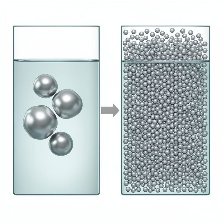
Enzyme Concentration vs. Particle Size
AI-generated conceptual illustration (silver nanoparticles in an aqueous solution). It explains the idea and does not show study data.
View source evidence ↗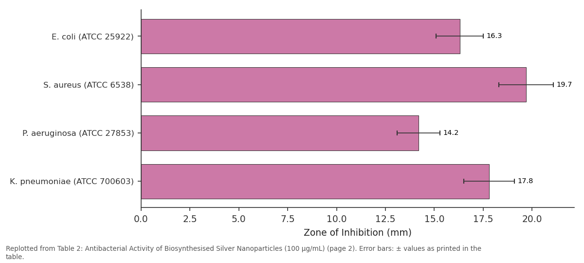
Zone of Inhibition (mm): E. coli (ATCC 25922) vs S. aureus (ATCC 6538) vs P. aeruginosa (ATCC... vs K. pneumoniae (ATCC...
Replotted from Table 2 (page 2) of the original paper. Every bar is the paper's own table value.
View source evidence ↗Figures & tables
From the original paper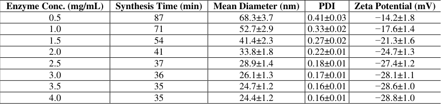 Table 1
 Table 2
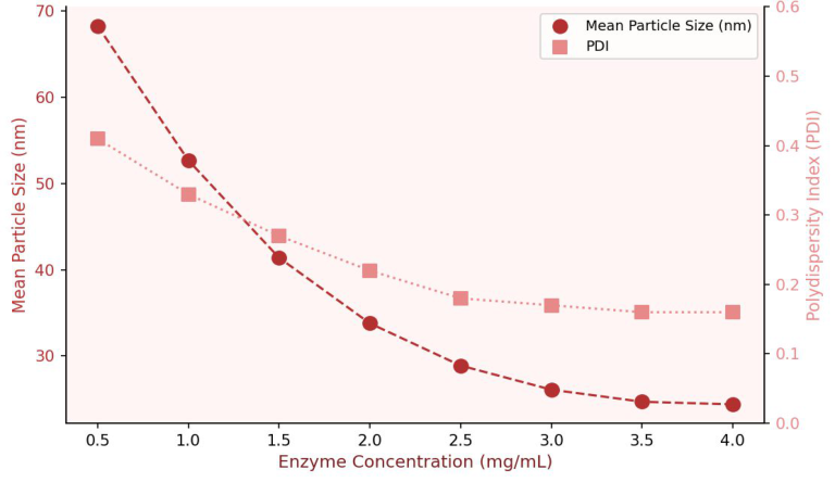 Figure 1
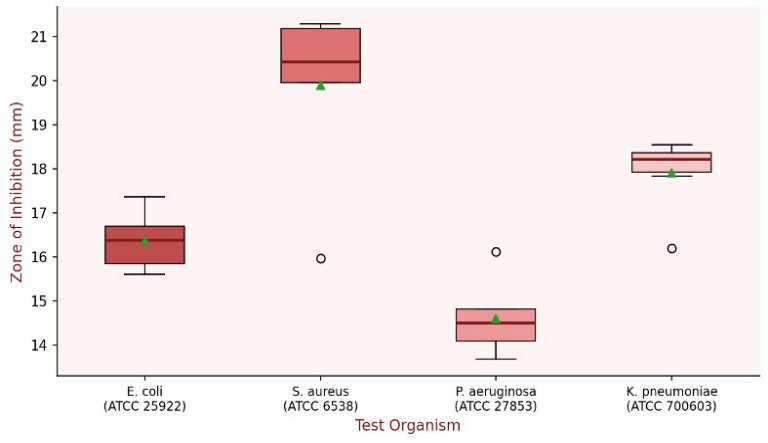 Figure 2
Introduction
Silver nanoparticles occupy a special position in nanomedicine and materials science because of the well-documented broad-spectrum antimicrobial activity of ionic and nanoparticulate silver combined with relatively low mammalian toxicity at therapeutic concentrations. Classical chemical synthesis routes-reduction with sodium borohydride or citrate-produce nanoparticles with controlled sizes but require toxic reducing agents, generate chemical waste, and often need separate capping steps to prevent aggregation. Biological approaches that harness the reducing capacity of microorganisms, plant extracts, or purified enzymes offer a path to greener production. Fungal systems are attractive for biosynthesis because they grow rapidly, tolerate high metal ion concentrations, and secrete extracellular enzymes that carry out reduction in the culture supernatant rather than intracellularly, simplifying downstream processing. Several groups have shown that NADH-dependent reductases are responsible for the conversion of Ag + to Ag0 in fungal supernatants, and partially purified preparations of these enzymes can be used directly as reducing agents, offering better control over reaction conditions than whole-cell or crude extract systems. Despite this potential, the enzyme-directed biosynthesis route has been characterised less rigorously than plant extract or whole-organism methods. Key process variables-enzyme concentration, substrate concentration, pH, temperature-have not been systematically optimised in parallel, and the relationship between enzyme loading and nanoparticle size distribution is not well documented. This research addresses these gaps by mapping the combined effect of enzyme concentration and reaction conditions on nanoparticle morphology and antibacterial performance, using a reductase partially purified from Aspergillus fumigatus.
Materials and Methods
Materials and Methods
Materials
Silver nitrate (AgNO 3 , 99.9%), analytical-grade solvents, NADH (disodium salt), and growth medium components were from Sigma-Aldrich, Mumbai, India. A. fumigatus MTCC 1811 was obtained from Microbial Type Culture Collection (IMTECH, Chandigarh, India). Mueller-Hinton agar, clinical bacterial strains (E. coli ATCC 25922, S. aureus ATCC 6538, P. aeruginosa ATCC 27853, K. pneumoniae ATCC 700603), and ampicillin discs (10 μg) were from HiMedia Laboratories, Mumbai. Research was conducted at the Verghese Kurien Institute of Food Technology, Anand, Gujarat, India, from March 2022 to November 2023.
Methods
A. fumigatus was cultivated in potato dextrose broth at 25 °C, 150 rpm, for 72 h. The mycelial mass was collected by filtration, washed, and disrupted by mortar-and-pestle grinding in liquid nitrogen. The homogenate was centrifuged (12,000 × g, 30 min, 4 °C) and the supernatant subjected to ammonium sulphate precipitation (40-80% saturation). The 40-80% fraction was dialysed (MWCO 12 kDa) against 50 mM phosphate buffer (pH 7.0) and used as the reductase preparation. For nanoparticle synthesis, 5 mL of 1 mM AgNO 3 was mixed with varying volumes of enzyme solution (0.5-4.0 mg/mL protein) and incubated in the dark. Reaction progress was followed by UV-Vis absorption at 420 nm. XRD was measured on a PANalytical X'Pert Pro (Cu Kα). Hydrodynamic size and PDI were measured by DLS (Malvern Zetasizer Nano ZS). TEM images were obtained on a JEOL JEM-2100F at 200 kV. Antibacterial testing used the disc diffusion method on Mueller-Hinton agar; nanoparticle suspensions (20 μL, 100 μg/mL) were loaded onto 6 mm discs and incubated at 37 °C for 24 h.
Reagents and Chemicals
AgNO 3 (Sigma-Aldrich, ≥99.9%, CAS 7761-88-8) was prepared as a 1 mM stock in deionised water and shielded from light at all times to prevent photoreduction. NADH (disodium salt, Sigma-Aldrich N8129) was dissolved in deionised water at 10 mM and used within 2 h of preparation to minimise oxidative degradation. Ammonium sulphate (enzyme grade, Sigma-Aldrich) was added as a finely ground powder to the crude enzyme extract over 30 min on ice. All buffer solutions were prepared from BDH AnalaR-grade reagents and filtered through 0.22 μm membranes before use. Mueller-Hinton agar (HiMedia M173) was prepared and sterilised according to the manufacturer's protocol. Ampicillin (10 μg) reference discs (HiMedia) were stored at -20 °C and equilibrated to room temperature for 1 h before use.
Quality Control and Calibration
The UV-Vis spectrophotometer (Shimadzu UV-1800) was calibrated daily with a certified neutral density filter before recording nanoparticle formation spectra. Zetasizer calibration used polystyrene latex standards (60 nm, Malvern DTS1235) before each measurement session; values outside±3 nm of the certified standard triggered instrument recalibration. XRD peak positions were validated against a NIST silicon standard (SRM 640e). For antibacterial assays, each plate included positive (ampicillin) and negative (deionised water) control discs; plates where the ampicillin zone of inhibition fell outside the CLSI acceptance range (16-22 mm for E. coli) were discarded. All syntheses and assays were performed in triplicate (n = 3) and results are expressed as means ± standard deviation.
Results
UV-Vis spectra of synthesis reaction mixtures showed the development of a surface plasmon resonance peak near 420 nm within 30-90 min, confirming the formation of silver nanoparticles. The intensity of this peak increased with enzyme concentration up to 2.5 mg/mL and plateaued thereafter, suggesting enzyme saturation of the available Ag + substrate. DLS measurements revealed that mean particle diameter decreased from 68.3 nm at 0.5 mg/mL enzyme to 28.9 nm at 2.5 mg/mL, with PDI falling from 0.41 to 0.18 over the same range (Figure 1). XRD patterns showed diffraction peaks at 2θ = 38.1°, 44.3°, 64.5°, and 77.4°, corresponding to the (111), ( 200), (220), and (311) planes of face-centred cubic silver (JCPDS 04-0783). The Scherrer equation applied to the (111) reflection gave a crystallite size of 24.3 nm, consistent with DLS data. FTIR spectra of dried nanoparticle preparations displayed amide I (1647 cm-1) and amide II (1541 cm-1) bands, confirming protein capping by the enzyme or its degradation products.
Discussion
The relationship between enzyme concentration and nanoparticle size observed here-larger particles at low enzyme loading, smaller and more monodisperse particles as enzyme concentration increases-fits a classical nucleationand-growth model. At low reductase levels, the rate of Ag0 atom generation is slow, so existing nuclei have time to aggregate before new nucleation events occur, resulting in larger and more polydisperse particles. Higher enzyme concentrations generate a burst of nuclei simultaneously, which compete for available Ag + and thereby limit individual particle growth. This
Conclusion
This research demonstrates that NADH-dependent reductase from A. fumigatus provides a reproducible, single-step enzymatic route to silver nanoparticles with controlled size and narrow size distribution. Optimal synthesis at 2.5 mg/mL enzyme concentration produced nanoparticles with a mean diameter of 28.9 nm, PDI of 0.18, and zeta potential of -27.4 mV, confirmed as face-centred cubic silver by XRD. Protein capping conferred colloidal stability and the nanoparticles exhibited clear antibacterial activity against four clinically relevant pathogens. These results establish enzymatic biosynthesis as a viable alternative to chemical methods for producing silver nanoparticles at small to moderate scale, and future research should address scale-up, stability on prolonged storage, and in vitro cytotoxicity profiling against mammalian cell lines.
Acknowledgement
Acknowledgements Funding Sources
Figures and tables from the paper
| Enzyme Conc. (mg/mL) | Synthesis Time (min) | Mean Diameter (nm) | PDI | Zeta Potential (mV) |
|---|---|---|---|---|
| 0.5 | 87 | 68.3±3.7 | 0.41±0.03 | −14.2±1.8 |
| 1.0 | 71 | 52.7±2.9 | 0.33±0.02 | −17.6±1.4 |
| 1.5 | 54 | 41.4±2.3 | 0.27±0.02 | −21.3±1.6 |
| 2.0 | 41 | 33.8±1.8 | 0.22±0.01 | −24.7±1.3 |
| 2.5 | 37 | 28.9±1.4 | 0.18±0.01 | −27.4±1.2 |
| 3.0 | 36 | 26.1±1.3 | 0.17±0.01 | −28.1±1.1 |
| 3.5 | 35 | 24.7±1.2 | 0.16±0.01 | −28.6±1.0 |
| 4.0 | 35 | 24.4±1.2 | 0.16±0.01 | −28.8±1.0 |
Table text extracted from the original PDF; superscript letters are shown inline. The original table image is below.
View original table image
| Organism | Zone of Inhibition (mm) | Ampicillin Control (mm) |
|---|---|---|
| E. coli (ATCC 25922) | 16.3±1.2 | 22.4±0.9 |
| S. aureus (ATCC 6538) | 19.7±1.4 | 24.7±1.1 |
| P. aeruginosa (ATCC 27853) | 14.2±1.1 | 19.6±0.8 |
| K. pneumoniae (ATCC 700603) | 17.8±1.3 | 21.3±0.9 |
Table text extracted from the original PDF; superscript letters are shown inline. The original table image is below.
View original table image
References
14 in the original paper- Ahmed S, Ahmad M, Swami BL, Ikram S. A review on plants extract mediated synthesis of silver nanoparticles for antimicrobial applications. J Adv Res. 2016;7(1):1728.
- Gade AK, Bonde P, Ingle AP, Marcato PD, Duran N, Rai MK. Exploitation of Aspergillus niger for synthesis of silver nanoparticles. J Biobased Mater Bioenergy. 2008;2(3):243-247.
- Mandal D, Bolander ME, Mukhopadhyay D, Sarkar G, Mukherjee P. The use of microorganisms for the formation of metal nanoparticles and their application. Appl Microbiol Biotechnol. 2006;69(5):485-492.
- Shankar SS, Ahmad A, Pasricha R, Sastry M. Bioreduction of chloroaurate ions by geranium leaves and its endophytic fungus yields gold and silver nanoparticles with different shapes. J Mater Chem. 2003;13(7):1822-1826.
- Ingle A, Gade A, Pierrat S, Sonnichsen C, Rai M. Mycosynthesis of silver nanoparticles using the fungus Fusarium acuminatum and its activity against some human pathogenic bacteria. Curr Nanosci. 2008;4(2):141-144.
- Birla SS, Tiwari VV, Gade AK, Ingle AP, Yadav AP, Rai MK. Fabrication of silver nanoparticles by Phoma glomerata and its combined effect against Escherichia coli, Pseudomonas aeruginosa and Staphylococcus aureus. Lett Appl Microbiol. 2009;48(2):173-179.
- Gericke M, Pinches A. Biological synthesis of metal nanoparticles. Hydrometallurgy. 2006;83(1-4):132-140.
- Klaus T, Joerger R, Olsson E, Granqvist CG. Silverbased crystalline nanoparticles, microbially fabricated. Proc Natl Acad Sci USA. 1999;96(24):13611-13614.
- Shahverdi AR, Minaeian S, Shahverdi HR, Jamalifar H, Nohi AA. Rapid synthesis of silver nanoparticles using culture supernatants of Enterobacteria. Process Biochem. 2007;42(5):919-923.
- Rai M, Yadav A, Gade A. Silver nanoparticles as a new generation of antimicrobials. Biotechnol Adv. 2009;27(1):76-83.
- Kalishwaralal K, Deepak V, Pandian SRK, Kottaisamy M, BarathManiKanth S, Kartikeyan B, et al. Biosynthesis of silver and gold nanoparticles using Brevibacterium casei. Colloids Surf B Biointerfaces. 2010;77(2):257-262.
- Mukherjee P, Ahmad A, Mandal D, Senapati S, Sainkar SR, Khan MI, et al. Bioreduction of AuCl₄⁻ ions by the fungus Verticillium sp. and surface trapping of the gold nanoparticles formed. Angew Chem Int Ed. 2001;40(19):3585-3588.
- Duran N, Marcato PD, Conti R, Alves OL, Costa FTM, Brocchi M. Potential use of silver nanoparticles on pathogenic bacteria, their toxicity and possible mechanisms of action. J Braz Chem Soc. 2010;21(6):949-959.
- Morones JR, Elechiguerra JL, Camacho A, Holt K, Kouri JB, Ramirez JT, et al. The bactericidal effect of silver nanoparticles. Nanotechnology. 2005;16(10):2346-2353.