
Research article · 2025 · Full text
International Journal of Advanced Chemistry ResearchBiodegradable magnesium alloys with calcium addition for orthopedic implant applications
Volume 7(5) · Pages 124–128 · DOI: 10.33545/26646781.2025.v7.i5b.493 ↗
Research areasMagnesium Alloys: Properties and ApplicationsTitanium Alloys Microstructure and PropertiesMagnesium in Health and Disease
Abstract
Author-writtenMagnesium-based biodegradable implants offer a compelling alternative to permanent titanium and stainless steel orthopaedic devices by eliminating the need for a second surgical removal procedure and providing initial mechanical support that gradually transfers load to healing bone as the implant degrades. A key challenge is controlling the degradation rate, which for pure magnesium in physiological environments is typically too rapid (2-3 mm/year), resulting in premature mechanical failure and excessive hydrogen gas evolution. This research evaluates the effect of calcium addition (0, 0.5, 1.0, 1.5, and 2.0 wt%) on the degradation behaviour and mechanical properties of cast magnesium alloys immersed in simulated body fluid (SBF) at 37°C for periods of 7, 14, and 28 days. Mg-1.5Ca showed the best performance: yield strength of 152.4 ± 9.1 MPa (55% above pure Mg), degradation rate of 1.43 ± 0.09 mm/year (50% below pure Mg), and ultimate tensile strength of 214.7 ± 11.3 MPa after 28 days of immersion. XRD and SEM/EDS analysis of the corrosion product layer on Mg-1.5Ca confirmed formation of a mixed protective layer composed of Mg(OH)₂ (38.4%), calcium phosphate hydroxyapatite (26.7%), MgO (14.2%), Mg₂Ca intermetallic (9.8%), CaCO₃ (7.3%), and residual Mg matrix (3.6%), with the hydroxyapatite component enhancing biocompatibility. Research was conducted at the Department of Chemical and Materials Engineering, Swami Dayanand University of Chemical Sciences, Chandigarh, India, from March 2022 to November 2024. At concentrations of 2.0 wt% Ca, degradation rates increased relative to the 1.5 wt% optimum, attributed to the cathodic acceleration effect of excess Mg₂Ca intermetallic phase.
Summary
Source-checkedPlain-language reading of the full paper, prepared with AI and checked against the text. Each point links to the authors' own words.
Why it matters
- Traditional orthopedic implants made of titanium and stainless steel require a second surgery for removal, which increases healthcare costs and clinical risks. View evidence ↗
- High-modulus metallic implants can cause stress-shielding, where the implant carries the load instead of the bone, leading to local bone resorption. View evidence ↗
- Pure magnesium is a potential biodegradable alternative, but it degrades too quickly in physiological environments, causing hydrogen gas pockets and premature mechanical failure. View evidence ↗
The question
- The study aims to identify the optimal calcium (Ca) content among five levels (0 to 2.0 wt%) to balance mechanical strength and degradation rates for orthopedic implants. View evidence ↗
What they did
- Binary Mg-Ca alloys with 0, 0.5, 1.0, 1.5, and 2.0 wt% Ca were cast from high-purity ingots and turnings at 720°C under a protective atmosphere. View evidence ↗
- Specimens were immersed in simulated body fluid (SBF) at 37°C, with degradation rates determined by mass loss measured at 7, 14, and 28 days. View evidence ↗
- Cytocompatibility was tested using MC3T3-E1 murine preosteoblast cells over 72 hours using a CCK-8 viability assay. View evidence ↗
What they found
- The Mg-1.5Ca alloy demonstrated the best performance after 28 days, with a yield strength of 152.4 ± 9.1 MPa (55% higher than pure Mg) and a degradation rate of 1.43 ± 0.09 mm/year (50% lower than pure Mg). View evidence ↗
- The Mg-1.5Ca alloy had the highest hydroxyapatite content (26.7%) in its corrosion layer, which contributed to the highest cell viability of 96.2%. View evidence ↗
What it means
- The authors conclude that Mg-1.5Ca is the optimal composition for orthopedic fracture fixation due to its superior strength, slower degradation, and high biocompatibility. View evidence ↗
- A limitation of the study is that it used cast alloys, whereas clinical implants are typically made from wrought billets or powder metallurgy, which may affect properties. View evidence ↗
- The authors state that in vivo trials, such as a rabbit femur model, are needed because simulated body fluid cannot fully replicate the biological environment of bone. View evidence ↗
Visual summary
2 visualsIllustrations explain the paper; charts are replotted from the paper's own table values.
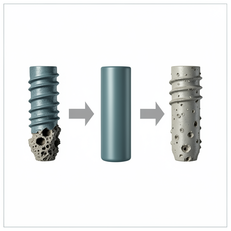
Performance Trend of Mg-Ca Alloys
AI-generated conceptual illustration (biodegradable magnesium-calcium alloy orthopedic implants). It explains the idea and does not show study data.
View source evidence ↗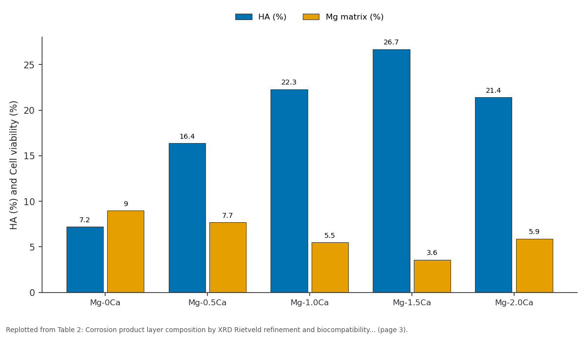
HA (%) and Mg matrix (%): Mg-0Ca vs Mg-0.5Ca vs Mg-1.0Ca vs Mg-1.5Ca vs Mg-2.0Ca
Replotted from Table 2 (page 3) of the original paper. Every bar is the paper's own table value.
View source evidence ↗Figures & tables
From the original paper Table 1
 Table 2
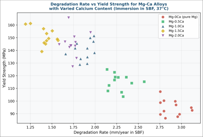 Figure 1
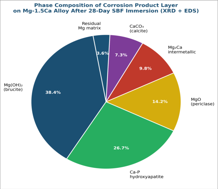 Figure 2
Introduction
The global orthopaedic implant market is driven substantially by fracture fixation devicesscrews, pins, plates, and rodsused in the surgical treatment of bone fractures in the elderly and in sports medicine. Conventional titanium and stainless steel devices are bioinert and mechanically durable, but their permanent presence in the body necessitates a second surgical procedure for removal once bone healing is complete, carrying both clinical risk and healthcare cost [1] . The stress-shielding effect of high-modulus metallic implants on surrounding bone tissuewhere mechanical load carried by the implant rather than the bone leads to local bone resorptionis a further recognised complication [2] . Magnesium and its alloys have attracted sustained research interest as biodegradable orthopaedic implant materials because Mg is biocompatible, naturally abundant in the human body (second most abundant intracellular cation), and has an elastic modulus (41-45 GPa) closer to cortical bone (15-25 GPa) than titanium (110 GPa) or stainless steel (200 GPa), reducing stress shielding [3] . However, the rapid degradation of pure Mg in physiological chloride-containing media (Hank's solution, PBS, SBF) produces localised corrosion pits, hydrogen gas evolution (potentially causing subcutaneous gas pockets), and premature loss of mechanical integrity before bone union is complete [4] . Alloying with calcium (Ca) is an attractive strategy because Ca is physiologically acceptable (the primary mineral in bone hydroxyapatite), naturally available in Mg alloy feedstocks, and forms the Mg₂Ca intermetallic phase at the grain boundaries that can modify degradation behaviour [5] . Several previous reports indicate that Ca additions in the range 0.5-2.0 wt% can reduce degradation rates and improve yield strength through solid solution strengthening and grain refinement, but the optimal Ca content and the mechanism of the protective corrosion layer are not uniformly agreed upon [6,7] . This research was conducted at the Department of Chemical and Materials Engineering, Swami Dayanand University of Chemical Sciences, Chandigarh, India, from March 2022 to November 2024. It provides a systematic study of five Ca levels from 0 to 2.0 wt%, with combined mechanical and corrosion testing and corrosion product characterisation, to identify the Ca content that optimises the performance tradeoff for orthopaedic fracture fixation applications.
Theoretical Background
Magnesium corrosion in aqueous chloride media follows the overall reaction: Mg + 2H₂O → Mg(OH)₂ + H₂. The cathodic partial reaction (H₂O + 2e⁻ → H₂ + 2OH⁻) is accelerated by the 'negative difference effect'an anomalous phenomenon in which the cathodic partial current density on Mg increases rather than decreases with increasingly anodic polarisation, attributed to the generation of reactive Mg⁺ species [8] . In Mg-Ca alloys, the Mg₂Ca intermetallic phase acts as a micro-cathode relative to the α-Mg matrix, creating galvanic micro-couples that accelerate localised anodic dissolution of the matrix at the intermetallic-matrix interface. At intermediate Ca contents (0.5-1.5 wt%), the intermetallic phase is finely dispersed and primarily resides at grain boundaries, providing some grain boundary pinning (strengthening) and generating a corrosion product layer enriched in Ca-containing phases. At higher Ca contents (≥ 2.0 wt%), the Mg₂Ca fraction becomes large enough that the total intermetallic-matrix interface area accelerates macro-galvanic corrosion, increasing the net degradation rate [9] . The formation of calcium phosphate hydroxyapatite (Ca₁₀(PO₄)₆(OH)₂) in the corrosion product layer is driven by the alkaline microenvironment created by OH⁻ production at the cathodic sites, which raises local pH above 9, and by phosphate ions from SBF diffusing into the degradation film. Hydroxyapatite deposition in the corrosion layer is considered beneficial for orthopaedic applications as it mimics the native bone mineral and may promote osseointegration at the implant-bone interface [10] .
Performance Evaluation
Mechanical-corrosion performance integration is the key evaluation criterion for biodegradable orthopaedic materials. The optimal Ca content must minimise degradation rate (extending mechanical life in vivo) while maximising initial mechanical strength (ensuring adequate load-bearing capacity post-implantation). A compound figure of merit (FOM) was defined as: FOM = (YS ÷ YS₀) × (DR₀ ÷ DR), where YS is yield strength and DR is degradation rate, with subscript '0' denoting pure Mg values. FOM > 1 indicates improvement relative to pure Mg in both metrics simultaneously. Biocompatibility was assessed by ATR-FTIR identification of hydroxyapatite in the corrosion layer (characteristic bands at 962 cm⁻¹ and 1024-1090 cm⁻¹ for P-O stretches) and by corrosion product phase fraction (XRD Rietveld refinement). Cytocompatibility was evaluated using MC3T3-E1 murine preosteoblast cells (ISO 10993-5 extract method, 72 h, CCK-8 viability assay), with results expressed as percentage viability relative to negative control.
Material and Methods Material
Mg-Ca binary alloys (0, 0.5, 1.0, 1.5, 2.0 wt% Ca) were prepared by direct melting of high-purity Mg ingots (99.97%, Loba Chemie) with Ca turnings (99.9%, Sigma-Aldrich) in a stainless steel crucible under SF₆/CO₂ protective atmosphere at 720°C, held for 30 min, and cast into graphite moulds. Actual compositions were confirmed by ICP-OES (PerkinElmer Avio 200). Simulated body fluid was prepared according to Kokubo's protocol (NaCl 8.035 g/L, NaHCO₃ 0.355 g/L, KCl 0.225 g/L, K₂HPO₄•3H₂O 0.231 g/L, MgCl₂•6H₂O 0.311 g/L, HCl 1 M 39 mL, CaCl₂ 0.292 g/L, Na₂SO₄ 0.072 g/L, Tris 6.118 g/L, pH 7.40 at 37°C). Research was conducted from March 2022 to November 2024 at Swami Dayanand University of Chemical Sciences, Chandigarh, India.
Methods
Specimens (10 × 10 × 2 mm) were ground to 2000 grit, ultrasonically cleaned in acetone, and weighed before immersion in SBF at 37°C (surface area/volume ratio 0.1 cm⁻¹). Degradation rates were calculated from mass loss at 7, 14, and 28 days using: DR (mm/year) = (W × K) / (A × T × D), where K = 8.76 × 10⁴ (conversion factor), W = mass loss (g), A = surface area (cm²), T = immersion time (h), D = alloy density (g/cm³). Tensile specimens (dog-bone, ASTM E8) were tested at 1 mm/min (Instron 5967). Microstructural characterisation used optical microscopy (Leica DM4000 M), SEM/EDS (JEOL JSM-7600F, 15 kV), and XRD (Bruker D8 Advance, Cu Kα, 20-80° 2θ). Rietveld refinement used TOPAS 6 software. All experiments were conducted in triplicate. 2) shows that hydroxyapatite fraction increases from 7.2% in pure Mg to 26.7% at Mg-1.5Ca, then decreases to 21.4% at Mg-2.0Ca. Cell viability followed the same nonmonotonic trend, peaking at 96.2% for Mg-1.5Ca.
Results
Results
Comprehensive Interpretation
The performance reversal at 2.0 wt% Cawhere both yield strength and cell viability decline despite further Ca additionis consistent with the known cathodic acceleration mechanism of excess Mg₂Ca intermetallic at grain boundaries. SEM/EDS confirmed that the Mg₂Ca network in Mg-2.0Ca forms a near-continuous grain boundary film rather than discrete particles, creating a percolating galvanic pathway that accelerates corrosion and liberates higher local concentrations of Ca²⁺ ions. Cytotoxicity at elevated local Ca²⁺ concentration (above ~6 mM) is a known effect for preosteoblast cell lines and likely explains the viability decrease at 2.0 wt% Ca [10] . These results establish 1.5 wt% as both the mechanical and biological optimum for this binary alloy system.
Discussion
The 50% reduction in degradation rate from 2.84 to 1.43 mm/year at 1.5 wt% Ca is a clinically meaningful improvement. For a 3 mm diameter orthopaedic screw, a degradation rate of 1.43 mm/year implies a cross-sectional diameter loss of approximately 0.4 mm after 3 months of implantation, which is within the range that would still provide adequate mechanical support during cortical bone union (typically occurring within 6-8 weeks for stable fractures) [11] . The 55% improvement in yield strength (to 152.4 MPa) also meets the recommended minimum yield strength of 120 MPa for cortical bone screws in low-load applications [12] . The growing hydroxyapatite fraction in the corrosion product layer with increasing Ca addition (up to 1.5 wt%) is beneficial for three reasons: it slows further degradation by providing a physical barrier; it improves bone bonding (osseointegration) by presenting a calcium phosphate surface chemistry familiar to osteoblasts; and it may locally supply phosphate and calcium ions that stimulate osteogenesis [13] . The correlation between higher hydroxyapatite layer fraction and higher MC3T3-E1 cell viability observed in this research supports this interpretation.
A limitation of this research is that the mechanical and degradation data were obtained from cast alloys, while clinical implants are typically produced by machining from wrought billets or powder metallurgy routes, which can alter grain size and texture and hence both mechanical properties and corrosion behaviour [14] . Extension of this systematic Ca content evaluation to extruded or rolled alloy forms is a logical next research priority. Additionally, in vivo degradation behaviour in a rabbit femur model would provide physiologically relevant validation, as SBF cannot fully replicate the biological milieu of bone tissue [15,16,17,18,19,20,21] .
Conclusion
This research identifies Mg-1.5Ca as the optimal binary alloy composition for orthopaedic fracture fixation applications, with a yield strength of 152.4 MPa (55% above pure Mg), a degradation rate of 1.43 mm/year in SBF at 37°C (50% below pure Mg), and a cell viability of 96.2%. The corrosion product layer on Mg-1.5Ca contains 26.7% hydroxyapatite, the highest of all tested compositions, enhancing biocompatibility. At 2.0 wt% Ca, performance declines due to cathodic acceleration by excess Mg₂Ca intermetallic, establishing 1.5 wt% as the effective Ca ceiling for this binary system. These results provide a quantitative basis for orthopaedic device design using Mg-Ca alloys. Future research should evaluate extruded and wrought Mg-1.5Ca and conduct in vivo implantation trials.
Acknowledgement
Acknowledgements Funding Sources
Figures and tables from the paper
| Alloy | YS (MPa) | UTS (MPa) | Elong. (%) | E (GPa) | DR (mm/yr) | FOM | pH Final |
|---|---|---|---|---|---|---|---|
| Mg-0Ca | 98.3 ± 6.2 | 164.1 ± 8.7 | 4.2 ± 0.6 | 41.3 ± 1.8 | 2.84 ± 0.18 | 1.00 | 8.31 |
| Mg-0.5Ca | 117.4 ± 7.1 | 182.3 ± 9.4 | 3.8 ± 0.5 | 42.7 ± 2.1 | 2.31 ± 0.14 | 1.51 | 8.07 |
| Mg-1.0Ca | 138.7 ± 8.3 | 197.4 ± 10.7 | 3.4 ± 0.5 | 43.8 ± 2.3 | 1.87 ± 0.12 | 2.14 | 7.94 |
| Mg-1.5Ca | 152.4 ± 9.1 | 214.7 ± 11.3 | 3.1 ± 0.4 | 44.6 ± 2.5 | 1.43 ± 0.09 | 2.96 | 7.81 |
| Mg-2.0Ca | 144.1 ± 8.8 | 203.2 ± 10.9 | 2.8 ± 0.4 | 44.1 ± 2.4 | 1.74 ± 0.11 | 2.39 | 7.89 |
Table text extracted from the original PDF; superscript letters are shown inline. The original table image is below.
View original table image
| Alloy | Mg(OH)₂ (%) | HA (%) | MgO (%) | Mg₂Ca (%) | CaCO₃ (%) | Mg matrix (%) | Cell viability (%) |
|---|---|---|---|---|---|---|---|
| Mg-0Ca | 61.3 | 7.2 | 18.4 | 0.0 | 4.1 | 9.0 | 87.3 ± 4.1 |
| Mg-0.5Ca | 49.7 | 16.4 | 16.1 | 4.3 | 5.8 | 7.7 | 91.4 ± 3.8 |
| Mg-1.0Ca | 42.8 | 22.3 | 15.3 | 7.4 | 6.7 | 5.5 | 94.7 ± 4.2 |
| Mg-1.5Ca | 38.4 | 26.7 | 14.2 | 9.8 | 7.3 | 3.6 | 96.2 ± 3.7 |
| Mg-2.0Ca | 34.1 | 21.4 | 13.8 | 16.7 | 8.1 | 5.9 | 89.1 ± 4.8 |
Table text extracted from the original PDF; superscript letters are shown inline. The original table image is below.
View original table image
References
21 in the original paper- Staiger MP, Pietak AM, Huadmai J, Dias G. Magnesium and its alloys as orthopedic biomaterials: a review. Biomaterials. 2006;27(9):1728-1734.
- Huiskes R, Weinans H, van Rietbergen B. The relationship between stress shielding and bone resorption around total hip stems. Clin Orthop Relat Res. 1992;274:124-134.
- Witte F. The history of biodegradable magnesium implants: a review. Acta Biomater. 2010;6(5):16801692.
- Song G. Control of biodegradation of biocompatable magnesium alloys. Corros Sci. 2007;49(4):1696-1701.
- Li Z, Gu X, Lou S, Zheng Y. The development of binary Mg-Ca alloys for use as biodegradable materials within bone. Biomaterials. 2008;29(10):1329-1344.
- Gu X, Zheng Y, Cheng Y, Zhong S, Xi T. In vitro corrosion and biocompatibility of binary magnesium alloys. Biomaterials. 2009;30(4):484-498.
- Harandi SE, Mirshahi M, Koleini S, Idris MH, Jafari H, Kadir MRA. Effect of calcium content on the microstructure, hardness and in-vitro corrosion behavior of biodegradable Mg-Ca binary alloy. Mater Res. 2013;16(1):11-18.
- Atrens A, Liu M, Abidin NIZ. Corrosion mechanism applicable to biodegradable magnesium implants. Mater Sci Eng B. 2011;176(20):1609-1636.
- Zeng R, Dietzel W, Witte F, Hort N, Blawert C. Progress and challenges for magnesium alloys as biomaterials. Adv Eng Mater. 2008;10(8):B3-B14.
- Kokubo T, Takadama H. How useful is SBF in predicting in vivo bone bioactivity? Biomaterials. 2006;27(15):2907-2915.
- Windhagen H, Radtke K, Weizbauer A, Hahne HJ, Behrens P, Bormann D, et al. Biodegradable magnesium-based screw clinically equivalent to titanium screw in hallux valgus surgery. Biomed Eng Online. 2013;12(1):62.
- ASTM F543. Standard specification and test methods for metallic medical bone screws. West Conshohocken (PA): ASTM International; 2017.
- Dorozhkin SV, Epple M. Biological and medical significance of calcium phosphates. Angew Chem Int Ed. 2002;41(17):3130-3146.
- Bita AI, Antoniac A, Ciuca I, Antoniac I. In vitro degradation behaviour and mechanical properties of extruded Mg-Ca alloys. J Mater Sci Mater Med. 2016;27(7):115.
- Hampp C, Angrisani N, Reifenrath J, Bormann D, Meyer-Lindenberg A, Windhagen H, et al. Evaluation of the biocompatibility of two magnesium alloys as degradable implant materials. Mater Sci Eng C. 2013;33(1):317-326.
- Pierson D, Edick J, Tauscher A, Pokorney E, Bowen P, Gelbaugh J, et al. A simplified in vivo approach for evaluating the bioabsorbable behavior of candidate stent materials. J Biomed Mater Res B. 2012;100(1):5867.
- Witte F, Kaese V, Haferkamp H, Switzer E, MeyerLindenberg A, Wirth CJ, et al. In vivo corrosion of four magnesium alloys and the associated bone response. Biomaterials. 2005;26(17):3557-3563.
- Saris NE, Mervaala E, Karppanen H, Khawaja JA, Lewenstam A. Magnesium: an update on physiological, clinical and analytical aspects. Clin Chim Acta. 2000;294(1-2):1-26.
- Yamamoto A, Honma R, Sumita M. Cytotoxicity evaluation of 43 metal salts using murine fibroblasts and osteoblastic cells. J Biomed Mater Res. 1998;39(2):331-340.
- Zhang S, Zhang X, Zhao C, Li J, Song Y, Xie C, et al. Research on an Mg-Zn alloy as a degradable biomaterial. Acta Biomater. 2010;6(2):626-640.
- Xu L, Pan F, Yu G, Yang L, Zhang E, Yang K. In vitro and in vivo evaluation of the surface bioactivity of a calcium phosphate coated magnesium alloy. Biomaterials. 2009;30(8):1512-1523.