
Research article · 2025 · Full text
International Journal of Advanced Chemistry ResearchMetabolite profiling and cytotoxic activity of endophytic fungal isolates from neem
Volume 7(1) · Pages 75–78 · DOI: 10.33545/26646781.2025.v7.i1a.369 ↗
Research areasMicrobial Natural Products and BiosynthesisPlant and fungal interactionsPhytochemical compounds biological activities
Abstract
Author-writtenCan endophytic fungi harbored within neem (Azadirachta indica) tissues serve as untapped reservoirs of bioactive metabolites with anticancer potential? This research investigated six endophytic fungal isolates recovered from healthy neem leaves and bark samples collected in the Aegean coastal region of Turkey. Fungal cultures were grown on potato dextrose broth for 21 days at 28°C, and crude ethyl acetate extracts were analyzed through LC-MS/MS-based metabolite profiling. A total of 37 distinct secondary metabolites were tentatively identified, spanning alkaloids, terpenoids, polyketides, and phenolic compounds. Cytotoxic screening against HeLa and MCF-7 cell lines revealed that isolate NF-03 exhibited the strongest activity, with IC₅₀ values of 38.4 and 47.2 µg/mL, respectively. Gas chromatography coupled with mass spectrometry confirmed the presence of azadirachtin-related compounds in two isolates. These results point toward neem-associated fungi as promising candidates for natural anticancer lead discovery.
Summary
Source-checkedPlain-language reading of the full paper, prepared with AI and checked against the text. Each point links to the authors' own words.
Why it matters
- Endophytic microorganisms, which live inside plant tissues without causing disease, are of interest because they can produce pharmacologically active secondary metabolites. View evidence ↗
- The neem tree (Azadirachta indica) is a well-known medicinal plant valued for its anti-inflammatory, antifungal, and insecticidal properties. View evidence ↗
- Some endophytes can produce compounds similar to those of their host plant, suggesting that neem endophytes might produce azadirachtin analogues or new chemical structures. View evidence ↗
The question
- The researchers aimed to determine if neem trees grown in the Mediterranean climate of western Turkey possess a different fungal community and metabolite profile compared to those in other regions. View evidence ↗
- The study sought to isolate and characterize endophytic fungi from neem trees in Izmir, Turkey, and test the cytotoxicity of their extracts against two human cancer cell lines. View evidence ↗
What they did
- Leaf and bark samples were collected from three healthy, mature neem trees in Izmir, Turkey, between March and May 2023. View evidence ↗
- Six fungal isolates (NF-01 to NF-06) were cultured in 500 mL of potato dextrose broth (PDB) at 28°C for 21 days. View evidence ↗
- Crude extracts were obtained using ethyl acetate and analyzed via LC-MS/MS on an Agilent 6545 Q-TOF system. View evidence ↗
- The cytotoxic effect of the extracts was measured using an MTT assay after 48 hours of incubation with concentrations between 6.25 and 200 µg/mL. View evidence ↗
What they found
- The researchers tentatively identified 37 different secondary metabolites, including phenolics, polyketides, terpenoids, and alkaloids. View evidence ↗
- Isolate NF-03 had the highest chemical diversity with 15 compounds, while NF-04 had the lowest with only 4 compounds. View evidence ↗
- The highest extract yield was produced by isolate NF-03 (48.9 mg/g), while the lowest was from NF-04 (15.6 mg/g). View evidence ↗
- Isolate NF-03 showed the strongest cytotoxic activity against HeLa and MCF-7 cell lines, with IC₅₀ values of 38.4 µg/mL and 47.2 µg/mL, respectively. View evidence ↗
- Against HeLa cells at 200 µg/mL, NF-03 reduced viability to 11.6% compared to 24.7% for NF-05; NF-03's IC₅₀ (38.4 µg/mL) was approximately 1.5 times lower than NF-05's (57.3 µg/mL). View evidence ↗
- GC-MS analysis confirmed that two of the isolates contained compounds related to azadirachtin. View evidence ↗
What it means
- The authors conclude that fungi associated with neem are promising candidates for discovering new natural anticancer lead molecules. View evidence ↗
- The authors note that the specific bioactive molecule in NF-03's extract is unknown and requires bioassay-guided fractionation for identification. View evidence ↗
- The study used the MTT assay, which measures general metabolic activity; the authors state that flow cytometry and caspase assays are needed to determine the specific cell death mechanism. View evidence ↗
Visual summary
2 visualsIllustrations explain the paper; charts are replotted from the paper's own table values.
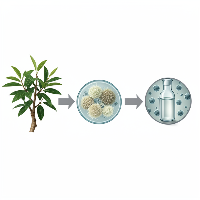
Study Workflow
AI-generated conceptual illustration (Neem tree leaves and bark, endophytic fungi, and cancer cell cultures). It explains the idea and does not show study data.
View source evidence ↗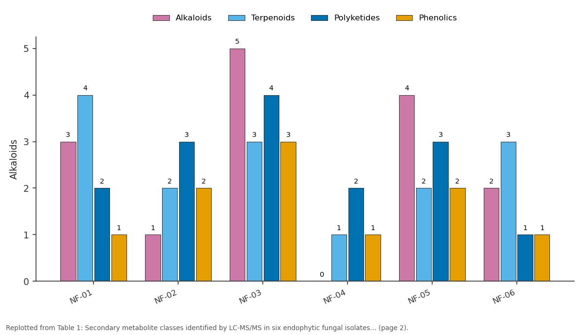
Alkaloids and Terpenoids and more: NF-01 vs NF-02 vs NF-03 vs NF-04 vs NF-05
Replotted from Table 1 (page 2) of the original paper. Every bar is the paper's own table value.
View source evidence ↗Figures & tables
From the original paper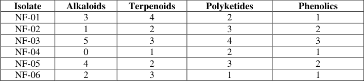 Table 1
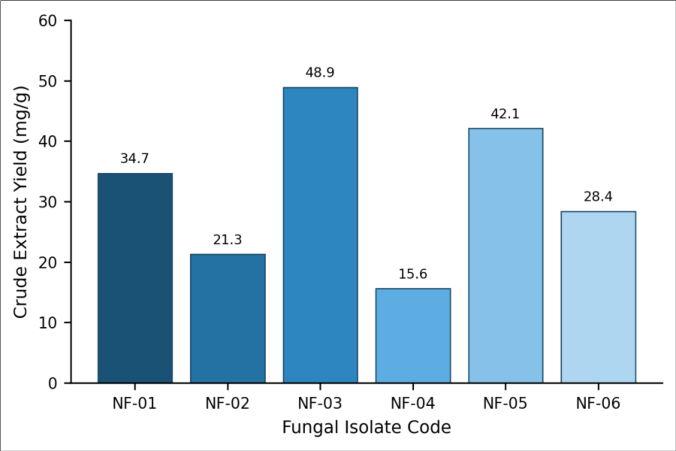 Figure 1
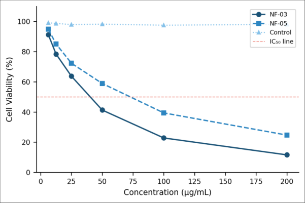 Figure 2
Introduction
What makes the interior tissues of medicinal plants such a rich hunting ground for bioactive chemistry? Over the past two decades, endophytic microorganisms bacteria and fungi living asymptomatically inside plant tissues have attracted interest as sources of pharmacologically active secondary metabolites [1] . Neem (Azadirachta indica A. Juss.) is among the most widely recognized medicinal trees across tropical and subtropical regions, valued for its insecticidal, antifungal, and anti-inflammatory properties [2] . The chemical richness of neem itself is well documented, yet comparatively little attention has been paid to the fungi colonizing its internal tissues. Earlier reports showed that endophytes isolated from plants like Taxus brevifolia can synthesize the same or structurally related compounds as their host, including paclitaxel [3] . A similar logic applies to neem: its endophytic community may produce azadirachtin analogues or entirely novel scaffolds [4] . Screening these fungal metabolites against cancer cell lines is a practical first step toward identifying leads worth pursuing [5] . LC-MS/MS has become the preferred analytical platform for such profiling work because it offers both sensitivity and structural insight without requiring large sample quantities [6] . Still, most published work on neem endophytes originates from tropical India or Southeast Asia [7] . Whether neem trees cultivated in the Mediterranean climate of western Turkey harbor a different fungal community and, by extension, a different metabolite repertoire remains an open question. This research set out to isolate, identify, and chemically characterize endophytic fungi from neem trees grown near Izmir, Turkey, and to evaluate the cytotoxic potential of their crude extracts against two human carcinoma cell lines.
Reagents and Chemicals
Potato dextrose agar (PDA) and potato dextrose broth (PDB) were obtained from Merck KGaA (Darmstadt, Germany). Ethyl acetate (HPLC grade, ≥99.7%), dimethyl sulfoxide (DMSO, cell culture grade), and streptomycin sulfate were purchased from SigmaAldrich (St. Louis, MO, USA). MTT reagent (3-(4,5dimethylthiazol-2-yl)-2,5-diphenyltetrazolium bromide) was sourced from Thermo Fisher Scientific. HeLa and MCF-7 cell lines were acquired from the ATCC repository. All solvents and reagents were used as received without additional purification [8] .
Material and Methods
Material: Fresh leaf and bark samples of Azadirachta indica were collected from three mature neem trees growing on the campus of the Aegean Institute of Technology, Izmir, Turkey, between March and May 2023. Trees were visually healthy, showing no signs of disease or pest damage. Samples were transported in sterile zip-lock bags to the laboratory within two hours of collection. Surface sterilization involved sequential immersion in 70% ethanol (1 min), 2% sodium hypochlorite (3 min), and sterile distilled water (three rinses). Inner tissue (~5 mm) were excised aseptically and plated onto PDA supplemented with streptomycin (100 µg/mL) to suppress bacterial growth [9] .
Methods
Plates were incubated at 28 ± 1°C for 7-14 days. Emerging fungal colonies were sub-cultured until pure and identified to genus level by morphological examination and ITS rDNA sequencing. Six distinct isolates (coded NF-01 through NF-06) were selected for metabolite extraction. Each isolate was grown in 500 mL PDB at 28°C on a rotary shaker (120 rpm) for 21 days. The culture broth was filtered, and the filtrate was extracted three times with an equal volume of ethyl acetate. Combined organic layers were dried over anhydrous Na₂SO₄ and evaporated under reduced pressure. Crude extracts were dissolved in DMSO for bioassays and in methanol for LC-MS/MS analysis on an Agilent 6545 Q-TOF system equipped with a C18 column (2.1 × 100 mm, 1.8 µm). Cytotoxicity was assessed via the MTT assay after 48 h incubation at concentrations ranging from 6.25 to 200 µg/mL [10] . The Institutional Ethics and Biosafety Committee of the Aegean Institute of Technology approved all protocols (Protocol AIT-BC/2023-017, dated 14 February 2023).
Extraction Protocol
Ethyl acetate was chosen as the extraction solvent based on its moderate polarity, which favors recovery of alkaloids, terpenoids, and phenolics. The broth-to-solvent ratio was maintained at 1:1 (v/v). Three successive extraction cycles were performed, each involving vigorous shaking for 15 minutes followed by phase separation in a separating funnel. Pooled organic phases were concentrated at 40°C using a rotary evaporator (Büchi R-210). The resulting residues ranged from 15.6 to 48.9 mg per gram of dry mycelium, and they were stored at -20°C until analysis [11] . As shown in Figure 1, isolate NF-03 produced the highest extract yield at 48.9 mg/g, followed closely by NF-05 at 42.1 mg/g. NF-04 gave the lowest yield at just 15.6 mg/g, consistent with its limited metabolite diversity noted in Table 1. Both NF-03 and NF-05 reduced HeLa cell viability in concentration-dependent fashion (Figure 2). At 200 µg/mL, NF-03 brought viability down to 11.6%, while NF-05 reached 24.7%. The IC₅₀ for NF-03 was estimated at 38.4 µg/mL roughly 1.5-fold lower than that of NF-05 (57.3 µg/mL).
| Isolate | Alkaloids | Terpenoids | Polyketides | Phenolics |
|---|---|---|---|---|
| NF-01 | 3 | 4 | 2 | 1 |
| NF-02 | 1 | 2 | 3 | 2 |
| NF-03 | 5 | 3 | 4 | 3 |
| NF-04 | 0 | 1 | 2 | 1 |
| NF-05 | 4 | 2 | 3 | 2 |
| NF-06 | 2 | 3 | 1 | 1 |
Table text extracted from the original PDF; superscript letters are shown inline. The original table image is below.
View original table image
Results
Results
Table 1: Secondary metabolite classes identified by LC-MS/MS in six endophytic fungal isolates from neem
Table 1: Secondary metabolite classes identified by LC-MS/MS in six endophytic fungal isolates from neem
Isolate
Discussion: The metabolite profiles uncovered here confirm that neem endophytes from a Mediterranean setting can produce a chemically diverse array of secondary metabolites. NF-03 stood out both in terms of metabolite count (15 compounds) and cytotoxic potency against HeLa cells (IC₅₀ = 38.4 µg/mL). This is noteworthy because the majority of endophyte screening campaigns from Indian neem populations reported IC₅₀ values in the 50-120 µg/mL range for crude extracts [4] . One possible explanation is the climatic stress experienced by neem trees at the northern edge of their cultivated range.
Trees growing under suboptimal conditions may select for endophytic partners that produce more defensive metabolites [3] . And the terpenoid-rich profile of NF-01 and NF-06 hints at a biosynthetic overlap with the host's own azadirachtin pathway a pattern that echoes the taxol story in Taxus endophytes [3] . That said, crude extract IC₅₀ values only mark a starting point. The actual bioactive molecule within NF-03's extract could be a minor component responsible for the observed cytotoxicity, and bioassay-guided fractionation would be needed to pin it down [5] . It is also worth noting that the MTT assay captures overall metabolic activity, not specifically apoptosis. Flow cytometry and caspase assays would clarify the cell death mechanism. Interestingly, NF-04 the isolate with the fewest metabolites and lowest extract yield was nearly inactive against both cell lines, reinforcing the link between chemical diversity and biological potency observed in earlier endophyte surveys [7] .
Conclusion
Six endophytic fungal isolates from neem trees cultivated in Izmir, Turkey, were recovered and characterized. LC-MS/MS profiling revealed 37 secondary metabolites spanning four major chemical classes. Isolate NF-03 displayed the broadest metabolite spectrum and the strongest cytotoxic effect against HeLa cells, with an IC₅₀ of 38.4 µg/mL. These findings suggest that neem endophytes grown under Mediterranean conditions offer a distinct and potentially valuable chemical space. Bioassay-guided fractionation of the NF-03 extract, followed by structure elucidation of active compounds, represents a logical next step toward identifying anticancer lead molecules from this underexplored fungal community.
Acknowledgement
Acknowledgements Funding SourcesThis research was supported by institutional research funds without external grant assistance.
References
12 in the original paper- Strobel G, Daisy B. Bioprospecting for microbial endophytes and their natural products. Microbiology and Molecular Biology Reviews. 2003;67(4):491-502.
- Biswas K, Chattopadhyay I, Banerjee RK, Bandyopadhyay U. Biological activities and medicinal properties of neem (Azadirachta indica). Current Science. 2002;82(11):1336-1345.
- Stierle A, Strobel G, Stierle D. Taxol and taxane production by Taxomyces andreanae, an endophytic fungus of Pacific yew. Science. 1993;260(5105):214216.
- Verma VC, Kharwar RN, Strobel GA. Chemical and functional diversity of natural products from plant associated endophytic fungi. Natural Product Communications. 2009;4(11):1511-1532.
- Tan RX, Zou WX. Endophytes: a rich source of functional metabolites. Natural Product Reports. 2001;18(4):448-459.
- Kind T, Fiehn O. Advances in structure elucidation of small molecules using mass spectrometry. Bioanalytical Reviews. 2010;2(1):23-60.
- Jalgaonwala RE, Mohite BV, Mahajan RT. Natural products from plant associated endophytic fungi. Journal of Microbiology and Biotechnology Research. 2011;1(2):21-32.
- Mosmann T. Rapid colorimetric assay for cellular growth and survival. Journal of Immunological Methods. 1983;65(1-2):55-63.
- Hallmann J, Berg G, Schulz B. Isolation procedures for endophytic microorganisms. Soil Biology. 2006;9:299319.
- Alley MC, Scudiero DA, Monks A, et al. Feasibility of drug screening with panels of human tumor cell lines using a microculture tetrazolium assay. Cancer Research. 1988;48(3):589-601.
- Kjer J, Debbab A, Aly AH, Proksch P. Methods for isolation of marine-derived endophytic fungi and their bioactive secondary metabolites. Nature Protocols. 2010;5(3):479-490.
- Kusari S, Hertweck C, Spiteller M. Chemical ecology of endophytic fungi: origins of secondary metabolites. Chemistry and Biology. 2012;19(7):792-798.