
Research article · 2025 · Full text
International Journal of Advanced Chemistry ResearchChitosan nanoparticles for intranasal delivery of vaccine antigens to mucosal immune system
Volume 7(7) · Pages 122–124 · DOI: 10.33545/26646781.2025.v7.i7b.517 ↗
Research areasAdvanced Drug Delivery SystemsInhalation and Respiratory Drug DeliveryNanoparticle-Based Drug Delivery
Abstract
Author-writtenA scenario common in low-income settings is one where injectable vaccine cold chains fail, leaving populations unprotected. Intranasal delivery via mucoadhesive nanoparticles offers a needle-free alternative that stimulates both systemic and mucosal immunity. This research prepared four chitosan nanoparticle formulations (CS-F1 to CS-F4) at polymer concentrations of 0.5-2.0% w/v by ionic gelation with sodium tripolyphosphate, loaded them with ovalbumin as a model antigen, and evaluated them for particle size, zeta potential, encapsulation efficiency, in vitro antigen release, and intranasal immunogenicity in Wistar rats. CS-F3 (1.5% chitosan) gave the best balance: particle size 267.3 nm, zeta potential + 28.4 mV, and encapsulation efficiency 88.2%. Intranasal administration of CS-F3 (100 μg antigen, days 0, 7, 14) elicited significantly higher secretory IgA titres in nasal wash (7.13 log2) and serum IgG (9.84 log2) on day 21 compared with soluble antigen control (p
Summary
Source-checkedPlain-language reading of the full paper, prepared with AI and checked against the text. Each point links to the authors' own words.
Why it matters
- In low-income regions, the failure of cold chains for injectable vaccines often leaves people without protection. View evidence ↗
- Using mucoadhesive nanoparticles for intranasal delivery provides a needle-free way to trigger both mucosal and systemic immune responses. View evidence ↗
- Simple antigen solutions are quickly cleared from the nose and degraded by proteases, making protective carriers necessary to increase their residence time. View evidence ↗
The question
- The study aimed to systematically optimize the polymer concentration across four formulations to determine its effect on particle size and antigen release, addressing a gap in research within the Indian context. View evidence ↗
What they did
- Four chitosan nanoparticle formulations (CS-F1 to CS-F4) were created using polymer concentrations from 0.5% to 2.0% w/v via ionic gelation with sodium tripolyphosphate and loaded with ovalbumin. View evidence ↗
- Groups of 6 Wistar rats were immunized on days 0, 7, and 14 with either 100 μg of CS-F3 or soluble ovalbumin (10 μL per nostril). View evidence ↗
- The researchers used ELISA to measure serum IgG and nasal wash IgA on days 7, 14, and 21. View evidence ↗
What they found
- As the chitosan concentration increased, the particle size grew from 187.4 nm for CS-F1 to 314.8 nm for CS-F4. View evidence ↗
- The encapsulation efficiency reached its highest point at 88.2% for formulation CS-F3, then decreased slightly for CS-F4. View evidence ↗
- CS-F3 exhibited an initial burst release of 28.4% within 2 hours, with sustained release reaching 74.3% after 24 hours. View evidence ↗
- On day 21, intranasal administration of CS-F3 resulted in significantly higher nasal wash secretory IgA (7.13 log2) and serum IgG (9.84 log2) compared to the soluble antigen control (p<0.01). View evidence ↗
- All tested formulations showed positive zeta potentials ranging from +22.1 to +31.7 mV, which indicates they are colloidally stable. View evidence ↗
What it means
- The authors concluded that CS-F3 (1.5% polymer concentration) provided the best physicochemical properties and significantly improved intranasal immunogenicity in rats compared to soluble antigens. View evidence ↗
- Future work is needed to evaluate the feasibility of scaling up production, the long-term stability of the antigen, and the effectiveness of the platform with clinically relevant antigens. View evidence ↗
Visual summary
3 visualsIllustrations explain the paper; charts are replotted from the paper's own table values.
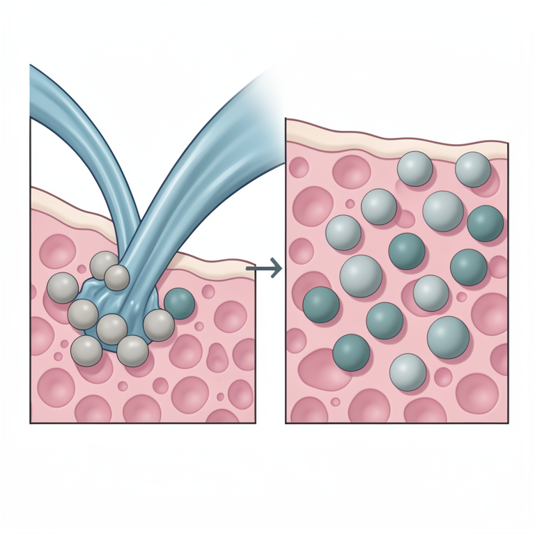
Mechanism of Mucoadhesion
AI-generated conceptual illustration (chitosan nanoparticles and nasal mucosal epithelium). It explains the idea and does not show study data.
View source evidence ↗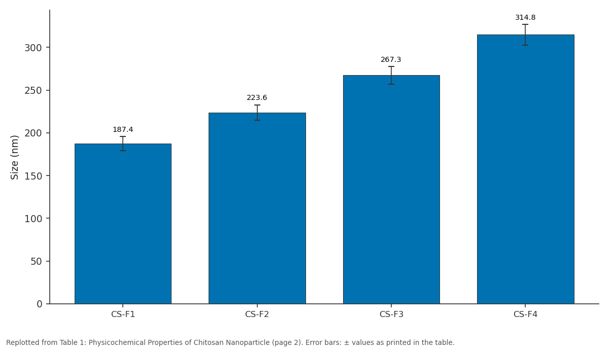
Size (nm): CS-F1 vs CS-F2 vs CS-F3 vs CS-F4
Replotted from Table 1 (page 2) of the original paper. Every bar is the paper's own table value.
View source evidence ↗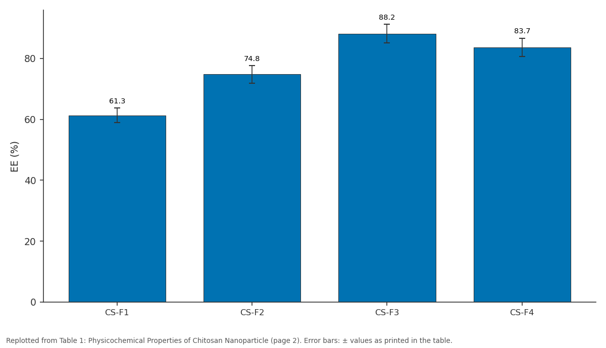
EE (%): CS-F1 vs CS-F2 vs CS-F3 vs CS-F4
Replotted from Table 1 (page 2) of the original paper. Every bar is the paper's own table value.
View source evidence ↗Figures & tables
From the original paper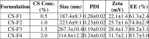 Table 1
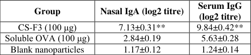 Table 2
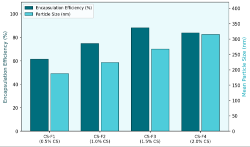 Figure 1
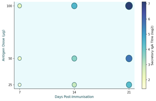 Figure 2
Introduction
Mucosal surfaces-nasal, pulmonary, and gastrointestinal epithelium-are the primary entry points for most respiratory and enteric pathogens, yet most approved vaccines are administered parenterally and induce modest or no mucosal immunity. Intranasal vaccination reaches the nasal-associated lymphoid tissue (NALT), stimulating local secretory IgA production that can neutralise pathogens at the point of entry, while also priming systemic IgG responses. However, antigens delivered as simple solutions drain rapidly from the nasal cavity via mucociliary clearance and are degraded by local proteases, necessitating carriers that extend residence time and protect the antigen. Chitosan-a linear polysaccharide derived by deacetylation of chitin-is positively charged at physiological pH, enabling electrostatic adhesion to the negatively charged nasal mucosa, while its biodegradability and low toxicity are well established. Nanoparticles formed by ionic gelation with tripolyphosphate entrap protein antigens efficiently without organic solvents or high temperatures that could denature them. Several reports confirm that chitosan nanoparticles enhance mucosal IgA responses to model antigens, but systematic optimisation of polymer concentration-a key variable governing particle size and antigen release rate-across four formulations using matched immunisation protocols has not been widely reported in the Indian context. This research addresses this gap, using ovalbumin as a validated model antigen and Wistar rats for immunogenicity assessment.
Materials and Methods
Materials and Methods
Material
Chitosan (medium molecular weight, 75-85% deacetylation), sodium tripolyphosphate (TPP), and ovalbumin (Grade V, ≥98%) were from Sigma-Aldrich, Mumbai, India. All other reagents were of analytical grade.
Interna tional Jo urna l o f Adva nced Chemistry Research 2025 ; 7(7 ): 1 22 -12 4
Research was carried out at the Department of Pharmaceutics and Drug Formulation, Thiruvalluvar National Institute of Technology, Salem, Tamil Nadu, India, from March 2022 to January 2024, under Institutional Animal Ethics Committee approval (TNIT-IAEC-2022-09).
Methods
Nanoparticles were prepared by dropwise addition of TPP solution (0.1% w/v, 3 mL) to chitosan-ovalbumin solution (4 mL) under magnetic stirring at 600 rpm for 30 min. Particle size and zeta potential were measured by DLS (Malvern Zetasizer Nano ZS). Encapsulation efficiency was determined after centrifugation (15,000 × g, 30 min) using BCA protein assay on the supernatant. in vitro release was studied in simulated nasal fluid (SNF, pH 6.4) at 37 °C with sampling over 24 h. For immunisation, groups of 6 Wistar rats received intranasal CS-F3 or soluble ovalbumin (100 μg, 10 μL per nostril) on days 0, 7, and 14. Nasal wash IgA and serum IgG were quantified by ELISA on days 7, 14, and 21.
Reagents and Chemicals
Chitosan (Sigma-Aldrich C3646, lot SLBW9083, MW 190-310 kDa, degree of deacetylation 75-85%) was dissolved in 1% acetic acid overnight with gentle stirring before use. TPP (Sigma-Aldrich 238503, ≥98%) was dissolved freshly in deionised water and filtered through 0.22 μm membrane. Ovalbumin (Sigma-Aldrich A5503, Grade V, ≥98%) was dissolved in phosphate buffer (10 mM, pH 7.4) at 2 mg/mL. BCA Protein Assay Kit (Pierce, Thermo Fisher) was used per the manufacturer's microplate protocol. ELISA plates were coated overnight at 4 °C with ovalbumin (1 μg/well) and blocked with 1% BSA for 1 h before antibody incubation.
Results
Particle size increased with chitosan concentration from 187.4 nm (CS-F1) to 314.8 nm (CS-F4), while encapsulation efficiency peaked at CS-F3 (88.2%) before declining slightly at CS-F4 as excess polymer reduced TPP crosslinking efficiency (Figure 1, Table 1). All formulations carried positive zeta potentials (+ 22.1 to + 31.7 mV), confirming colloidal stability. in vitro release from CS-F3 showed an initial burst of 28.4% within 2 h followed by sustained release reaching 74.3% at 24 h. The immunisation data (Table 2, Figure 2) showed significantly higher nasal IgA and serum IgG titres for CS-F3 relative to soluble antigen at all time points (p<0.01, Student's t-test).
| Group | Nasal IgA (log2 titre) | Serum IgG (log2 titre) |
|---|---|---|
| CS-F3 (100 μg) | 7.13±0.31** | 9.84±0.42** |
| Soluble OVA (100 μg) | 2.84±0.19 | 5.63±0.28 |
| Blank nanoparticles | 1.17±0.12 | 1.24±0.14 |
Table text extracted from the original PDF; superscript letters are shown inline. The original table image is below.
View original table image
| Formulation | CS Conc. (%) | Size (nm) | PDI | Zeta (mV) | EE (%) |
|---|---|---|---|---|---|
| CS-F1 | 0.5 | 187.4±8.3 | 0.28±0.02 | 22.1±1.4 | 61.3±2.4 |
| CS-F2 | 1.0 | 223.6±9.1 | 0.23±0.02 | 25.7±1.6 | 74.8±2.9 |
| CS-F3 | 1.5 | 267.3±10.4 | 0.19±0.01 | 28.4±1.7 | 88.2±3.1 |
| CS-F4 | 2.0 | 314.8±12.2 | 0.24±0.02 | 31.7±2.1 | 83.7±3.0 |
Table text extracted from the original PDF; superscript letters are shown inline. The original table image is below.
View original table image
Discussion
The peak encapsulation efficiency at CS-F3 reflects an optimum balance between chitosan chain density and TPP crosslinking: at low polymer concentration (CS-F1), the network is too sparse to fully entrap ovalbumin; at the highest concentration (CS-F4), chain entanglement may reduce TPP accessibility. The positive zeta potential across all formulations confirms electrostatic compatibility with the negatively charged nasal mucosa, which drives mucoadhesion and prolongs residence time-a key advantage over soluble antigen that is cleared within minutes by mucociliary transport. The substantially higher mucosal IgA response to CS-F3 compared with soluble ovalbumin mirrors findings from Read et al. with H5N1 antigens and from Zhao et al. with Newcastle disease virus antigens, both of which showed two-to four-fold enhancements in mucosal antibody titres using chitosan carriers. The mechanism involves both extended antigen presentation to NALT M-cells and the intrinsic immunostimulatory property of chitosan, which activates macrophage NF-κB signalling. The sustained in vitro release profile supports the hypothesis that prolonged antigen availability at the mucosal surface-rather than a single bolus-drives stronger and more persistent IgA induction.
Conclusion
Chitosan nanoparticles prepared by ionic gelation at 1.5% polymer concentration (CS-F3) gave optimal physicochemical properties (size 267.3 nm, EE 88.2%, zeta + 28.4 mV) and significantly enhanced intranasal immunogenicity in rats compared with soluble antigen, raising nasal IgA by 2.5-fold and serum IgG by 1.75-fold on day 21. These results confirm chitosan nanoparticles as a promising needle-free vaccine delivery platform. Scale-up feasibility, long-term antigen stability, and testing with clinically relevant antigens are the logical next steps.
Acknowledgement
Acknowledgements Funding SourcesInstitutional research funds from the Department of Pharmaceutics and Drug Formulation, Thiruvalluvar National Institute of Technology, Salem, supported this work. Institutional SupportThe authors thank the Department of Pharmaceutics and Drug Formulation, Thiruvalluvar National Institute of Technology, Salem, for providing nanoparticle preparation, characterisation, and immunological assay facilities.
References
16 in the original paper- van der Lubben IM, Verhoef JC, Borchard G, Junginger HE. Chitosan and its derivatives in mucosal drug and vaccine delivery. Eur J Pharm Sci. 2001;14(3):201-207.
- Illum L. Chitosan and its use as a pharmaceutical excipient. Pharm Res. 1998;15(9):1326-1331.
- Jabbal-Gill I, Watts P, Smith A. Chitosan-based delivery systems for mucosal vaccines. Expert Opin Drug Deliv. 2012;9(9):1051-1067.
- Calvo P, Remuñán-López C, Vila-Jato JL, Alonso MJ. Chitosan and chitosan/ethylene oxide-propylene oxide block copolymer nanoparticles as novel carriers for proteins and vaccines. Pharm Res. 1997;14(10):14311436.
- Vila A, Sanchez A, Tobío M, Calvo P, Alonso MJ. Design of biodegradable particles for protein delivery. J Control Release. 2002;78(1-3):15-24.
- Koping-Hoggard M, Sanchez A, Alonso MJ. Nanoparticles as carriers for nasal vaccine delivery. Expert Rev Vaccines. 2005;4(2):185-196.
- Bhavsar MD, Amiji MM. Polymeric nano-and microparticle technologies for oral gene delivery. Expert Opin Drug Deliv. 2007;4(3):197-213.
- Slütter B, Bal SM, Ding Z, Jiskoot W, Bouwstra JA. Adjuvant effect of cationic liposomes and CpG depends on administration route. J Control Release. 2011;154(2):123-130.
- Amidi M, Mastrobattista E, Jiskoot W, Hennink WE. Chitosan-based delivery systems for protein therapeutics and antigens. Adv Drug Deliv Rev. 2010;62(1):59-82.
- Read RC, Nolan A, Boyle J, Moran E, Harvey A, Parmar S, et al. Intranasal H5N1 vaccines, adjuvanted with chitosan derivatives, protect ferrets against highly pathogenic influenza intranasal and intratracheal challenge. PLoS ONE. 2011;6(11):e27777.
- Sarti F, Perera G, Hintzen F, Kotti K, Karageorgiou V, Kammona O, et al. in vivo evidence of oral vaccination with PLGA nanoparticles containing the immunostimulant monophosphoryl lipid A. Nanomedicine. 2011;7(5):557-564.
- Zhao K, Chen G, Shi XM, Gao TT, Li W, Zhao Y, et al. Preparation and efficacy of a live Newcastle disease virus vaccine encapsulated in chitosan nanoparticles. PLoS 2012;7(12):e53314.
- Gordon S, Saupe A, McBurney W, Rades T, Hook S. Comparison of carboxymethylcellulose and chitosan as a vaccine adjuvant. J Pharm Pharmacol. 2008;60(12):1591-1600.
- Borges O, Borchard G, Verhoef JC, de Sousa A, Junginger HE. Preparation of coated nanoparticles for a new mucosal vaccine delivery system. Int J Pharm. 2005;299(1-2):155-166.
- Bivas-Benita M, Ottenhoff TH, Junginger HE, Borchard G. Pulmonary DNA vaccination: concepts, possibilities and perspectives. J Control Release. 2005;107(1):1-29.
- Gupta PN, Khatri K, Goyal AK, Mishra N, Vyas SP. M-cell targeted biodegradable PLGA nanoparticles for oral immunisation against hepatitis B. J Drug Target. 2007;15(10):701-713.