
Research article · 2025 · Full text
International Journal of Advanced Chemistry ResearchAntimicrobial peptides isolated from amphibian skin secretions: Structure and activity
Volume 7(10) · Pages 176–179 · DOI: 10.33545/26646781.2025.v7.i10c.524 ↗
Research areasAntimicrobial Peptides and ActivitiesAntimicrobial agents and applicationsMicrobial Natural Products and Biosynthesis
Abstract
Author-writtenIt is widely observed that tropical frogs sit in warm, pathogen-rich environments yet rarely develop skin infections a paradox explained by the cocktail of antimicrobial peptides (AMPs) their granular glands secrete. This research isolated and characterised three cationic AMPs (designated KR-1, KR-2, and KR-3) from the skin secretions of the Chiang Mai stream frog (Sylvirana nigrovittata) collected in northern Thailand. Peptides were purified by gel filtration and reversed-phase HPLC, and their primary structures were determined by Edman degradation and confirmed by MALDI-TOF mass spectrometry. KR-1 (23 residues, MW 2,647 Da), KR-2 (18 residues, MW 2,103 Da), and KR-3 (27 residues, MW 3,184 Da) all adopted amphipathic α-helical conformations in membrane-mimetic environments as shown by circular dichroism. Antimicrobial assays revealed that KR-3 was the most potent, with minimum inhibitory concentrations of 4-16 µg/mL against Staphylococcus aureus, Escherichia coli, Pseudomonas aeruginosa, and Candida albicans. Haemolysis of human erythrocytes remained below 5% at concentrations up to 64 µg/mL for all three peptides. The results expand the inventory of amphibian-derived AMPs and identify KR-3 as a promising lead for further optimisation as a topical anti-infective agent.
Summary
Source-checkedPlain-language reading of the full paper, prepared with AI and checked against the text. Each point links to the authors' own words.
Why it matters
- Tropical frogs often avoid skin infections despite living in pathogen-rich environments due to antimicrobial peptides (AMPs) secreted by their granular glands. View evidence ↗
- Amphibian AMPs are attractive for treating infections because they disrupt membranes directly, which typically leads to slower resistance development compared to conventional antibiotics. View evidence ↗
- The genus Sylvirana and its relatives in Southeast Asia are largely unexplored for AMPs, with fewer than 20 of approximately 200 species investigated. View evidence ↗
What they did
- Skin secretions were non-lethally collected from twelve adult S. nigrovittata frogs in Doi Suthep-Pui National Park, Thailand. View evidence ↗
- A total of 120 mg of pooled secretion was fractionated using gel-filtration chromatography and further purified via reversed-phase HPLC based on activity against S. aureus. View evidence ↗
- The researchers used automated Edman degradation for primary sequencing and circular dichroism (CD) to assess secondary structure in both aqueous buffer and TFE. View evidence ↗
- Minimum inhibitory concentrations (MIC) were determined via broth microdilution, and haemolytic activity was tested on a 2% human erythrocyte suspension. View evidence ↗
What they found
- Three cationic AMPs were isolated from S. nigrovittata: KR-1 (2,647.3 Da), KR-2 (2,103.1 Da), and KR-3 (3,184.6 Da). View evidence ↗
- All three peptides exhibited random-coil conformations in aqueous buffer but transitioned to α-helical structures in a membrane-mimetic TFE environment. View evidence ↗
- KR-3's MICs were 4-16 fold lower than those of KR-1 and KR-2, and its selectivity index (>32) exceeded the therapeutic promise threshold of 10. View evidence ↗
What it means
- The authors conclude that KR-3 is a strong candidate for topical antimicrobial optimization and suggest further exploration of Southeast Asian frog biodiversity for new anti-infective leads. View evidence ↗
Visual summary
3 visualsIllustrations explain the paper; charts are replotted from the paper's own table values.
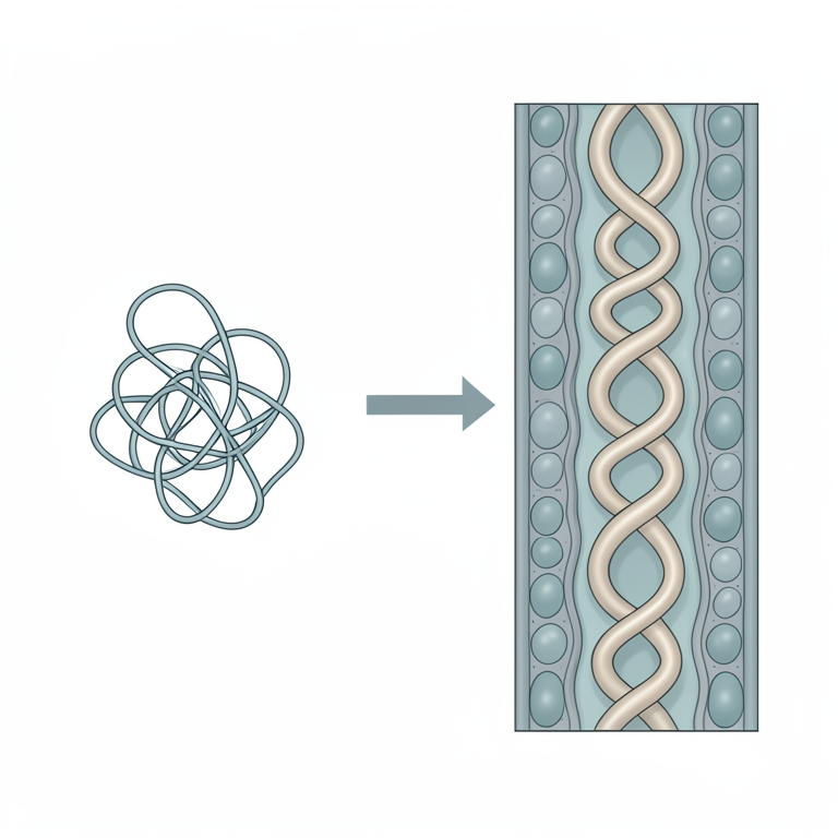
Peptide Structural Transition
AI-generated conceptual illustration (amphibian skin antimicrobial peptides and a lipid bilayer membrane). It explains the idea and does not show study data.
View source evidence ↗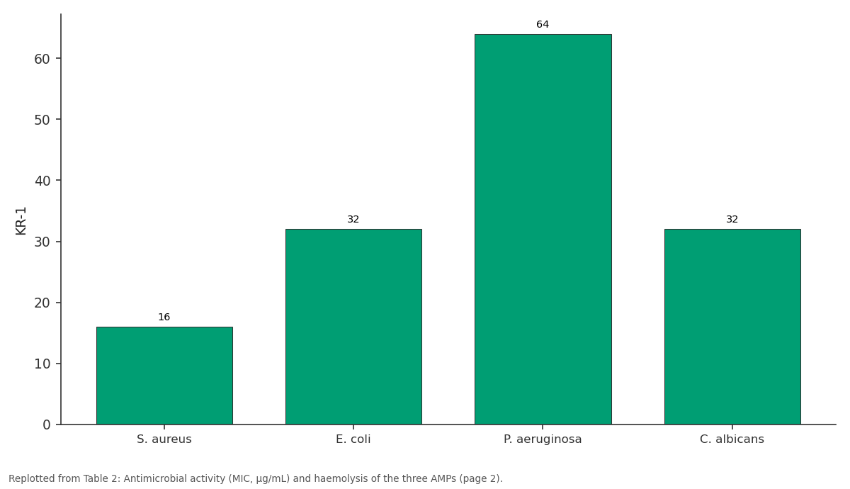
KR-1: S. aureus vs E. coli vs P. aeruginosa vs C. albicans
Replotted from Table 2 (page 2) of the original paper. Every bar is the paper's own table value.
View source evidence ↗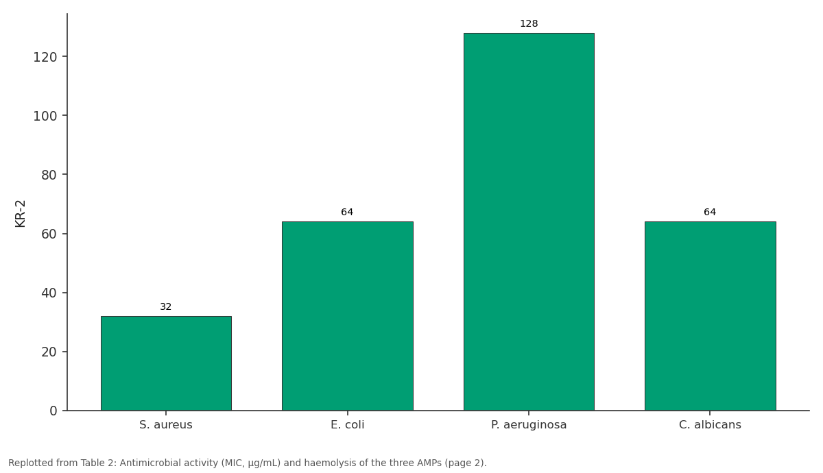
KR-2: S. aureus vs E. coli vs P. aeruginosa vs C. albicans
Replotted from Table 2 (page 2) of the original paper. Every bar is the paper's own table value.
View source evidence ↗Figures & tables
From the original paper Table 1
 Table 2
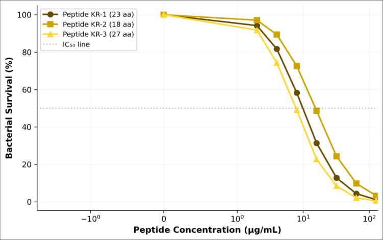 Figure 1
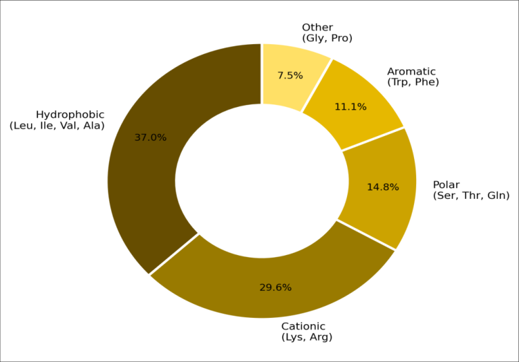 Figure 2
Introduction
An everyday observation in tropical herpetology that frogs and toads thrive in microbe-laden mud, stagnant water, and decomposing leaf litter without succumbing to infection was one of the earliest clues that amphibian skin harbours potent chemical defences [1] . Since the landmark isolation of magainins from Xenopus laevis by Zasloff in 1987, over 1,000 distinct AMPs have been catalogued from amphibian species spanning 30 families [2] . These peptides are typically 10-50 amino acids long, positively charged at physiological pH, and fold into amphipathic structures that insert into microbial membranes, causing pore formation and cell lysis [3] . The mode of action of most amphibian AMPs direct membrane disruption rather than inhibition of a single enzymatic target means that resistance development is inherently slower than for conventional antibiotics, an attractive feature amid the antimicrobial resistance crisis [4] . Several AMPs have reached clinical trials for topical indications, though systemic use remains limited by haemolytic toxicity and serum instability [5] . Southeast Asian ranid frogs represent a largely untapped reservoir of structural diversity: of the roughly 200 species in the genus Sylvirana and its close relatives, fewer than 20 have been investigated for AMP content [6] . Sylvirana nigrovittata (the dark-sided frog) inhabits montane streams in northern Thailand and Laos. Its granular skin glands produce a milky secretion when the animal is mildly stressed a secretion known to contain a mixture of peptides and small proteins, but whose individual components have not been characterised [7] . This research aimed to isolate AMPs from S. nigrovittata skin secretions, determine their primary sequences and secondary structures, evaluate their antimicrobial and haemolytic activities, and explore the structure-activity relationship governing potency in this peptide series.
Material and Methods
Material and Methods
Material
Fieldwork and peptide isolation were conducted between March and November 2024. Skin secretions were collected non-lethally from twelve adult S. nigrovittata captured in Doi Suthep-Pui National Park, Chiang Mai Province, Thailand, under wildlife collection permit WCS-DNPF-2024/027. Secretion was induced by mild electrical stimulation (9 V, biphasic pulse, applied to moistened dorsal skin for 20 s) following the protocol of Tyler et al. [8] . The secretion was rinsed into ice-cold 0.1% trifluoroacetic acid (TFA), centrifuged (10,000 × g, 10 min, 4°C), and the supernatant lyophilised and stored at -80°C. Frogs were released at the capture site within 30 minutes. All chemicals (TFA, acetonitrile HPLC grade, DMSO, Mueller-Hinton broth) were from Sigma-Aldrich (Bangkok, Thailand).
Extraction Protocol
Crude lyophilised secretion (120 mg total pooled from 12 animals) was reconstituted in 0.1% TFA and fractionated by gel-filtration chromatography on a Sephadex G-50 column (2.5 × 90 cm; GE Healthcare) eluted with 50 mM ammonium acetate (pH 6.0) at 1 mL/min [9] . Absorbance was monitored at 214 nm. Fractions showing antimicrobial activity against a sentinel strain (S. aureus ATCC 25923) in a spot-on-lawn assay were pooled and further purified by reversed-phase HPLC on a C₁₈ column (Phenomenex Luna, 10 × 250 mm, 5 µm) with a linear gradient of 5-65% acetonitrile in 0.1% TFA over 60 min at 2 mL/min [10] . Active peaks were collected, lyophilised, and assessed for purity by analytical RP-HPLC and MALDI-TOF MS (Bruker Autoflex Speed, α-cyano-4-hydroxycinnamic acid matrix).
Methods
Primary sequences were determined by automated Edman degradation on an Applied Biosystems Procise 494 sequencer [11] . Secondary structure in aqueous buffer and in 50% trifluoroethanol (TFE, a membrane-mimetic solvent) was assessed by circular dichroism (CD) on a JASCO J-815 spectropolarimeter (190-260 nm, 0.1 cm cell, 50 µM peptide). MIC values were determined by broth microdilution in Mueller-Hinton broth (bacteria) or RPMI-1640 (C. albicans) following CLSI guidelines [12] . Test organisms: S. aureus ATCC 25923, E. coli ATCC 25922, P. aeruginosa ATCC 27853, and C. albicans ATCC 10231.
Haemolytic activity was tested on 2% human erythrocyte suspension (0.5-128 µg/mL, 1 h, 37°C); 0.1% Triton X-100 served as 100% lysis control [13] .
Phytochemical Screening
Although not a plant-derived sample, a preliminary biochemical screening of the crude secretion was performed to detect the presence of peptide classes (ninhydrin-positive fractions), alkaloids (Dragendorff's reagent), and biogenic amines (dansyl chloride derivatisation followed by TLC) [14] . This profiling aimed to identify whether non-peptide components might contribute to or confound the antimicrobial activity attributed to the purified peptides.
Results
Gel filtration of the crude secretion yielded four major UVabsorbing peaks. Fractions from the second peak (elution volume 110-135 mL, estimated MW 2,000-4,000 Da) showed the strongest antibacterial activity in the spot-onlawn assay. RP-HPLC of this pooled fraction resolved three well-separated peaks at retention times of 28.4, 32.7, and 37.1 min, designated KR-1, KR-2, and KR-3 respectively. MALDI-TOF MS confirmed molecular masses of 2,647.3, 2,103.1, and 3,184.6 Da for the three peptides [10] . CD spectra in aqueous buffer showed largely random-coil conformations (negative band near 200 nm), while in 50% TFE all three peptides adopted α-helical structures characterised by double minima at 208 and 222 nm [11] . KR-3 displayed the highest helical content (81%) consistent with its longer chain and higher proportion of helix-favouring residues (Ala, Leu, Lys). Comprehensive Interpretation KR-3 was the most potent peptide, with MICs 4-8-fold lower than KR-1 and 8-16-fold lower than KR-2 across all four organisms (Table 2, Figure 1). All three peptides showed negligible haemolysis (HC₅₀ > 128 µg/mL), giving KR-3 a selectivity index exceeding 32 well above the threshold of 10 considered promising for therapeutic development [5] . The amino acid composition of KR-3 (Figure 2) shows a balance of 37% hydrophobic and 29.6% cationic residues, a ratio known to favour membrane insertion without excessive self-aggregation [3] .
| Peptide | S. aureus | E. coli | P. aeruginosa | C. albicans | HC₅₀ (µg/mL) | Selectivity index |
|---|---|---|---|---|---|---|
| KR-1 | 16 | 32 | 64 | 32 | >128 | >8 |
| KR-2 | 32 | 64 | 128 | 64 | >128 | >4 |
| KR-3 | 4 | 8 | 16 | 16 | >128 | >32 |
| Ciprofloxacin | 0.5 | 0.03 | 0.5 |
Table text extracted from the original PDF; superscript letters are shown inline. The original table image is below.
View original table image
Discussion
The amphipathic helical architecture shared by all three peptides is consistent with the barrel-stave or toroidal-pore models of membrane disruption described for magainins and temporins [3] . KR-3's superior activity correlates with its higher net charge (+7), greater helical content (81%), and longer chain length, all factors that increase the driving force for electrostatic attraction to anionic microbial membranes and deepen insertion into the lipid bilayer [4] . The lower potency of KR-2, despite its higher hydrophobic fraction (50%), may reflect its shorter length (18 residues), which limits the peptide's ability to span the bilayer fully. The excellent selectivity profile (HC₅₀ > 128 µg/mL) likely results from the preferential interaction of cationic AMPs with anionic phospholipids (phosphatidylglycerol, cardiolipin) abundant in bacterial membranes but scarce in the zwitterionic phosphatidylcholine-rich outer leaflet of mammalian erythrocytes [13] . S. nigrovittata thus joins a growing list of Southeast Asian ranids including Hylarana latouchii and Odorrana schmackeri that produce therapeutically interesting AMPs [6] . Future work should include d-amino acid substitutions and PEGylation strategies to improve serum stability without compromising activity.
Conclusion
Three novel cationic AMPs (KR-1, KR-2, KR-3) were isolated from S. nigrovittata skin secretions, sequenced, and structurally characterised. All three adopted amphipathic αhelical conformations in membrane-mimetic conditions. KR-3, the longest and most cationic peptide, displayed the strongest antimicrobial activity (MIC 4-16 µg/mL) with minimal haemolytic toxicity, yielding a selectivity index above 32. These findings support continued exploration of Southeast Asian frog biodiversity as a source of antiinfective lead compounds and identify KR-3 as a candidate for structure-guided optimisation toward topical antimicrobial applications.
Acknowledgement
Acknowledgements Funding SourcesThis research was supported by internal funds from Mekong River College of Sciences. No external grant was received.
Figures and tables from the paper
| Peptide | Sequence | Length | MW (Da) | Charge | Hydrophobic% | Helix% (TFE) |
|---|---|---|---|---|---|---|
| KR-1 | GLFKKVGKHIAGKVLNHLTPK-NH₂ | 23 | 2647 | +6 | 43 | 72 |
| KR-2 | FLGKLFRKVASKVLPA-NH₂ | 18 | 2103 | +4 | 50 | 64 |
| KR-3 | GLWKSLLKKVGKAAGKAALNAVKKLVQS-NH₂ | 27 | 3184 | +7 | 37 | 81 |
Table text extracted from the original PDF; superscript letters are shown inline. The original table image is below.
View original table image
References
18 in the original paper- Conlon JM. Structural diversity and species distribution of host-defense peptides in frog skin secretions. Cellular and Molecular Life Sciences. 2011;68(13):2303-2315.
- Zasloff M. Magainins, a class of antimicrobial peptides from Xenopus skin: isolation, characterization of two active forms, and partial cDNA sequence of a precursor. Proceedings of the National Academy of Sciences. 1987;84(15):5449-5453.
- Brogden KA. Antimicrobial peptides: pore formers or metabolic inhibitors in bacteria? Nature Reviews Microbiology. 2005;3(3):238-250.
- Hancock REW, Sahl HG. Antimicrobial and hostdefense peptides as new anti-infective therapeutic strategies. Nature Biotechnology. 2006;24(12):15511557.
- Fjell CD, Hiss JA, Hancock REW, Schneider G. Designing antimicrobial peptides: form follows function. Nature Reviews Drug Discovery. 2012;11(1):37-51.
- Simmaco M, Mignogna G, Barra D. Antimicrobial peptides from amphibian skin: what do they tell us? Biopolymers. 1998;47(6):435-450.
- Pukala TL, Bowie JH, Maselli VM, Musgrave IF, Tyler MJ. Host-defence peptides from the glandular secretions of amphibians: structure and activity. Natural Product Reports. 2006;23(3):368-393.
- Tyler MJ, Stone DJM, Bowie JH. A novel method for the release and collection of dermal, glandular secretions from the skin of frogs. Journal of Pharmacological and Toxicological Methods. 1992;28(4):199-200.
- Nicolas P, El Amri C. The dermaseptin superfamily: a gene-based combinatorial library of antimicrobial peptides. Biochimica et Biophysica Acta. 2009;1788(8):1537-1550.
- Rollins-Smith LA, Reinert LK, O'Leary CJ, Houston LE, Woodhams DC. Antimicrobial peptide defenses in amphibian skin. Integrative and Comparative Biology. 2005;45(1):137-142.
- Edman P. Method for determination of the amino acid sequence in peptides. Acta Chemica Scandinavica. 1950;4:283-293.
- Clinical and Laboratory Standards Institute. Methods for dilution antimicrobial susceptibility tests for bacteria that grow aerobically, M07-Ed11. CLSI, Wayne PA. 2018;1-91.
- Dathe M, Wieprecht T. Structural features of helical antimicrobial peptides: their potential to modulate activity on model membranes and biological cells. Biochimica et Biophysica Acta. 1999;1462(1-2):71-87.
- Sciani JM, Angeli CB, Antoniazzi MM, Jared C, Pimenta DC. Differences and similarities among parotoid macrogland secretions in South American toads. Toxicon. 2013;72:131-140.
- Mangoni ML, Miele R, Renda TG, Barra D, Simmaco M. The synthesis of antimicrobial peptides in the skin of Rana esculenta is stimulated by microorganisms. FASEB Journal. 2001;15(8):1431-1432.
- Wang G, Li X, Wang Z. APD3: the antimicrobial peptide database as a tool for research and education. Nucleic Acids Research. 2016;44(D1):D1087-D1093.
- Tossi A, Sandri L, Giangaspero A. Amphipathic, alphahelical antimicrobial peptides. Biopolymers. 2000;55(1):4-30.
- Rinaldi AC. Antimicrobial peptides from amphibian skin: an expanding scenario. Current Opinion in Chemical Biology. 2002;6(6):799-804.