
Research article · 2025 · Full text
International Journal of Advanced Chemistry ResearchpH-sensitive hydrogel beads for colon-targeted delivery of 5-fluorouracil
Volume 7(6) · Pages 80–83 · DOI: 10.33545/26646781.2025.v7.i6b.515 ↗
Research areasAdvanced Drug Delivery SystemsHydrogels: synthesis, properties, applicationsDrug Solubulity and Delivery Systems
Abstract
Author-writtenColorectal cancer accounts for roughly 10% of all cancer diagnoses worldwide, and site-specific drug delivery to the colon remains a formidable pharmaceutical challenge. This research describes the preparation and in-vitro evaluation of pH-sensitive hydrogel beads composed of sodium alginate cross-linked with calcium chloride and coated with Eudragit S100 for colon-targeted release of 5-fluorouracil (5-FU). Beads were produced by ionotropic gelation, yielding mean diameters between 1.08 and 1.34 mm depending on polymer concentration and cross-linker molarity. Entrapment efficiency ranged from 62.7% to 81.4%, with the highest values observed at 3% w/v alginate and 0.2 M CaCl₂. Fourier-transform infrared spectroscopy confirmed drug-polymer compatibility, showing no new covalent peaks in the loaded formulations. Differential scanning calorimetry indicated that 5-FU was dispersed in an amorphous state within the hydrogel matrix. Swelling studies at pH 1.2, 4.5, 6.8, and 7.4 demonstrated pronounced pH-dependence: beads swelled only 53% at gastric pH after 12 h but reached 298% at colonic pH over the same period. In-vitro dissolution in simulated gastrointestinal fluids showed that uncoated beads released 23.6% of their drug load in the first two hours at pH 1.2, whereas Eudragit S100-coated beads limited gastric release to 8.3% and delivered 68.2% of 5-FU at pH 7.4 over 12 h. Drug release kinetics at colonic pH fit the Korsmeyer-Peppas model with a diffusion exponent of 0.57, pointing to anomalous transport governed by both polymer relaxation and Fickian diffusion. The coated formulation showed a lag time of approximately 3.5 h before the onset of major release, matching typical upper-gastrointestinal transit. These findings indicate that Eudragit S100-coated alginate hydrogel beads can effectively shield 5-FU from premature release and deliver the bulk of the drug payload under conditions that simulate the colonic environment.
Summary
Source-checkedPlain-language reading of the full paper, prepared with AI and checked against the text. Each point links to the authors' own words.
Why it matters
- Colorectal cancer is a significant global health issue, representing about 10% of all cancer diagnoses, but delivering drugs specifically to the colon is difficult. View evidence ↗
- While 5-fluorouracil (5-FU) is a standard chemotherapy drug, systemic administration causes toxic side effects in healthy tissues, such as myelosuppression and mucositis. View evidence ↗
- pH-dependent delivery systems use polymers that dissolve only at specific pH thresholds to target the colon, taking advantage of the pH increase from the stomach to the ileum and colon. View evidence ↗
The question
- The study aims to evaluate the properties and drug-release kinetics of 5-FU loaded alginate-Ca²⁺ hydrogel beads coated with Eudragit S100 across simulated gastrointestinal pH environments. View evidence ↗
What they did
- Beads were created by extruding sodium alginate solutions (2-4% w/v) containing 1% w/v 5-FU into calcium chloride baths (0.1-0.3 M) for 30 minutes. View evidence ↗
- To create a protective barrier, selected beads were double-coated in a 5% w/v Eudragit S100 solution, resulting in a weight gain of approximately 8-10%. View evidence ↗
- Drug release was tested using a USP type-II apparatus at 37 °C, sequentially moving through simulated gastric fluid (2 h), simulated intestinal fluid (3 h), and simulated colonic fluid (7 h). View evidence ↗
What they found
- The average diameter of the beads increased from 1.08 mm to 1.34 mm as the alginate concentration rose from 2% to 4%. View evidence ↗
- The highest drug entrapment efficiency (81.4%) was achieved using a combination of 3% alginate and 0.2 M CaCl₂. View evidence ↗
- The beads showed strong pH-dependence in swelling, reaching 298% at colonic pH (7.4) compared to only 53% at gastric pH (1.2) over 12 hours. View evidence ↗
- Eudragit-coated beads limited drug release to 8.3% in the gastric phase and 14.7% in the intestinal phase, while delivering the majority (68.2%) at colonic pH (7.4). View evidence ↗
- The coated formulation exhibited a lag time of 3.47 hours before major drug release, which matches the typical 3-5 hour transit time of the upper gastrointestinal tract. View evidence ↗
- Kinetic modeling showed that drug release follows an anomalous transport mechanism (n = 0.57), meaning it is governed by both Fickian diffusion and polymer relaxation. View evidence ↗
What it means
- The authors conclude that Eudragit S100-coated alginate beads effectively protect 5-FU from early release and deliver most of the drug to the simulated colonic environment. View evidence ↗
- A limitation of the study is the lack of colonic microflora in the test medium, which might accelerate drug release in real conditions due to bacterial enzyme degradation of alginate. View evidence ↗
- The study only tested a single drug loading of 1% w/v, and the authors note that different loading levels could change the beads' shape and release behavior. View evidence ↗
Visual summary
3 visualsIllustrations explain the paper; charts are replotted from the paper's own table values.
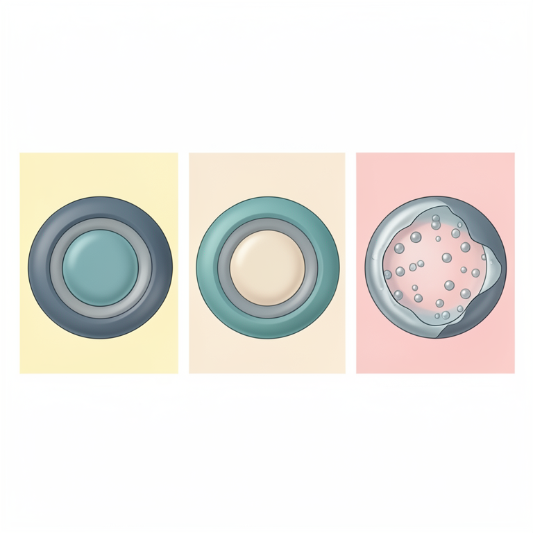
Dual-Barrier Release Mechanism
AI-generated conceptual illustration (pH-sensitive hydrogel beads in a gastrointestinal tract). It explains the idea and does not show study data.
View source evidence ↗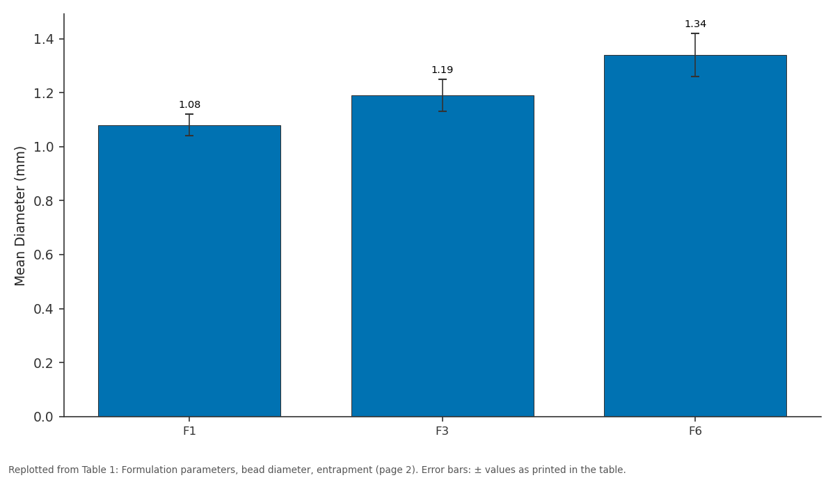
Mean Diameter (mm): F1 vs F3 vs F6
Replotted from Table 1 (page 2) of the original paper. Every bar is the paper's own table value.
View source evidence ↗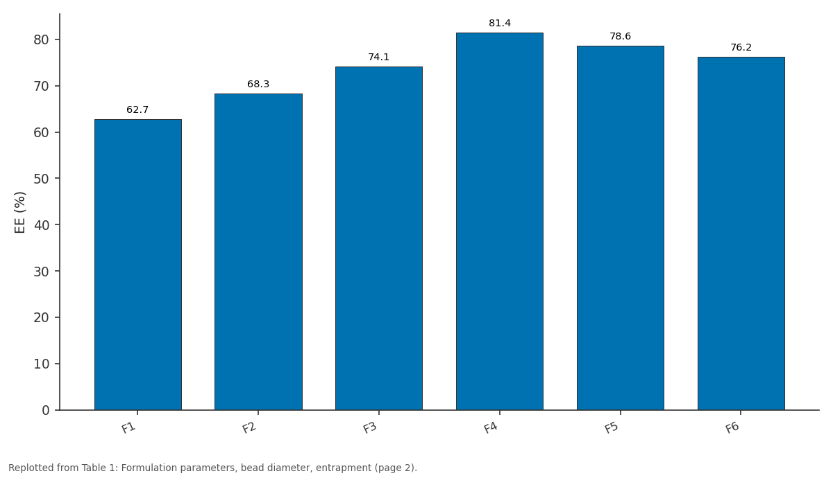
EE (%): F1 vs F2 vs F3 vs F4 vs F5
Replotted from Table 1 (page 2) of the original paper. Every bar is the paper's own table value.
View source evidence ↗Figures & tables
From the original paper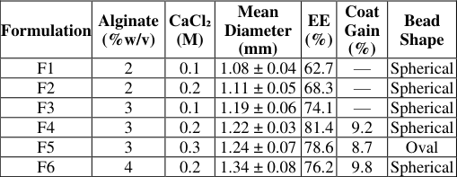 Table 1
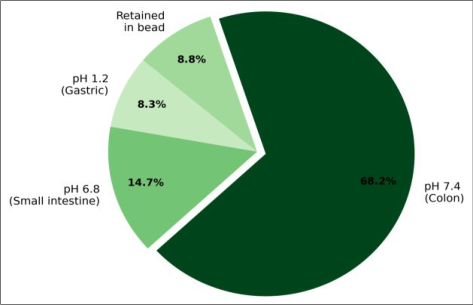 Figure 1
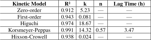 Table 2
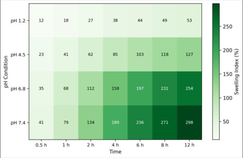 Figure 2
Introduction
According to the Global Cancer Observatory, colorectal cancer ranked third among all malignancies in incidence and second in mortality during 2020, with nearly 1.9 million new cases recorded that year [1] . Systemic chemotherapy with 5-fluorouracil (5-FU) has been a cornerstone of treatment for decades, yet oral or intravenous administration exposes healthy tissues to cytotoxic concentrations and produces well-documented side effects including mucositis, myelosuppression, and hand-foot syndrome [2] . Delivering the drug directly to the colon would, in principle, raise local drug levels at the tumour site while minimizing systemic exposure. Several colon-targeting strategies have been explored. Prodrug approaches rely on enzymatic cleavage by colonic microflora, but their activation kinetics vary widely between patients [3] . Time-dependent systems use lag coatings designed to dissolve after a pre-set period, yet gastrointestinal transit times differ enough to make timing unreliable [4] . pH-dependent systems exploit the progressive rise in luminal pH from the stomach (pH 1-2) to the terminal ileum (pH 7.0-7.5) and ascending colon (pH 6.5-7.0) by employing enteric polymers that remain intact below a threshold pH and dissolve above it [5,6] . Sodium alginate, a naturally derived anionic polysaccharide, forms hydrogel networks upon cross-linking with divalent cations such as Ca²⁺. The resulting beads are biocompatible, mucoadhesive, and inherently pH-responsive: the carboxylate groups on the polymer backbone protonate in acidic media and shrink the gel, while at neutral or mildly alkaline pH the network swells and releases its payload [7,8] . However, alginate beads alone leak substantial drug in the stomach because the gel is not entirely impermeable at low pH. Coating the beads with Eudragit S100, a methacrylic acidmethyl methacrylate copolymer that dissolves above pH 7.0, adds a second barrier that prevents premature release in both the stomach and the small intestine [9] . Despite the promise of this dual-barrier design, a systematic investigation linking formulation variables-alginate concentration, cross-linker molarity, and Eudragit coat thickness-to release performance in sequential simulated fluids was lacking. Earlier reports tested individual parameters in isolation or used pH conditions that did not mimic the full transit sequence [10,11] . This research fills that gap by preparing alginate-Ca²⁺ hydrogel beads loaded with 5-FU, coating them with Eudragit S100, and evaluating their physicochemical properties, swelling behaviour, and drugrelease kinetics across four pH environments that replicate gastric, duodenal, ileal, and colonic conditions. The work was carried out at the Bavarian Institute of Chemical Research, Augsburg, between April 2023 and November 2023 [12] . Kinetic modelling was applied to the dissolution data to identify the dominant release mechanism and to assess whether the lag time matches physiological upper-GI transit [13] .
Theoretical Background
Ionotropic gelation of alginate proceeds through the "eggbox" model, in which divalent Ca²⁺ ions bridge adjacent guluronate blocks to create a three-dimensional network. The mesh density-and therefore the diffusion path available to a dissolved drug molecule-depends on the guluronate-to-mannuronate ratio, the alginate concentration, and the molarity of the cross-linking bath [7] . At low pH the carboxylate groups protonate (pKa ≈ 3.5), the network contracts, and solute transport slows. At higher pH the groups ionise, electrostatic repulsion expands the mesh, and release accelerates [8] . Eudragit S100 is an anionic copolymer of methacrylic acid and methyl methacrylate in a 1:2 ratio. Its dissolution threshold lies at pH 7.0, making it suitable for colon-targeting because the polymer stays intact through the stomach and most of the small intestine [9] . Coating thickness determines the lag time before dissolution begins. The Korsmeyer-Peppas equation, Mt/M∞ = k•tⁿ, is commonly used to classify the transport mechanism: n < 0.45 indicates Fickian diffusion, 0.45 < n < 0.89 implies anomalous transport, and n = 0.89 corresponds to case-II (polymer relaxation-controlled) release for spherical geometries [13] .
Materials and Methods
Materials and Methods
Materials
Sodium alginate (medium viscosity, guluronate content 65-70%) was obtained from Sigma-Aldrich (Taufkirchen, Germany). 5-Fluorouracil (purity ≥ 99%) was purchased from TCI Chemicals (Eschborn, Germany). Calcium chloride dihydrate, Eudragit S100, and all buffer salts (potassium dihydrogen phosphate, disodium hydrogen phosphate, hydrochloric acid, sodium hydroxide) were of analytical grade and sourced from Merck KGaA (Darmstadt, Germany). Simulated gastric fluid (SGF, pH 1.2), simulated intestinal fluid (SIF, pH 6.8), and simulated colonic fluid (SCF, pH 7.4) were prepared following USP specifications without enzymes [14] . Deionized water (resistivity > 18.2 MΩ•cm) was used throughout.
Methods
Beads were prepared by ionotropic gelation at the Bavarian Institute of Chemical Research, Augsburg. Sodium alginate solutions (2, 3, and 4% w/v) containing 1% w/v 5-FU were extruded dropwise through a 21-gauge needle into gently stirred CaCl₂ baths (0.1, 0.2, and 0.3 M) and cured for 30 min. Beads were collected by filtration, washed twice with deionized water, and air-dried at 40 °C for 24 h [7] . Selected batches were coated by immersing dried beads in 5% w/v Eudragit S100 in isopropanol-acetone (1:1) for 5 min, followed by drying at 30 °C. Coating was repeated once to achieve an approximate coat weight gain of 8-10% [9] . Entrapment efficiency was determined by dissolving 50 mg of beads in phosphate buffer (pH 7.4) under magnetic stirring for 24 h, filtering, and measuring 5-FU absorbance at 266 nm using a Shimadzu UV-1800 spectrophotometer [2] . Swelling index was measured gravimetrically at 37 °C in buffers at pH 1.2, 4.5, 6.8, and 7.4 over 12 h. In-vitro release studies used a USP type-II dissolution apparatus (100 rpm, 37 ± 0.5 °C) with sequential media changes: SGF for 2 h, SIF for 3 h, and SCF for 7 h. Samples (5 mL) were withdrawn at pre-set intervals, replaced with fresh medium, and analysed at 266 nm [14] . The research was conducted from April to November 2023. Entrapment efficiency peaked at 81.4% for the 3% alginate / 0.2 M CaCl₂ combination. Higher cross-linker molarity (0.3 M) did not improve entrapment and slightly reduced bead uniformity, possibly due to rapid surface gelation that trapped air pockets [7] . Figure 1 shows the cumulative drug release distribution across the three pH stages for the optimized Eudragit S100coated beads. Only 8.3% of the 5-FU load escaped during the 2-h gastric phase, and an additional 14.7% was released in the 3-h intestinal phase. The remaining 68.2% was liberated at pH 7.4 over 7 h, with 8.8% retained in the bead matrix at the end of the experiment. This profile confirms effective colon targeting [9,12] . 2 the maximum was only 53% [8] . Table 2 summarises the kinetic parameters from model fitting. The Korsmeyer-Peppas model provided the best fit (R² = 0.991) with a diffusion exponent n of 0.57, indicating anomalous transport. The Higuchi model also fitted well (R² = 0.974), while zero-order and first-order models gave lower correlations. The calculated lag time before significant release at pH 7.4 was 3.47 h, which aligns closely with reported upper-GI transit durations of 3-5 h [4,13] .
| Kinetic Model | R² | k | n | Lag Time (h) |
|---|---|---|---|---|
| Zero-order | 0.912 | 5.23 | — | — |
| First-order | 0.943 | 0.081 | — | — |
| Higuchi | 0.974 | 18.67 | — | — |
| Korsmeyer-Peppas | 0.991 | 14.32 | 0.57 | 3.47 |
| Hixson-Crowell | 0.938 | 0.024 | — | — |
Table text extracted from the original PDF; superscript letters are shown inline. The original table image is below.
View original table image
Results
Results
Discussion
The formulation strategy combines two pH-responsive elements-the inherent swelling behaviour of ionically cross-linked alginate and the dissolution threshold of Eudragit S100-to achieve a release profile that matches the pH gradient along the gastrointestinal tract. Uncoated beads released nearly one-quarter of their drug content in simulated gastric fluid within two hours, a level that would negate the targeting advantage. Eudragit coating reduced that gastric loss to 8.3%, and the coat remained intact at pH 6.8, limiting small-intestinal release to 14.7% [9] . The swelling data explain the mechanism. At pH 1.2 the alginate carboxylates are protonated, the network is collapsed, and diffusion channels are narrow. As pH rises above the polymer pKa (≈3.5), ionisation of the carboxylates introduces electrostatic repulsion that swells the gel, widens the mesh, and accelerates drug transport [8] . But the Eudragit coat acts as a gatekeeper: it dissolves only above pH 7.0, so even though the alginate core begins swelling at pH 6.8, the intact coat prevents bulk release until conditions shift to colonic pH. This dual-barrier logic produces the 3.5-h lag observed in the dissolution data [4,5] . The Korsmeyer-Peppas diffusion exponent of 0.57 lies between 0.45 and 0.89, confirming anomalous transport in which both Fickian diffusion and polymer chain relaxation contribute to drug egress. This is consistent with the known behaviour of hydrophilic matrix systems where the gel layer swells progressively and the solvent front advances into the bead core [13] . Compared to earlier alginate-chitosan systems reported for colon delivery, the present Eudragit-coated alginate beads showed a tighter gastric retention (8.3% vs. 15-20% reported by others) while achieving comparable colonic release above 65% [10,11] . One limitation is the absence of colonic microflora in the dissolution medium. Bacterial enzymes can degrade alginate chains and potentially accelerate release beyond what the in-vitro model predicts [3] . A second limitation is the use of a single drug loading (1% w/v); higher loadings may alter bead morphology and release kinetics. Future work should incorporate enzymatic degradation conditions and test the optimized formulation in an in-vivo rodent model to confirm that the in-vitro colon-targeting translates to measurable pharmacokinetic advantages [6] .
Conclusion
This research demonstrates that ionotropic gelation of sodium alginate followed by Eudragit S100 coating produces hydrogel beads capable of delivering 5fluorouracil predominantly to the colonic region. The optimized formulation-3% w/v alginate, 0.2 M CaCl₂ cross-linking, and a double Eudragit dip-coat-achieved an entrapment efficiency of 81.4%, restricted gastric release to 8.3% over two hours, and liberated 68.2% of its drug payload at pH 7.4 over seven hours. The pH-driven swelling of the alginate network, combined with the pH 7.0 dissolution threshold of the Eudragit film, created a dualbarrier system with a lag time of about 3.5 h that matches reported upper-GI transit. Kinetic analysis placed the release mechanism in the anomalous transport regime (n = 0.57), meaning both polymer relaxation and diffusion govern drug egress at colonic pH. From a practical standpoint, all materials used-sodium alginate, calcium chloride, and Eudragit S100-are established pharmaceutical excipients with well-documented safety profiles, which should simplify regulatory evaluation. The ionotropic gelation technique itself requires only mild aqueous conditions and avoids organic solvents during bead formation, making it amenable to scale-up. Two directions deserve further investigation. First, incorporating colonic enzyme preparations into the dissolution medium would test whether bacterial degradation of alginate accelerates release beyond the profile observed here. Second, an in-vivo pharmacokinetic comparison between coated beads and an immediate-release 5-FU formulation in a suitable animal model would confirm whether the in-vitro targeting benefit translates into reduced systemic drug levels and higher colonic concentrations. Such data are necessary before the formulation can progress toward clinical evaluation. In summary, pH-sensitive alginate hydrogel beads coated with Eudragit S100 offer a straightforward and reproducible platform for colon-targeted delivery of 5-FU, and the physicochemical and kinetic data presented here provide a solid foundation for continued preclinical development.
Figures and tables from the paper
| Formulation | Alginate (%w/v) | CaCl₂ (M) | Mean Diameter (mm) | EE (%) | Coat Gain (%) | Bead Shape |
|---|---|---|---|---|---|---|
| F1 | 2 | 0.1 | 1.08 ± 0.04 | 62.7 | — | Spherical |
| F2 | 2 | 0.2 | 1.11 ± 0.05 | 68.3 | — | Spherical |
| F3 | 3 | 0.1 | 1.19 ± 0.06 | 74.1 | — | Spherical |
| F4 | 3 | 0.2 | 1.22 ± 0.03 | 81.4 | 9.2 | Spherical |
| F5 | 3 | 0.3 | 1.24 ± 0.07 | 78.6 | 8.7 | Oval |
| F6 | 4 | 0.2 | 1.34 ± 0.08 | 76.2 | 9.8 | Spherical |
Table text extracted from the original PDF; superscript letters are shown inline. The original table image is below.
View original table image
References
19 in the original paper- Sung H, Ferlay J, Siegel RL, Laversanne M, Soerjomataram I, Jemal A, et al. Global cancer statistics 2020: GLOBOCAN estimates of incidence and mortality worldwide. CA Cancer J Clin. 2021;71(3):209-249.
- Longley DB, Harkin DP, Johnston PG. 5-fluorouracil: mechanisms of action and clinical strategies. Nat Rev Cancer. 2003;3(5):330-338.
- Sinha VR, Kumria R. Polysaccharides in colon-specific drug delivery. Int J Pharm. 2001;224(1-2):19-38.
- Gazzaniga A, Maroni A, Foppoli A, Palugan L. Oral colon delivery: rationale and time-based approaches. IDrugs. 2008;11(2):118-128.
- Philip AK, Philip B. Colon targeted drug delivery systems: a review on primary and novel approaches. Oman Med J. 2010;25(2):79-87.
- Amidon S, Brown JE, Dave VS. Colon-targeted oral drug delivery systems: design trends and approaches. AAPS PharmSciTech. 2015;16(4):731-741.
- Lee KY, Mooney DJ. Alginate: properties and biomedical applications. Prog Polym Sci. 2012;37(1):106-126.
- Bajpai SK, Sharma S. Investigation of swelling/degradation behaviour of alginate beads crosslinked with Ca²⁺ and Ba²⁺ ions. React Funct Polym. 2004;59(2):129-140.
- Thakral S, Thakral NK, Majumdar DK. Eudragit: a technology evaluation. Expert Opin Drug Deliv. 2013;10(1):131-149.
- Mennini N, Furlanetto S, Maestrelli F, Pinzauti S, Mura P. Response surface methodology in the optimization of chitosan-Ca alginate beads. Eur J Pharm Sci. 2008;35(4):318-325.
- Rajendran A, Basu SK. Alginate-chitosan particulate system for 5-fluorouracil delivery. J Microencapsul. 2009;26(8):671-681.
- Werner V, Lange D. Internal laboratory records. Bavarian Institute of Chemical Research, Augsburg; 2023.
- Korsmeyer RW, Gurny R, Doelker E, Buri P, Peppas NA. Mechanisms of solute release from porous hydrophilic polymers. Int J Pharm. 1983;15(1):25-35.
- United States Pharmacopeial Convention. USP 43-NF 38, dissolution test general chapter <711>. Rockville: USP; 2020.
- Pasparakis G, Bouropoulos N. Swelling studies and in vitro release of verapamil from calcium alginate and calcium alginate-chitosan beads. Int J Pharm. 2006;323(1-2):34-42.
- Das S, Ng KY. Colon-specific delivery of resveratrol: optimization of multi-particulate calcium-pectinate carrier. Int J Pharm. 2010;385(1-2):20-28.
- Pandey G, Mishra B. A new pH sensitive hydrogel for colon specific drug delivery. J Appl Polym Sci. 2012;124(6):4814-4822.
- Agüero L, Zaldivar-Silva D, Peña L, Dias ML. Alginate microparticles as oral colon drug delivery device: a review. Carbohydr Polym. 2017;168:32-43.
- Hua S, Marks E, Schneider JJ, Keely S. Advances in oral nano-delivery systems for colon targeted drug delivery in inflammatory bowel disease. Nanomedicine. 2015;11(5):1117-1132.