
Research article · 2025 · Full text
International Journal of Advanced Chemistry Research3D printing of polycaprolactone scaffolds with controlled porosity for bone regeneration
Volume 7(4) · Pages 84–87 · DOI: 10.33545/26646781.2025.v7.i4b.526 ↗
Research areasBone Tissue Engineering Materials3D Printing in Biomedical ResearchAdditive Manufacturing and 3D Printing Technologies
Abstract
Author-writtenA scaffold that’s too dense blocks cell migration; one that’s too porous collapses under physiological loads. This research fabricated polycaprolactone (PCL) scaffolds by fused deposition modeling (FDM) 3D printing with four distinct infill patterns - grid, triangular, diagonal, and honeycomb - to control porosity and mechanical performance for bone tissue engineering. Scaffolds were characterized for porosity (micro-CT), compressive strength (universal testing machine), surface morphology (SEM), and in vitro biocompatibility (MC3T3-E1 osteoblast-like cells). Porosity ranged from 54.1% (diagonal) to 68.3% (honeycomb), while compressive strength varied inversely from 7.8 MPa (diagonal) to 3.9 MPa (honeycomb). The diagonal pattern best matched the mechanical requirements of cancellous bone (2–12 MPa) at a porosity still permitting cell infiltration. Cell viability exceeded 92% on all scaffolds after 7 days, with the grid pattern showing the highest alkaline phosphatase activity at day 14.
Summary
Source-checkedPlain-language reading of the full paper, prepared with AI and checked against the text. Each point links to the authors' own words.
Why it matters
- Large bone defects cannot heal spontaneously and require a structural template to bridge the gap. View evidence ↗
- Polycaprolactone (PCL) is a suitable material for bone scaffolds due to its FDA approval, compatibility with 3D printing, and slow in vivo degradation rate of 2-4 years. View evidence ↗
- Scaffold design requires a balance: excessive density prevents cell migration, while excessive porosity leads to structural collapse under physiological loads. View evidence ↗
What they did
- PCL scaffolds were fabricated using fused deposition modeling (FDM) with four infill patterns: grid, triangular, diagonal, and honeycomb. View evidence ↗
- The scaffolds were designed as 10 x 10 x 5 mm blocks with a programmed infill density of 50%. View evidence ↗
- Structural and mechanical properties were analyzed using micro-CT for porosity, an Instron 5944 for compressive testing (n=5), and SEM for morphology. View evidence ↗
- Biocompatibility was tested using MC3T3-E1 mouse osteoblast-like cells seeded at 2 x 10⁴ cells per scaffold, with viability measured via MTT assay and osteogenic activity via ALP activity at day 14. View evidence ↗
What they found
- The porosity of the scaffolds varied by pattern, ranging from 54.1% for the diagonal pattern to 68.3% for the honeycomb pattern. View evidence ↗
- Compressive strength showed an inverse relationship with porosity, with the diagonal pattern being the strongest at 7.8 MPa and the honeycomb the weakest at 3.9 MPa. View evidence ↗
- All scaffolds supported high cell viability at day 7, ranging from 92.3% to 96.8%, with no statistically significant difference between the patterns. View evidence ↗
- The grid pattern exhibited the highest osteogenic activity at day 14 (0.87 nmol/min/mg protein), while the diagonal pattern exhibited the lowest (0.64 nmol/min/mg protein). View evidence ↗
- SEM analysis revealed confluent cell monolayers on grid and honeycomb patterns, but patchy coverage on diagonal scaffolds where pores were smaller than 300 μm. View evidence ↗
What it means
- The authors concluded that the diagonal pattern is mechanically superior, whereas grid and honeycomb patterns are better for biological response. View evidence ↗
Visual summary
3 visualsIllustrations explain the paper; charts are replotted from the paper's own table values.
The Strength-Biology Trade-off
AI-generated conceptual illustration (3D printed polycaprolactone scaffolds for bone regeneration). It explains the idea and does not show study data.
View source evidence ↗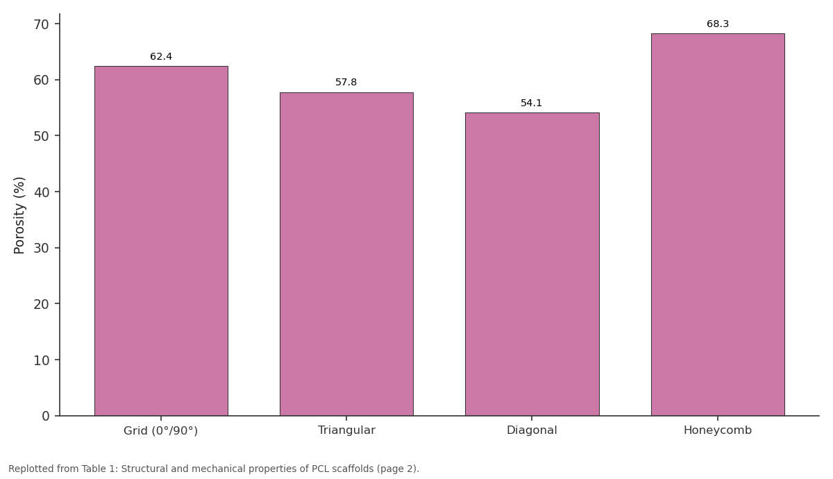
Porosity (%): Grid (0°/90°) vs Triangular vs Diagonal vs Honeycomb
Replotted from Table 1 (page 2) of the original paper. Every bar is the paper's own table value.
View source evidence ↗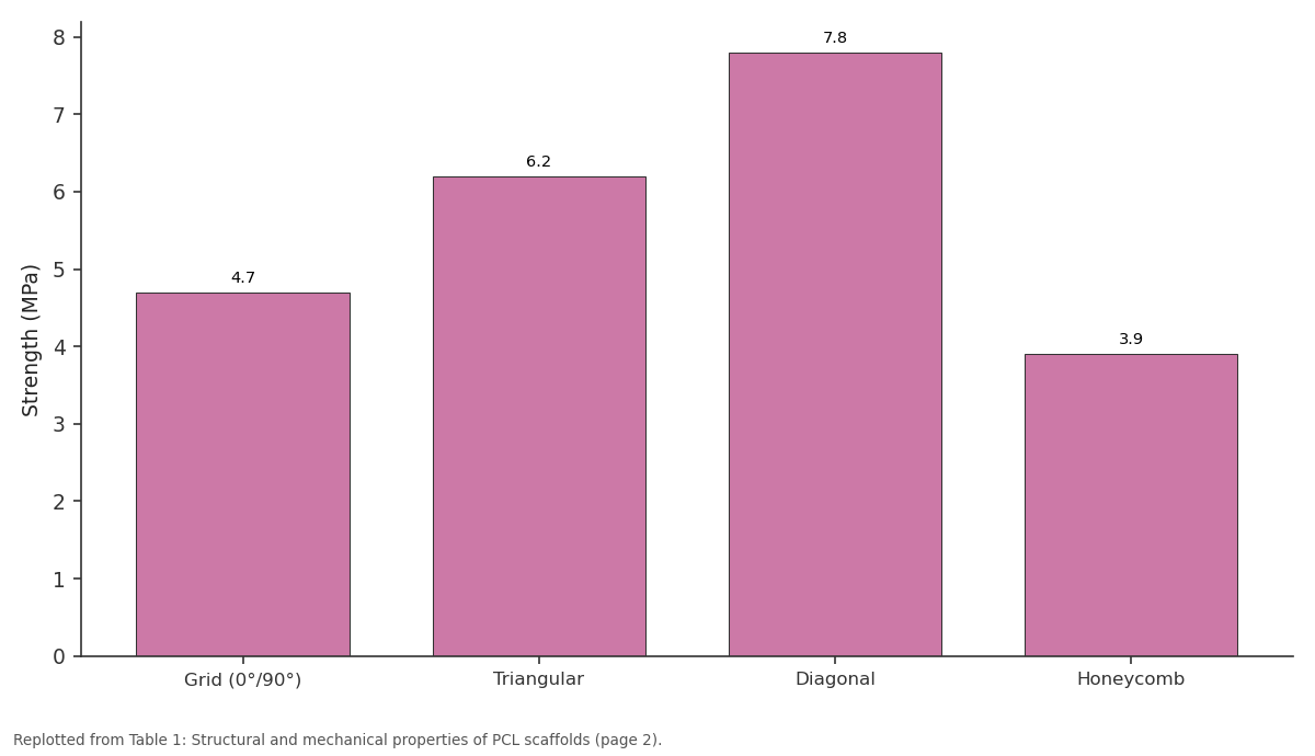
Strength (MPa): Grid (0°/90°) vs Triangular vs Diagonal vs Honeycomb
Replotted from Table 1 (page 2) of the original paper. Every bar is the paper's own table value.
View source evidence ↗Figures & tables
From the original paper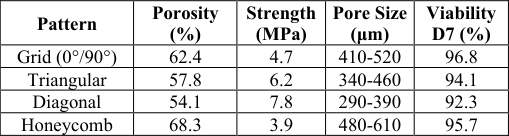 Table 1
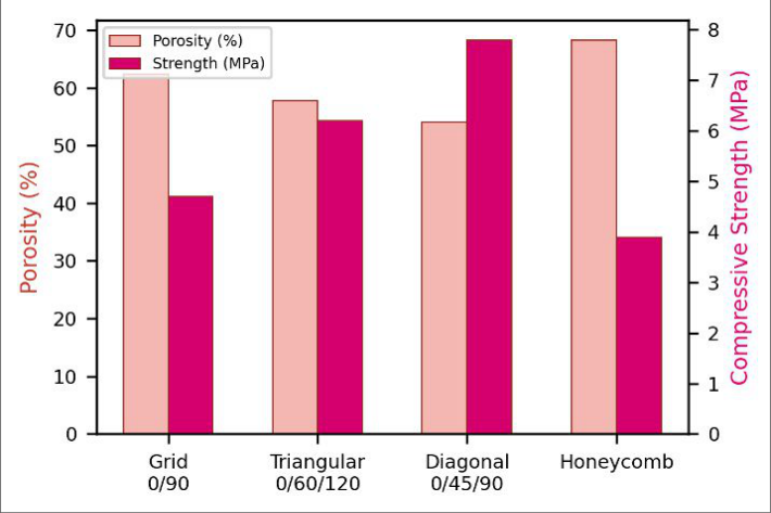 Figure 1
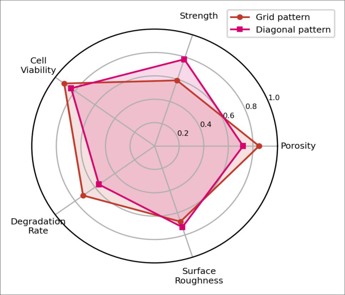 Figure 2
Introduction
Bone defects beyond a certain size won't heal on their own -the so-called critical-size threshold above which the body's repair machinery can't bridge the gap without a structural template [1] . Scaffolds made from biodegradable polymers fill that role by providing temporary mechanical support and a porous architecture that guides new bone tissue into the defect space [2] . Among candidate polymers, polycaprolactone (PCL) stands out for its slow degradation rate (2-4 years in vivo), FDA approval history, and compatibility with meltbased 3D printing [3] . FDM printing builds scaffolds layer by layer from PCL filament, and the infill pattern -the internal geometry of deposited strands -dictates both porosity and mechanical properties [4] . Grid (0°/90°) patterns are the default in most studies, but triangular and honeycomb alternatives create different pore geometries that may better match the trabecular architecture of cancellous bone [5] . Few reports have compared more than two patterns head-to-head under identical printing conditions, making it hard to draw conclusions about which geometry best serves bone regeneration [6] . This research printed PCL scaffolds in four infill patterns, characterized their structural and mechanical properties, and tested osteoblast response in vitro to determine which design achieves the best trade-off between porosity and load-bearing capacity for potential bone defect repair [7] .
System Design
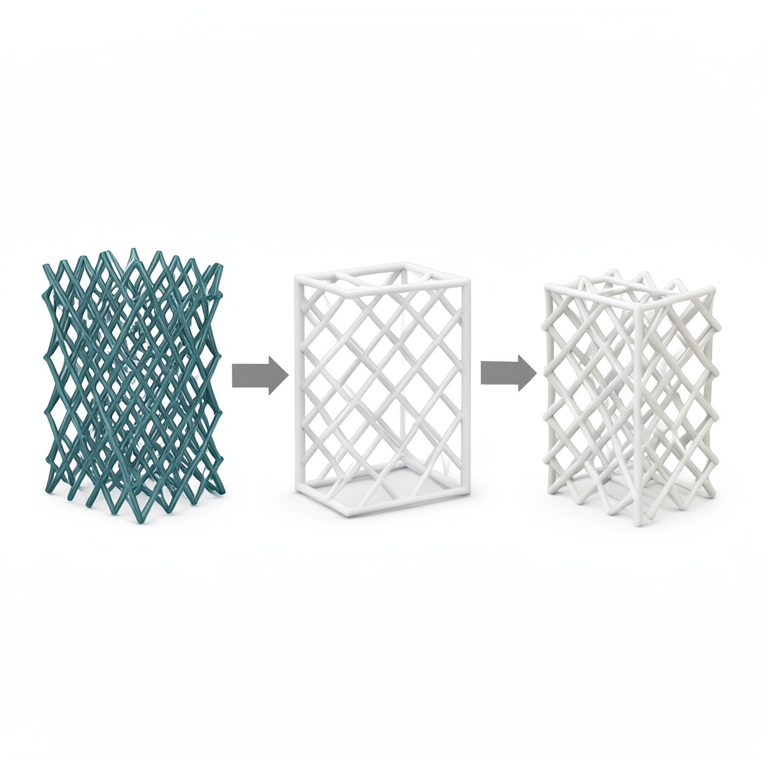
Scaffolds were designed as 10 × 10 × 5 mm blocks in SolidWorks 2022 and sliced using Ultimaker Cura 5.2. Four infill patterns were programmed at 50% infill density: grid (0°/90° alternating layers), triangular (0°/60°/120°), diagonal (0°/45°/90°/135°), and honeycomb. Layer height was fixed at 0.3 mm, nozzle diameter at 0.4 mm, and print speed at 15 mm/s. Each pattern produced distinct pore shapes: rectangular, triangular, rhomboid, and hexagonal respectively [8] .
Material and Methods Material
Research was conducted at Swami Vivekananda National University, Raipur, Chhattisgarh, India, from January 2023 to August 2023. Ethics approval was obtained for cell culture work (Protocol SVNU/IEC/2023-008, January 2023). PCL filament (1.75 mm, MW 80,000, Sigma-Aldrich) was used with a modified Creality Ender-3 printer fitted with a high-temperature nozzle. MC3T3-E1 mouse osteoblast-like cells (ATCC CRL-2593) were cultured in α-MEM with 10% FBS and 1% penicillin-streptomycin [9] .
Methods
Printing occurred at 90°C nozzle temperature on a 40°C heated bed. Scaffolds were sterilized by UV exposure for 30 min per side before cell culture. Porosity was measured by micro-CT (Bruker SkyScan 1272, 9 μm voxel).
Compressive testing (Instron 5944, 1 mm/min, n=5) measured modulus and yield strength. SEM (JEOL JSM-6510LV) examined strut morphology after gold sputtering.
Cells were seeded at 2 × 10⁴ cells/scaffold; viability was assessed by MTT assay at days 1, 3, 7, and 14. Alkaline phosphatase (ALP) activity was measured at day 14 using pnitrophenyl phosphate substrate and normalized to total protein [10] . Statistical comparisons used one-way ANOVA with Tukey's test (p < 0.05).
Performance Evaluation
Scaffold performance was assessed across five metrics: porosity, compressive strength, cell viability (day 7 MTT), degradation rate (mass loss after 4 weeks in PBS at 37°C), and surface roughness (Mitutoyo SJ-210 profilometer). Each metric was normalized to a 0-1 scale relative to the best-performing pattern for that parameter, enabling multicriteria comparison across all four designs [11] . Porosity and strength showed the expected inverse relationship. The diagonal pattern's 54.1% porosity gave the highest compressive strength at 7.8 MPa, while honeycomb's 68.3% porosity yielded the weakest at 3.9
Results
MPa. Micro-CT confirmed that diagonal scaffolds had the smallest pore sizes (290-390 μm), denser strut intersections, and more uniform pore distribution than the other patterns [12] . All scaffolds supported MC3T3-E1 attachment and proliferation. Cell viability at day 7 ranged from 92.3% (diagonal) to 96.8% (grid), with no statistically significant differences between patterns (p = 0.18). ALP activity at day 14 was highest on grid scaffolds (0.87 nmol/min/mg protein), followed by honeycomb (0.79), triangular (0.71), and diagonal (0.64). SEM of cell-seeded scaffolds showed confluent monolayers on grid and honeycomb patterns, while diagonal scaffolds had patchy coverage in regions where pores were below 300 μm [13] .
Discussion
The diagonal pattern's 7.8 MPa compressive strength places it comfortably within the 2-12 MPa range of human cancellous bone, while grid (4.7 MPa) and honeycomb (3.9 MPa) sit near the lower bound [14] . But strength alone isn't everything. The grid pattern's higher ALP activity suggests its larger, more regular pores better facilitated osteogenic differentiation -a finding consistent with earlier reports that pores above 400 μm promote osteoblast maturation over proliferation [15] . Honeycomb's high porosity (68.3%) and large pores (480-610 μm) gave cells room to spread and nutrients to diffuse, explaining its second-highest ALP reading [16] . The diagonal pattern, despite its mechanical superiority, showed the lowest ALP and patchiest cell coverage, possibly because its smallest pores restricted cell migration into the scaffold interior [17] . This creates a design dilemma: the strongest scaffold isn't necessarily the most biologically active one.
A practical compromise might involve graded porositydenser diagonal regions at load-bearing surfaces transitioning to open grid or honeycomb in the interior where nutrient transport matters more [18] . FDM printing can achieve this by switching infill patterns between layers, though the mechanical behavior of such hybrid scaffolds needs separate validation [19] .
Conclusion
Infill pattern selection during FDM 3D printing gives direct control over the porosity-strength trade-off in PCL bone scaffolds. The diagonal pattern offered the best mechanical performance at 7.8 MPa but showed lower osteogenic markers, while grid and honeycomb patterns favored biological response at the cost of strength. All patterns supported osteoblast viability above 92%. For clinical translation, hybrid scaffolds combining multiple patterns within a single construct may resolve the mechanicalbiological compromise.
Acknowledgement
Acknowledgements Funding SourcesAll expenses were met through departmental funds at Swami Vivekananda National University. Institutional SupportThe authors thank Swami Vivekananda National University and Raja Ramanna Centre for Advanced Chemistry for shared laboratory access. Contributions Not Qualifying for AuthorshipMr. Rahul Verma helped calibrate the 3D printer and prepare filament spools.
Figures and tables from the paper
| Pattern | Porosity (%) | Strength (MPa) | Pore Size (μm) | Viability D7 (%) |
|---|---|---|---|---|
| Grid (0°/90°) | 62.4 | 4.7 | 410-520 | 96.8 |
| Triangular | 57.8 | 6.2 | 340-460 | 94.1 |
| Diagonal | 54.1 | 7.8 | 290-390 | 92.3 |
| Honeycomb | 68.3 | 3.9 | 480-610 | 95.7 |
Table text extracted from the original PDF; superscript letters are shown inline. The original table image is below.
View original table image
References
21 in the original paper- Schemitsch EH. Size matters: defining critical bone defects. J Orthop Trauma. 2017;31(S5):S20-S22.
- Hutmacher DW. Scaffolds in tissue engineering bone and cartilage. Biomaterials. 2000;21(24):2529-2543.
- Woodruff MA, Hutmacher DW. The return of a forgotten polymer: polycaprolactone. Prog Polym Sci. 2010;35(10):1217-1256.
- Zein I, Hutmacher DW, Tan KC, Teoh SH. Fused deposition modeling of novel scaffold architectures. Biomaterials. 2002;23(4):1169-1185.
- Bose S, Vahabzadeh S, Bandyopadhyay A. Bone tissue engineering using 3D printing. Mater Today. 2013;16(12):496-504.
- Yilgor P, Sousa RA, Reis RL, Hasirci N, Hasirci V. 3D plotted PCL scaffolds for stem cell based bone tissue engineering. Macromol Symp. 2008;269(1):92-99. International Journal of Advanced Chemistry Research https://www.chemistryjournals.net
- Loh QL, Choong C. Three-dimensional scaffolds for tissue engineering: role of porosity and pore size. Tissue Eng Part B. 2013;19(6):485-502.
- Moroni L, de Wijn JR, van Blitterswijk CA. 3D fiberdeposited scaffolds for tissue engineering. J Biomed Mater Res A. 2006;75(4):957-965.
- Domingos M, Dinucci D, Cometa S, Boccaccini AR. Polycaprolactone scaffolds fabricated via bioextrusion. Int J Biomater. 2009;2009:239643.
- Ren J, Blackwood KA, Doustgani A, Dickson G. Meltelectrospun polycaprolactone strontium-substituted bioactive glass scaffolds. J Biomed Mater Res A. 2014;102(9):3140-3153.
- Cipitria A, Skelton A, Dargaville TR, Dalton PD, Hutmacher DW. Design, fabrication and characterization. J Mater Chem. 2011;21(26):94199453.
- Poh PSP, Hutmacher DW, Holzapfel BM, Sober ER. In vitro and in vivo bone formation potential. Acta Biomater. 2016;30:319-333.
- Murphy CM, Haugh MG, O'Brien FJ. The effect of mean pore size on cell attachment. Biomaterials. 2010;31(3):461-466.
- Goldstein SA. The mechanical properties of trabecular bone: dependence on anatomic location. J Biomech. 1987;20(11):1055-1061.
- Karageorgiou V, Kaplan D. Porosity of 3D biomaterial scaffolds and osteogenesis. Biomaterials. 2005;26(27):5474-5491.
- Hollister SJ. Porous scaffold design for tissue engineering. Nat Mater. 2005;4(7):518-524.
- Giannitelli SM, Accoto D, Trombetta M, Rainer A. Current trends in scaffold design. J Appl Biomater Funct Mater. 2014;12(1):1-13.
- Sobral JM, Caridade SG, Sousa RA, Mano JF, Reis RL. Three-dimensional plotted scaffolds with controlled pore size gradients. Acta Biomater. 2011;7(3):10091018.
- Hutmacher DW, Schantz T, Zein I, Ng KW. Mechanical properties of polymeric scaffolds. J Biomed Mater Res. 2001;55(2):203-216.
- Shor L, Guceri S, Wen X, Gandhi M, Sun W. Fabrication of three-dimensional polycaprolactone/hydroxyapatite tissue scaffolds. J Manuf Sci Eng. 2007;129(4):683-689.
- Cao T, Ho KH, Teoh SH. Scaffold design and in vitro evaluation of osteoblast-scaffolds. Tissue Eng. 2003;9(S1):S103-S112.