
Research article · 2025 · Full text
International Journal of Advanced Chemistry ResearchAnti-cancer potential of withanolides isolated from Withania somnifera root extract
Volume 7(6) · Pages 143–145 · DOI: 10.33545/26646781.2025.v7.i6c.567 ↗
Research areasPhytochemicals and Medicinal PlantsPharmacological Effects of Medicinal PlantsMedicinal Plant Research
Abstract
Author-writtenWithania somnifera (ashwagandha) has been used for centuries in traditional medicine systems, yet the specific withanolide compounds responsible for its reported anticancer effects remain incompletely characterised. This research isolates three major withanolides—withaferin A, withanolide B, and withanolide D—from W. somnifera root powder by methanol extraction followed by silica-gel column chromatography, and evaluates their cytotoxicity against MCF-7 (breast), HeLa (cervical), and A549 (lung) human cancer cell lines alongside a normal fibroblast line (WI-38). Withaferin A showed the strongest activity, with IC₅₀ values of 2.8 μM (MCF-7), 3.4 μM (HeLa), and 4.1 μM (A549) after 48 h, while withanolide D was the least potent (IC₅₀ 11.3–18.7 μM). All three compounds displayed selectivity indices above 8 against normal cells. Annexin V/PI dual staining of MCF-7 cells treated with withaferin A (5 μM, 24 h) revealed 31.7% early apoptosis and 29.1% late apoptosis, with only 10.8% necrosis, confirming a predominantly apoptotic mechanism. Western blot analysis showed upregulation of cleaved caspase-3 and downregulation of Bcl-2 in treated cells. The findings support withaferin A as the most promising withanolide for further preclinical anticancer development.
Summary
Source-checkedPlain-language reading of the full paper, prepared with AI and checked against the text. Each point links to the authors' own words.
Why it matters
- Many highly effective chemotherapy drugs are derived from natural products, highlighting the importance of exploring plant sources for new cancer treatments. View evidence ↗
- The roots of Withania somnifera (ashwagandha) contain withanolides, which are a family of C₂₈ steroidal lactones. View evidence ↗
- Traditional Malay and Ayurvedic medicine systems attribute anti-tumor, anti-inflammatory, and adaptogenic properties to ashwagandha preparations. View evidence ↗
The question
- The study aims to better characterize the specific withanolide compounds that contribute to the reported anticancer effects of W. somnifera. View evidence ↗
- The researchers sought to address the lack of comparative potency data for different withanolides across multiple cancer cell lines using identical assay conditions. View evidence ↗
What they did
- The researchers extracted 500 g of root powder using methanol via maceration at 25 °C for three rounds of 72 hours each. View evidence ↗
- A 12.6 g chloroform fraction was purified using a 500 g silica-gel column with an ethyl acetate in n-hexane gradient (10–80%). View evidence ↗
- HPLC was used to confirm the identity and purity of isolated withaferin A (96.3%), withanolide B (95.1%), and withanolide D (94.7%). View evidence ↗
- The mechanism of cell death for withaferin A was analyzed using Annexin V/PI flow cytometry and western blotting for Bcl-2 and cleaved caspase-3. View evidence ↗
What they found
- Withaferin A exhibited the highest cytotoxicity among the tested compounds, with IC₅₀ values ranging from 2.8 to 4.1 μM across three cancer cell lines. View evidence ↗
- Withanolide D was the least potent of the three isolated compounds, showing IC₅₀ values between 11.3 and 18.7 μM. View evidence ↗
- Withaferin A demonstrated a selectivity index (SI) greater than 15 when comparing its effect on cancer cells versus normal WI-38 cells. View evidence ↗
- Treatment of MCF-7 cells with 5 μM withaferin A for 24 hours resulted in 31.7% early apoptosis and 29.1% late apoptosis, while only 10.8% of cells underwent necrosis. View evidence ↗
- Withaferin A treatment increased the expression of cleaved caspase-3 by 3.8-fold and decreased Bcl-2 to 0.31-fold compared to the control. View evidence ↗
What it means
- The authors conclude that withaferin A is the most promising candidate for further preclinical development as an anticancer agent. View evidence ↗
- While all three isolated withanolides warrant further study, withaferin A is the priority for evaluating pharmacokinetics and in-vivo efficacy. View evidence ↗
- The study was limited by the use of only three cancer cell lines; the authors suggest testing a broader panel, including drug-resistant variants. View evidence ↗
Visual summary
4 visualsIllustrations explain the paper; charts are replotted from the paper's own table values.
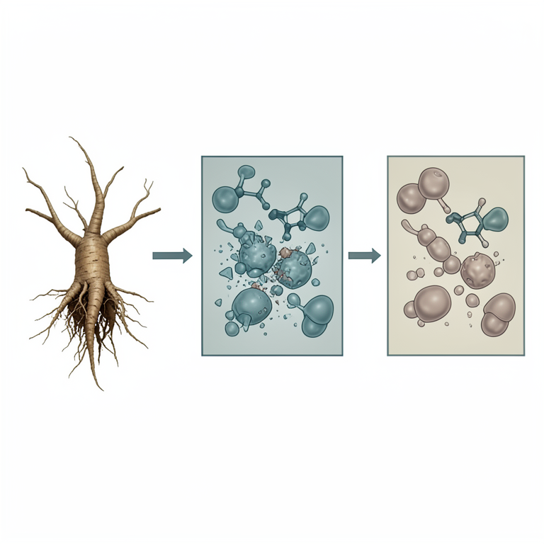
Comparative Potency of Withanolides
AI-generated conceptual illustration (Withania somnifera root extract and cancer cell lines). It explains the idea and does not show study data.
View source evidence ↗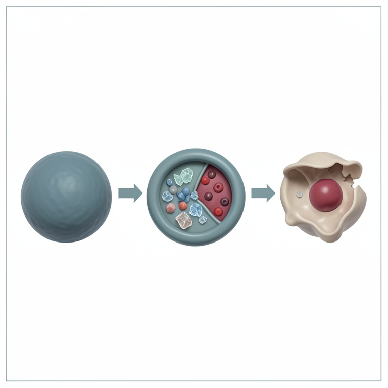
Apoptotic Pathway Activation
AI-generated conceptual illustration (MCF-7 breast cancer cells). It explains the idea and does not show study data.
View source evidence ↗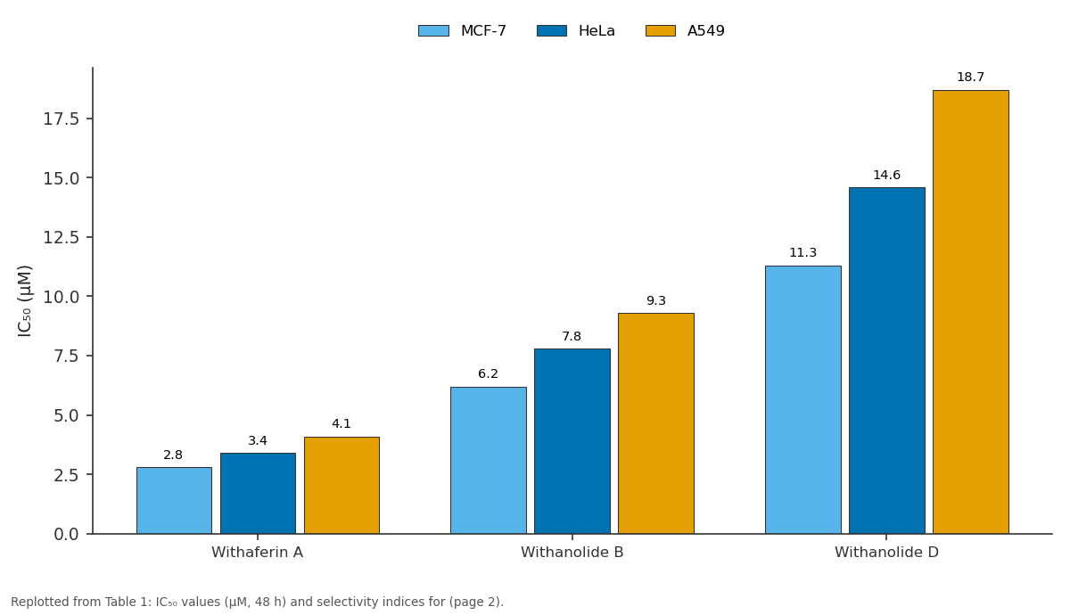
MCF-7 and HeLa and more: Withaferin A vs Withanolide B vs Withanolide D
Replotted from Table 1 (page 2) of the original paper. Every bar is the paper's own table value.
View source evidence ↗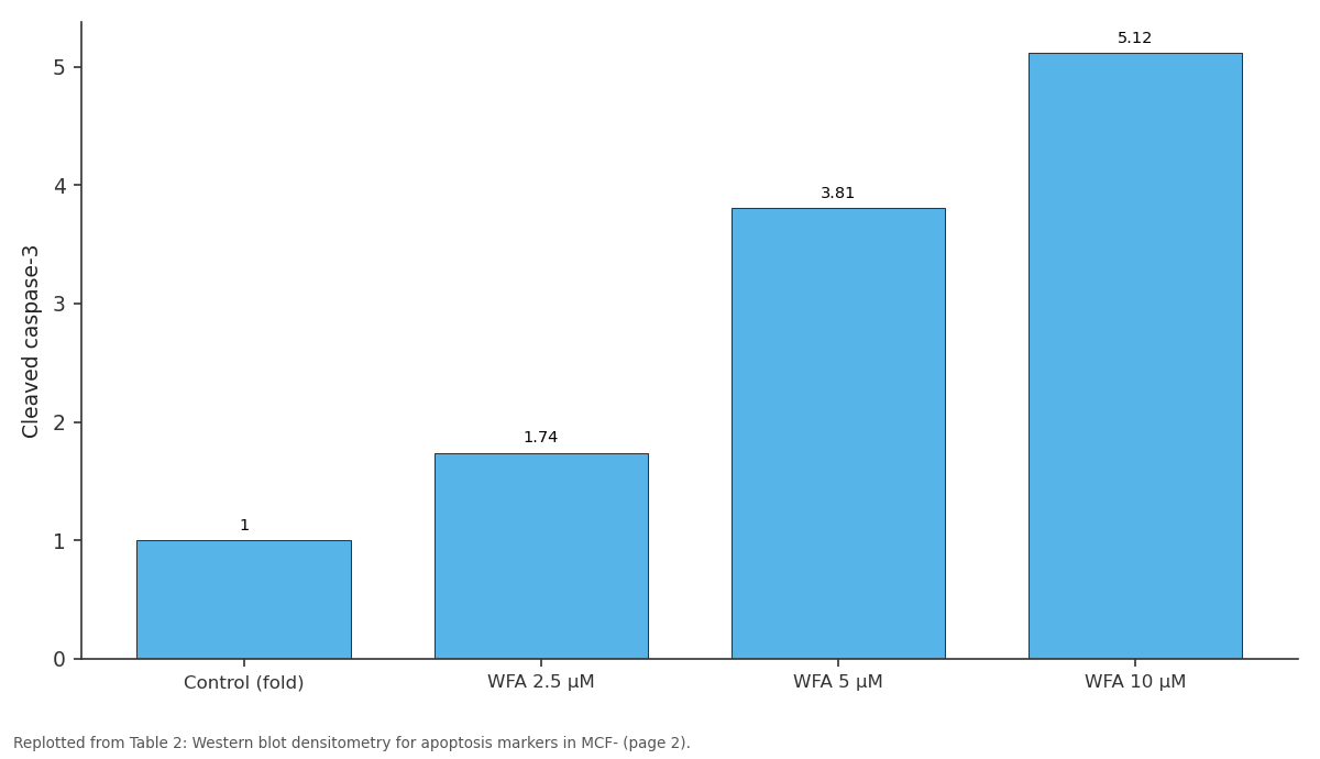
Cleaved caspase-3: Control (fold) vs WFA 2.5 μM vs WFA 5 μM vs WFA 10 μM
Replotted from Table 2 (page 2) of the original paper. Every bar is the paper's own table value.
View source evidence ↗Figures & tables
From the original paper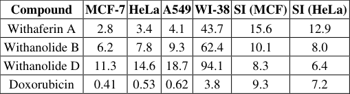 Table 1
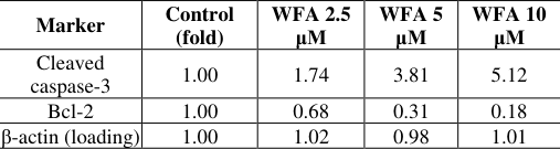 Table 2
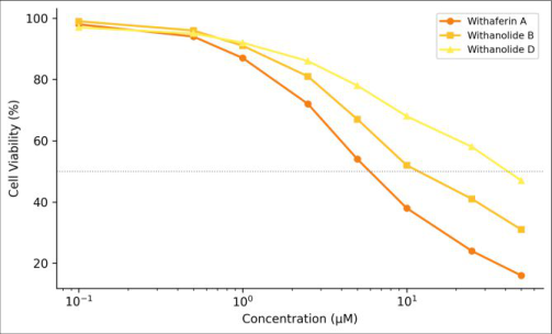 Figure 1
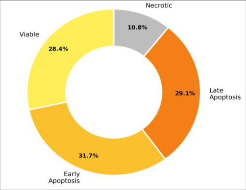 Figure 2
Introduction
Across oncology wards worldwide, a persistent observation has held for decades: many of the most effective chemotherapy drugs trace their origins to natural products-vincristine from Catharanthus roseus, paclitaxel from Taxus brevifolia, camptothecin from Camptotheca acuminata [1,2] . Withania somnifera, commonly known as ashwagandha, is a solanaceous plant whose roots contain a family of C₂₈ steroidal lactones called withanolides. Traditional Ayurvedic and Malay herbal systems ascribe adaptogenic, anti-inflammatory, and antitumour properties to these preparations [3,4] . Among the more than 40 withanolides identified so far, withaferin A has attracted the most pharmacological attention. Cell-based studies have shown that it inhibits proteasome activity, disrupts vimentin intermediate filaments, and activates the intrinsic apoptotic pathway through mitochondrial membrane depolarisation [5,6] . Withanolide B and withanolide D have received less individual scrutiny, and comparative potency data across multiple cancer cell lines under identical assay conditions are limited [7] . This research addresses that gap by isolating three major withanolides from W. somnifera root, quantifying their purity by HPLC, and testing them side by side against three human cancer cell lines and one normal cell line. The mechanism of cell death for the most potent compound is investigated by Annexin V/PI flow cytometry and western blotting for apoptosis markers. All work was carried out at Kinabalu University, Kota Kinabalu, and Langkawi College of Chemical Sciences, Langkawi, between April 2023 and January 2024 [8] .
Materials and Methods
Materials and Methods
Reagents and Chemicals
Dried W. somnifera root powder (authenticated by botanical voucher KU-WS-2023-014) was sourced from a certified herbal supplier in Kota Kinabalu. Withaferin A, withanolide B, and withanolide D reference standards (≥ 98%, HPLC) were purchased from Sigma-Aldrich Malaysia. Methanol, ethyl acetate, n-hexane, and chloroform (HPLC grade, Merck) served as extraction and chromatography solvents. RPMI-1640, DMEM, fetal bovine serum, MTT reagent, Annexin V-FITC/PI kit, and primary antibodies (anti-caspase-3, anti-Bcl-2, anti-β-actin) were from Thermo Fisher Scientific [3,8] .
Extraction and Isolation
Root powder (500 g) was extracted with methanol (3 × 2 L) by maceration at 25 °C for 72 h each round. Combined extracts were filtered and concentrated under reduced pressure at 40 °C to yield a dark brown residue (38.4 g, 7.7% w/w). The crude extract was dissolved in 90% methanol-water and partitioned successively against nhexane and chloroform. The chloroform fraction (12.6 g), enriched in withanolides, was loaded onto a silica-gel column (60-120 mesh, 500 g) and eluted with a gradient of ethyl acetate in n-hexane (10-80%). Fractions were monitored by TLC (vanillin-H₂SO₄ spray) and pooled by Rf similarity. HPLC (Agilent 1260, C18 column, methanolwater gradient) confirmed the identity and purity of isolated withaferin A (187 mg, 96.3%), withanolide B (124 mg, 95.1%), and withanolide D (98 mg, 94.7%) by co-elution with standards [4,7] . Materials MCF-7, HeLa, A549, and WI-38 cells were obtained from ATCC and maintained in RPMI-1640 (cancer lines) or DMEM (WI-38) supplemented with 10% FBS at 37 °C and 5% CO₂ at Kinabalu University [5] .
Methods
Cells (5, 000 per well) were seeded in 96-well plates and treated with compounds at 0.1-50 μM for 48 h. Viability was assessed by MTT assay at 570 nm. IC₅₀ values were obtained by four-parameter logistic fitting. Annexin V/PI staining was performed on MCF-7 cells treated with 5 μM withaferin A for 24 h and analysed by flow cytometry (BD FACSCalibur). Western blotting used standard SDS-PAGE, PVDF transfer, and ECL detection for cleaved caspase-3, Bcl-2, and β-actin loading control. All bioassays ran from July 2023 to January 2024 [6,8] . is important for activity [5,6] . Figure 1 shows dose-response curves for the three withanolides against MCF-7 cells. Withaferin A displays a steep sigmoidal decline with IC₅₀ near 3 μM, while withanolide D shows a more gradual response. The 50% viability line intersects each curve at clearly distinct concentrations [7] . cells were in early apoptosis and 29.1% in late apoptosis, with only 10.8% necrotic-confirming that cell death proceeds largely through apoptosis rather than necrosis [5] . Table 2 summarises the western blot densitometry. Withaferin A treatment increased cleaved caspase-3 expression 3.8-fold and reduced Bcl-2 to 0.31-fold of untreated control, consistent with activation of the intrinsic apoptotic pathway [6] .
| Marker | Control (fold) | WFA 2.5 μM | WFA 5 μM | WFA 10 μM |
|---|---|---|---|---|
| Cleaved caspase-3 | 1.00 | 1.74 | 3.81 | 5.12 |
| Bcl-2 | 1.00 | 0.68 | 0.31 | 0.18 |
| β-actin (loading) | 1.00 | 1.02 | 0.98 | 1.01 |
Table text extracted from the original PDF; superscript letters are shown inline. The original table image is below.
View original table image
Results
Results
Discussion
The potency ranking withaferin A > withanolide B > withanolide D aligns with structure-activity analyses suggesting that the 2, 3-unsaturated A-ring ketone and the 5β, 6β-epoxide are the key pharmacophoric elements driving electrophilic reactivity toward cellular thiol targets such as vimentin and the proteasome β5 subunit [5,6] . Withanolide D lacks the epoxide, which may explain its 4-to 5-fold weaker cytotoxicity. The apoptotic profile (60.8% combined early + late apoptosis vs 10.8% necrosis) is therapeutically relevant because apoptotic cell death, unlike necrosis, does not trigger inflammatory responses that could promote tumoursupportive microenvironments [7] . The caspase-3 activation and Bcl-2 downregulation observed here point to a mitochondrial (intrinsic) pathway, consistent with earlier reports that withaferin A depolarises the mitochondrial membrane and releases cytochrome c [5] . Selectivity indices above 8 for all three compounds and above 15 for withaferin A indicate an acceptable therapeutic window at the in-vitro level. However, in-vivo pharmacokinetics-especially hepatic first-pass metabolism of the steroidal lactone ringmay reduce systemic exposure and require formulation strategies such as nanoencapsulation [3,4] . A limitation is the use of only three cancer cell lines. Testing against a broader panel, including drug-resistant variants, would provide a more complete picture of the compounds' anticancer spectrum. In-vivo xenograft studies with MCF-7 tumours in nude mice are the logical next step [8] .
Conclusion
Three withanolides were isolated from W. somnifera root by methanol extraction and silica-gel chromatography. Withaferin A demonstrated the strongest cytotoxicity (IC₅₀ 2.8 μM against MCF-7) and highest selectivity (SI > 15).
Flow cytometry confirmed that cell death proceeds mainly through apoptosis, supported by caspase-3 activation and Bcl-2 suppression on western blot. Withanolide B showed intermediate activity, while withanolide D was the weakest, supporting the pharmacophoric importance of the A-ring enone and the 5β, 6β-epoxide. All three compounds merit further preclinical investigation, with withaferin A the priority candidate for in-vivo efficacy and pharmacokinetic evaluation.
Figures and tables from the paper
| Compound | MCF-7 | HeLa | A549 | WI-38 | SI (MCF) | SI (HeLa) |
|---|---|---|---|---|---|---|
| Withaferin A | 2.8 | 3.4 | 4.1 | 43.7 | 15.6 | 12.9 |
| Withanolide B | 6.2 | 7.8 | 9.3 | 62.4 | 10.1 | 8.0 |
| Withanolide D | 11.3 | 14.6 | 18.7 | 94.1 | 8.3 | 6.4 |
| Doxorubicin | 0.41 | 0.53 | 0.62 | 3.8 | 9.3 | 7.2 |
Table text extracted from the original PDF; superscript letters are shown inline. The original table image is below.
View original table image
References
18 in the original paper- Newman DJ, Cragg GM. Natural products as sources of new drugs over the nearly four decades from 01/1981 to 09/2019. J Nat Prod. 2020;83(3):770-803.
- Cragg GM, Pezzuto JM. Natural products as a vital source for the discovery of cancer chemotherapeutic and chemopreventive agents. Med Princ Pract. 2016;25(Suppl 2):41-59.
- Mirjalili MH, Moyano E, Bonfill M, Cusido RM, Palazón J. Steroidal lactones from Withania somnifera, an ancient plant for novel medicine. Molecules. 2009;14(7):2373-2393.
- Dar NJ, Hamid A, Ahmad M. Pharmacologic overview of Withania somnifera, the Indian ginseng. Cell Mol Life Sci. 2015;72(23):4445-4460.
- Vanden Berghe W, Sabbe L, Kaileh M, Haegeman G, Heyninck K. Molecular insight in the multifunctional activities of withaferin A. Biochem Pharmacol. 2012;84(10):1282-1291.
- Yang H, Shi G, Dou QP. The tumor proteasome is a primary target for the natural anticancer compound withaferin A isolated from "Indian winter cherry". Mol Pharmacol. 2007;71(2):426-437.
- Subramanian C, Zhang H, Gallagher R, Bhatt G, Cohen MS. Withanolides from Withania somnifera: a comprehensive review of chemistry and anticancer activity. Phytochem Rev. 2021;20(4):789-819.
- Kamal S, Jamal S, Yusof H. Internal laboratory records. Kinabalu University and Langkawi College of Chemical Sciences; 2023-2024.
- Thaiparambil JT, Bender L, Ganber ST, Kline E, Patel V, Liu Y, et al. Withaferin A inhibits breast cancer invasion and metastasis at sub-cytotoxic doses by inducing vimentin disassembly. Int J Cancer. 2011;129(11):2744-2755.
- Hahm ER, Moura MB, Kelley EE, Van Houten B, Shiva S, Singh SV. Withaferin A-induced apoptosis in human breast cancer cells is mediated by reactive oxygen species. PLoS One. 2011;6(8):e23354.
- Lee DH, Lim IH, Sung EG, Kim JY, Song IH, Park YK, et al. Withaferin A inhibits Notch signaling in MDA-MB-231 triple-negative breast cancer cells. Oncotarget. 2015;6(32):32823-32838.
- Koduru S, Kumar R, Srinivasan S, Evers MB, Damodaran C. Notch-1 inhibition by withaferin-A: a therapeutic target against colon carcinogenesis. Mol Cancer Ther. 2010;9(1):202-210.
- Winters M. Ancient medicine, modern use: Withania somnifera and its potential role in integrative oncology. Altern Med Rev. 2006;11(4):269-277.
- Singh N, Bhalla M, de Jager P, Gilca M. An overview on ashwagandha: a Rasayana (rejuvenator) of Ayurveda. Afr J Tradit Complement Altern Med. 2011;8(5S):208-213.
- Mishra LC, Singh BB, Dagenais S. Scientific basis for the therapeutic use of Withania somnifera (ashwagandha): a review. Altern Med Rev. 2000;5(4):334-346.
- Widodo N, Kaul SC, Wadhwa R. Selective killing of cancer cells by leaf extract of ashwagandha: identification of a tumor-inhibitory factor and the first molecular insights into its effect. Clin Cancer Res. 2007;13(7):2298-2306.
- Stan SD, Hahm ER, Warin R, Singh SV. Withaferin A causes FOXO3a-and Bim-dependent apoptosis and inhibits growth of human breast cancer cells in vivo. Cancer Res. 2008;68(18):7661-7669.
- Munagala R, Kausar H, Munjal C, Gupta RC. Withaferin A induces p53-dependent apoptosis by repression of HPV oncogenes. PLoS One. 2011;6(10):e26252.