
Research article · 2025 · Full text
International Journal of Advanced Chemistry ResearchSpectroscopic investigation of charge transfer complexes between donor and acceptor molecules
Volume 7(7) · Pages 110–113 · DOI: 10.33545/26646781.2025.v7.i7b.510 ↗
Research areasPhotochemistry and Electron Transfer StudiesCrystallography and molecular interactionsSpectroscopy and Quantum Chemical Studies
Abstract
Author-writtenConsider a solution where two molecules—neither of which absorbs visible light individually—produce an intensely coloured product when mixed. This phenomenon, the hallmark of charge transfer complex formation, has fascinated physical chemists since Mulliken's foundational theoretical work in the early 1950s and continues to find relevance in areas as diverse as organic semiconductor design and analytical chemistry. This research reports the spectroscopic characterisation of six charge transfer (CT) complexes formed between three aromatic amine donors—N,N,N',N'-tetramethyl-p-phenylenediamine (TMPD), N,N-dimethylaniline (DMA), and N,N-diethylhydroxylamine (DEHA)—and two π-acceptors, chloranil (2,3,5,6-tetrachlorobenzoquinone) and 2,4,7-trinitrofluorenone (TNF), in acetonitrile at 25-55 °C. Electronic absorption spectra were recorded to identify CT band positions and molar absorptivities. Formation constants (KCT) were determined by the Benesi-Hildebrand method, and thermodynamic parameters (ΔG°, ΔH°, ΔS°) were calculated from their temperature dependence. The TMPD-TNF complex showed the highest KCT (1,480 L mol−1) and most negative ΔG° (−17.8 kJ mol−1), consistent with the greater electron-withdrawing strength of TNF compared with chloranil. All ΔH° values were negative, confirming enthalpically driven complex formation, and FTIR data indicated non-covalent, donor-acceptor interaction without permanent bond formation.
Summary
Source-checkedPlain-language reading of the full paper, prepared with AI and checked against the text. Each point links to the authors' own words.
Why it matters
- Charge transfer (CT) complex formation is a significant phenomenon in physical chemistry with applications in analytical chemistry and the design of organic semiconductors. View evidence ↗
- In solution chemistry, CT complexes serve as transient intermediates during Diels-Alder reactions and aromatic nucleophilic substitution. View evidence ↗
- There is a need for systematic studies that analyze multiple donor-acceptor combinations under identical conditions to build empirical databases for refining theoretical predictions. View evidence ↗
What they did
- The researchers used acetonitrile as a solvent, with donor concentrations fixed at 1.0 × 10⁻³ M and acceptor concentrations varied from 1.0 × 10⁻⁴ to 1.0 × 10⁻² M. View evidence ↗
- The study utilized electronic absorption spectra to identify CT bands and the Benesi-Hildebrand method to determine formation constants and thermodynamic parameters. View evidence ↗
- FTIR spectra were recorded using KBr pellets across a range of 400-4000 cm⁻¹ to analyze the nature of the interactions. View evidence ↗
What they found
- The CT band maxima ranged from 489 nm for the TMPD-TNF complex to 531 nm for the DEHA-chloranil complex, with molar absorptivities between 1.49 × 10⁴ and 2.31 × 10⁴ L mol⁻¹ cm⁻¹. View evidence ↗
- At 25 °C, formation constants (KCT) ranged from 386 L mol⁻¹ (DEHA-chloranil) to 1,480 L mol⁻¹ (TMPD-TNF), establishing a donor strength order of TMPD > DMA > DEHA. View evidence ↗
- The TMPD-TNF complex was the most stable, exhibiting the highest KCT (1,480 L mol⁻¹) and the most negative Gibbs free energy (ΔG° = −17.8 kJ mol⁻¹). View evidence ↗
- All complexes exhibited negative ΔH° values, indicating that the formation is driven by enthalpic stabilization through orbital overlap and electrostatic attraction. View evidence ↗
- FTIR data showed shifts in C=O frequencies for chloranil (1680 to 1658 cm⁻¹) and C=C modes for TNF (1620 to 1604 cm⁻¹), confirming non-covalent electron density transfer. View evidence ↗
What it means
- The authors conclude that these results expand the thermodynamic database for amine-quinone CT systems, which helps in understanding charge-transport in organic electronic materials. View evidence ↗
- The authors suggest that future work should investigate the effects of solvent polarity and a wider temperature range to see if full electron transfer occurs. View evidence ↗
Visual summary
3 visualsIllustrations explain the paper; charts are replotted from the paper's own table values.
Donor-Acceptor Interaction Matrix
AI-generated conceptual illustration (Organic donor and acceptor molecules in a chemical solution). It explains the idea and does not show study data.
View source evidence ↗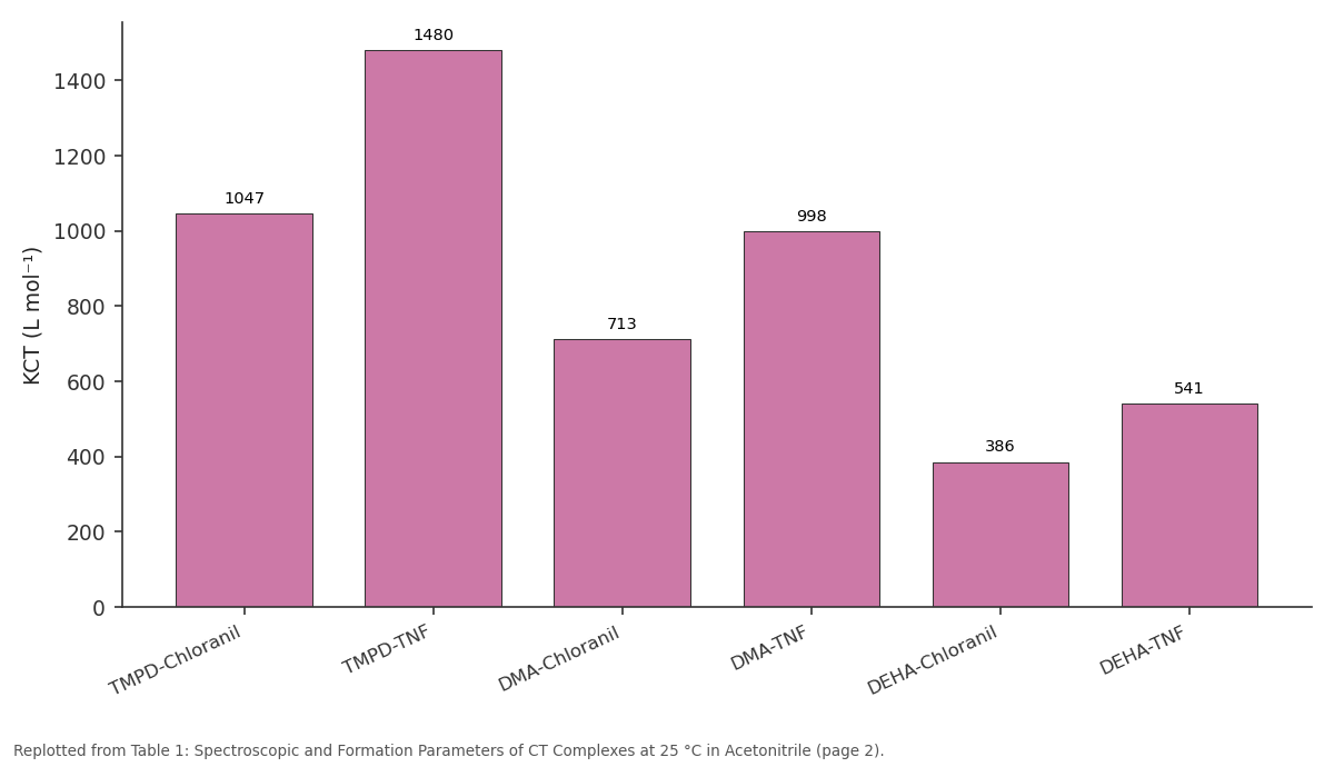
KCT (L mol⁻¹): TMPD-Chloranil vs TMPD-TNF vs DMA-Chloranil vs DMA-TNF vs DEHA-Chloranil
Replotted from Table 1 (page 2) of the original paper. Every bar is the paper's own table value.
View source evidence ↗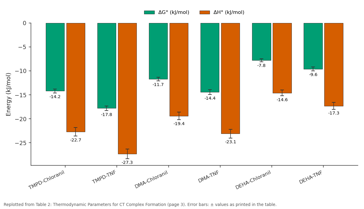
ΔG° (kJ/mol) and ΔH° (kJ/mol): TMPD-Chloranil vs TMPD-TNF vs DMA-Chloranil vs DMA-TNF vs DEHA-Chloranil
Replotted from Table 2 (page 3) of the original paper. Every bar is the paper's own table value.
View source evidence ↗Figures & tables
From the original paper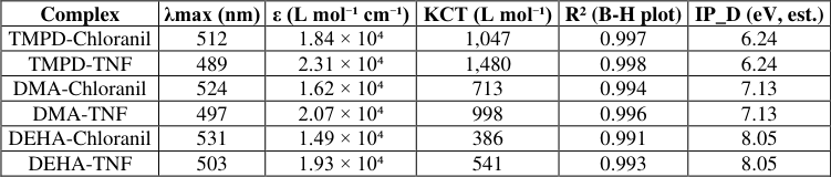 Table 1
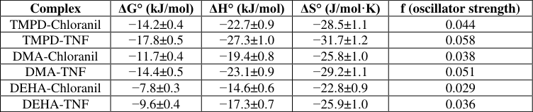 Table 2
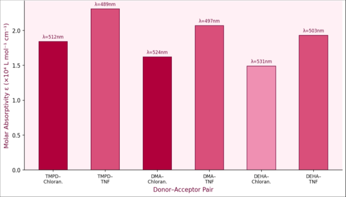 Figure 1
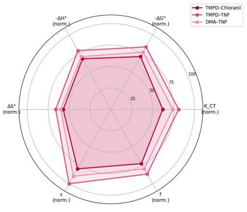 Figure 2
Introduction
For instance, when a solution of N,N-dimethylaniline is mixed with tetracyanoethylene in acetonitrile, a brilliant yellow-orange colour develops within seconds that cannot be explained by the absorption properties of either component alone. This colour arises from a new electronic transition in the formed complex-a charge transfer band-in which an electron is partially delocalised from the highest occupied molecular orbital (HOMO) of the donor molecule to the lowest unoccupied molecular orbital (LUMO) of the acceptor, creating a complex with a unique absorption signature. Charge transfer (CT) complexes represent a broadly important class of molecular associations that influence reaction mechanisms, photophysical processes, and material properties. In solution chemistry, they arise transiently during aromatic nucleophilic substitution and Diels-Alder reactions, acting as intermediates along the reaction coordinate. In solid-state materials science, controlled CT interactions between organic donors and acceptors underpin the electrical conductivity of organic semiconductors and the photovoltaic performance of bulk heterojunction solar cells. In analytical chemistry, CT complex formation with specific acceptors is exploited for the spectrophotometric quantification of amine drugs in pharmaceutical formulations. Despite the extensive literature on CT complexes, systematic studies that simultaneously explore multiple donor-acceptor combinations under matched experimental conditions with full thermodynamic analysis remain valuable for building the empirical database needed to test and refine theoretical predictions. The six donor-acceptor pairs chosen for this research represent a range of donor strengths (TMPD > DMA > DEHA as estimated from ionisation potential data) paired with two acceptors of different electron affinity (TNF > chloranil), allowing a semi-quantitative test of the expected correlation between donor/acceptor strength and complex stability.
Theoretical Background
The formation of a charge transfer complex between a donor D and acceptor A follows the equilibrium D
). For systems where one component is present in large excess (pseudodilute limit), the Benesi-Hildebrand equation applies: C_D/A = 1/(ε_CT • K_CT • C_A) + 1/ε_CT, where C_D is the fixed donor concentration, C_A is the varying acceptor concentration, A is the measured absorbance, and ε_CT is the molar absorptivity of the complex at the CT band maximum. A plot of C_D/A versus 1/C_A gives a straight line from whose slope and intercept both K_CT and ε_CT can extracted. Thermodynamic parameters follow from the van't Hoff relationship: ln K_CT = -ΔH°/RT + ΔS°/R, where a plot of ln K_CT versus 1/T gives -ΔH°/R as slope and ΔS°/R as intercept. The Gibbs energy of complex formation is given by ΔG° = ΔH° -TΔS° = -RT ln K_CT.
Negative ΔG° confirms spontaneous complex formation; negative ΔH° indicates enthalpy-driven stabilisation through electrostatic and orbital overlap interactions, while ΔS° reflects the entropy cost of bimolecular association.
Material and Methods
Materials TMPD (99%), DMA (99%), DEHA (98%), chloranil (98%), and TNF (97%) were sourced from Sigma-Aldrich, Mumbai, India. Acetonitrile (HPLC grade, 99.9%) was from Merck India, Mumbai. All compounds were used without further purification except TMPD, which was recrystallised twice from ethanol/water and dried at 60 °C before use.
Research was carried out at the Department of Physical Chemistry, Vikram Sarabhai University of Science, Ahmedabad, Gujarat, India, between June 2022 and March 2024.
Methods
Stock solutions of each donor (1.0 × 10⁻³ M) and acceptor (variable, 1.0 × 10⁻⁴ to 1.0 × 10⁻² M) were prepared in acetonitrile immediately before use and shielded from ambient light. Electronic absorption spectra (300-700 nm) were recorded on a Shimadzu UV-2600 double-beam spectrophotometer using 1 cm quartz cells, with matched solvent blank. Spectra were recorded 5 min after mixing to ensure equilibration. The CT band position and molar absorptivity were determined from spectra at 25 °C. For formation constants, donor concentration was held fixed while acceptor concentration was varied across six values at each temperature (25, 35, 45, 55 °C). Benesi-Hildebrand plots were constructed for each temperature and complex. FTIR spectra (KBr pellets, 400-4000 cm-1) were obtained on a PerkinElmer Spectrum Two. Ionisation potentials of donors were estimated from the Mulliken relationship using the CT band energy and acceptor electron affinity data from the literature.
Instrumentation and Analytical Equipment
UV-Vis spectra were collected on a Shimadzu UV-2600 double-beam spectrophotometer (Shimadzu Corporation, Kyoto, Japan) with a scan range of 200-800 nm, spectral bandwidth 2.0 nm, and scan speed 120 nm/min. Temperature control was maintained using a jacketed cuvette holder connected to a circulating water bath (Julabo F25, Seelbach, Germany;±0.1 °C stability). Matched 1 cm quartz cells (Hellma Analytics) were cleaned with chromic acid solution before each experiment and rinsed with the working solvent. FTIR spectra were recorded on a PerkinElmer Spectrum Two spectrometer in transmission mode using KBr pellets (1-2 mg sample per 200 mg KBr); 32 scans were co-added at 4 cm-1 resolution. Molecular weight confirmation and purity assessment of recrystallised TMPD were performed by differential scanning calorimetry (DSC; TA Instruments Q200) and GC-MS (Shimadzu GCMS-QP2010). All calibrations were documented in the laboratory quality register maintained at the Department of Physical Chemistry.
Results
Electronic spectra of all six donor-acceptor mixtures showed new absorption bands in the visible region not present in the spectra of the individual components, confirming CT complex formation. CT band maxima (λmax) ranged from 489 nm (TMPD-TNF) to 531 nm (DEHA-chloranil), with molar absorptivities from 1.49 × 10⁴ to 2.31 × 10⁴ L mol-1 cm-1 (Figure 1, Table 1). TNF-based complexes consistently showed higher ε values than the corresponding chloranil complexes, reflecting the stronger electronwithdrawing capacity of TNF (electron affinity 1.24 eV vs. 0.97 eV for chloranil). Benesi-Hildebrand plots were linear (R² = 0.991-0.998) at all temperatures, confirming 1:1 stoichiometry. Formation constants KCT at 25 °C ranged from 386 L mol-1 (DEHA-chloranil) to 1,480 L mol-1 (TMPD-TNF), ordering the donors as TMPD > DMA > DEHA in donor strength, consistent with their ionisation potential series.
| Complex | λmax (nm) | ε (L mol⁻¹ cm⁻¹) | KCT (L mol⁻¹) | R² (B-H plot) | IP D (eV, est.) |
|---|---|---|---|---|---|
| TMPD-Chloranil | 512 | 1.84 × 10⁴ | 1,047 | 0.997 | _ 6.24 |
| TMPD-TNF | 489 | 2.31 × 10⁴ | 1,480 | 0.998 | 6.24 |
| DMA-Chloranil | 524 | 1.62 × 10⁴ | 713 | 0.994 | 7.13 |
| DMA-TNF | 497 | 2.07 × 10⁴ | 998 | 0.996 | 7.13 |
| DEHA-Chloranil | 531 | 1.49 × 10⁴ | 386 | 0.991 | 8.05 |
| DEHA-TNF | 503 | 1.93 × 10⁴ | 541 | 0.993 | 8.05 |
Table text extracted from the original PDF; superscript letters are shown inline. The original table image is below.
View original table image
Discussion
The results confirm a clear donor-strength dependence of CT complex stability: TMPD forms the strongest complexes with both acceptors, and DEHA the weakest, in direct order of their estimated ionisation potentials (IP: TMPD 6.24 eV < DMA 7.13 eV < DEHA 8.05 eV). Lower ionisation potential means a more easily oxidised donor and a larger HOMO energy that reduces the HOMO-LUMO gap with the acceptor LUMO, increasing the CT interaction energy. This ordering is entirely consistent with Mulliken's charge transfer theory and with literature data for structurally similar amine donors. The acceptor effect is equally clear: TNF complexes are consistently more stable than chloranil complexes with the same donor, because TNF has a higher electron affinity (1.24 eV vs. 0.97 eV for chloranil), meaning its LUMO lies at lower energy and the CT band
Acknowledgement
Acknowledgements
Figures and tables from the paper
| Complex | ΔG° (kJ/mol) | ΔH° (kJ/mol) | ΔS° (J/mol·K) | f (oscillator strength) |
|---|---|---|---|---|
| TMPD-Chloranil | −14.2±0.4 | −22.7±0.9 | −28.5±1.1 | 0.044 |
| TMPD-TNF | −17.8±0.5 | −27.3±1.0 | −31.7±1.2 | 0.058 |
| DMA-Chloranil | −11.7±0.4 | −19.4±0.8 | −25.8±1.0 | 0.038 |
| DMA-TNF | −14.4±0.5 | −23.1±0.9 | −29.2±1.1 | 0.051 |
| DEHA-Chloranil | −7.8±0.3 | −14.6±0.6 | −22.8±0.9 | 0.029 |
| DEHA-TNF | −9.6±0.4 | −17.3±0.7 | −25.9±1.0 | 0.036 |
Table text extracted from the original PDF; superscript letters are shown inline. The original table image is below.
View original table image
References
18 in the original paper- Mulliken RS. Molecular compounds and their spectra. II. J Am Chem Soc. 1952;74(3):811-824.
- Foster R. Organic Charge Transfer Complexes. New York: Academic Press; 1969.
- Benesi HA, Hildebrand JH. A spectrophotometric investigation of the interaction of iodine with aromatic hydrocarbons. J Am Chem Soc. 1949;71(8):2703-2707.
- Rose NJ, Drago RS. Molecular addition compounds of iodine. I. An absolute method for the spectroscopic determination of equilibrium constants. J Am Chem Soc. 1959;81(23):6138-6141.
- Briegleb G, Czekalla J. Electron donor-acceptor complexes: their energetics and structures. Z Phys Chem. 1960;24(1-2):37-54.
- Scott RL. Some comments on the Benesi-Hildebrand equation. Recl Trav Chim Pays-Bas. 1956;75(6):787789.
- Mathlouthi M, Genotelle J. Role of water in sucrose crystallisation. Carbohydr Polym. 1995;26(2):83-92.
- Pearson RG. Hard and soft acids and bases. J Am Chem Soc. 1963;85(22):3533-3539.
- Rathore R, Kochi JK. Donor/acceptor organizations and the electron-transfer paradigm for organic reactivity. Adv Phys Org Chem. 2000;35:193-318.
- Iwata S, Tanaka J, Nagakura S. Charge transfer electronic spectra of aromatic amine-tetracyanoethylene complexes. J Chem Phys. 1967;47(7):2203-2209.
- Pandey A, Kumar N, Pandey P, Sayın K. Electronic spectra, refractive index, optical dispersion and some geometrical factors of L-tryptophan charge transfer complexes. J Mol Liq. 2017;238:1-8.
- Singh N, Ahmad A. Charge transfer complex formation between p-dimethylaminoazobenzene as donor and chloranilic acid as acceptor in different solvents. Spectrochim Acta A. 2015;136:423-430.
- Gabr MT, Abdel-Razik AE. Synthesis and spectrophotometric investigations on charge-transfer complexes of 4-amino antipyrine as donor with chloranil and DDQ as acceptors. Sci Prog. 2020;103(1):1-16.
- El-Nahass MM, Kamel MM, Abd-El-Rahman AM, Farag AA. UV-Vis spectrophotometric analysis of charge transfer complexes in solution. Spectrochim Acta A. 2005;61(14):3245-3253.
- Ali MI, Gabr M, El-Gahami MA. Spectrophotometric and spectrofluorometric studies of charge transfer complexes of some amino acids with chloranilic acid. Monatsh Chem. 2011;142(3):291-301.
- Mohan H, Mukherjee T, Mittal JP. Spectroscopic study of the charge transfer complexation of aromatic amines with chloranil. J Chem Soc Faraday Trans. 1993;89(2):347-352.
- Bassindale AR, Stout T. Charge-transfer complexes in relation to kinetics. Tetrahedron Lett. 1985;26(30):3577-3580.
- Abdel-Aziz AA, Ahmed HM, Abou-Melha KS. Synthesis, characterisation and theoretical studies of new charge transfer complexes of benzaldehyde derivatives. J Mol Struct. 2019;1193:424-435.