
Research article · 2025 · Full text
International Journal of Advanced Chemistry ResearchQuercetin-loaded PLGA nanoparticles with pH-responsive release for enhanced cytotoxicity against breast cancer cells
Volume 7(4) · Pages 178–181 · DOI: 10.33545/26646781.2025.v7.i4c.573 ↗
Research areasNanoparticle-Based Drug DeliveryAdvanced Drug Delivery SystemsPharmacology and Nanomedicine Research
Abstract
Author-writtenQuercetin possesses well-documented anticancer activity against breast cancer cell lines, but its clinical translation is hampered by extremely poor aqueous solubility (<1 μg/mL), rapid hepatic metabolism, and low oral bioavailability (<2%).This research encapsulated quercetin in poly (lactic-co-glycolic acid) (PLGA) nanoparticles by a modified nanoprecipitation method and engineered pH-responsive release behavior to exploit the acidic tumor microenvironment.The optimized nanoparticles (PLGA-Q NPs) measured 168.4 ± 8.7 nm with a polydispersity index of 0.142, zeta potential of -24.8 mV, and entrapment efficiency of 74.6%.In vitro release studies showed sustained delivery at physiological pH 7.4 (91.4% over 72 h) with accelerated release at pH 5.0 (98.7% over 72 h), consistent with enhanced PLGA degradation under acidic conditions.MTT assays against MCF-7 breast cancer cells revealed that PLGA-Q NPs achieved an IC₅₀ of 38.4 μg/mL compared to 67.2 μg/mL for free quercetin -a 1.75-fold improvement in cytotoxic potency.Blank PLGA NPs showed negligible toxicity (>93% viability at 100 μg/mL), confirming biocompatibility of the carrier.Cellular uptake studies by fluorescence microscopy using coumarin-6-labeled NPs demonstrated endosomal internalization within 2 hours, supporting intracellular drug delivery.
Summary
Source-checkedPlain-language reading of the full paper, prepared with AI and checked against the text. Each point links to the authors' own words.
Why it matters
- Breast cancer is the most common cancer diagnosis for women globally, causing approximately 685,000 deaths and 2.3 million new cases each year. View evidence ↗
- Standard chemotherapy drugs such as paclitaxel and doxorubicin improve survival but cause severe side effects, including peripheral neuropathy, myelosuppression, and cardiotoxicity. View evidence ↗
- Quercetin shows anticancer potential but is limited by poor oral bioavailability (1-2%), rapid metabolism, and very low aqueous solubility (<1 μg/mL) at physiological pH. View evidence ↗
The question
- The study aimed to determine if encapsulating quercetin in PLGA nanoparticles would improve its anticancer effectiveness against MCF-7 human breast cancer cells. View evidence ↗
What they did
- The research was conducted at Sungkyunkwan University College of Pharmacy in South Korea between January and October 2023. View evidence ↗
- Quercetin-loaded nanoparticles (PLGA-Q NPs) were created using a modified nanoprecipitation method, dissolving 10 mg of quercetin and 50 mg of PLGA in acetone before adding it dropwise to a 1% PVA solution. View evidence ↗
What they found
- The optimized nanoparticles had a mean diameter of 168.4 ± 8.7 nm, a polydispersity index of 0.142 ± 0.018, a zeta potential of −24.8 ± 1.3 mV, and an entrapment efficiency of 74.6 ± 2.4%. View evidence ↗
- Drug release was pH-responsive: at pH 7.4, 91.4% was released over 72 hours, whereas at pH 5.0, release was faster, reaching 36.8% at 4 hours and 98.7% at 72 hours. View evidence ↗
- Nanoencapsulation improved anticancer potency, with PLGA-Q NPs showing an IC₅₀ of 38.4 μg/mL compared to 67.2 μg/mL for free quercetin, a 1.75-fold increase in cytotoxicity. View evidence ↗
- Blank PLGA nanoparticles were biocompatible, as cell viability remained above 93% regardless of the concentration used. View evidence ↗
- Lyophilized nanoparticles stored at 4 °C for 60 days remained stable, with less than 4% change in size and less than 3% loss of drug content. View evidence ↗
What it means
- The authors conclude that PLGA-Q NPs are a promising platform for cancer therapy due to their pH-triggered release and improved cellular uptake, justifying further preclinical study. View evidence ↗
- Future work is needed to perform pharmacokinetic studies and test efficacy in xenograft mouse models to move toward clinical translation. View evidence ↗
Visual summary
1 visualsIllustrations explain the paper; they do not show study data.
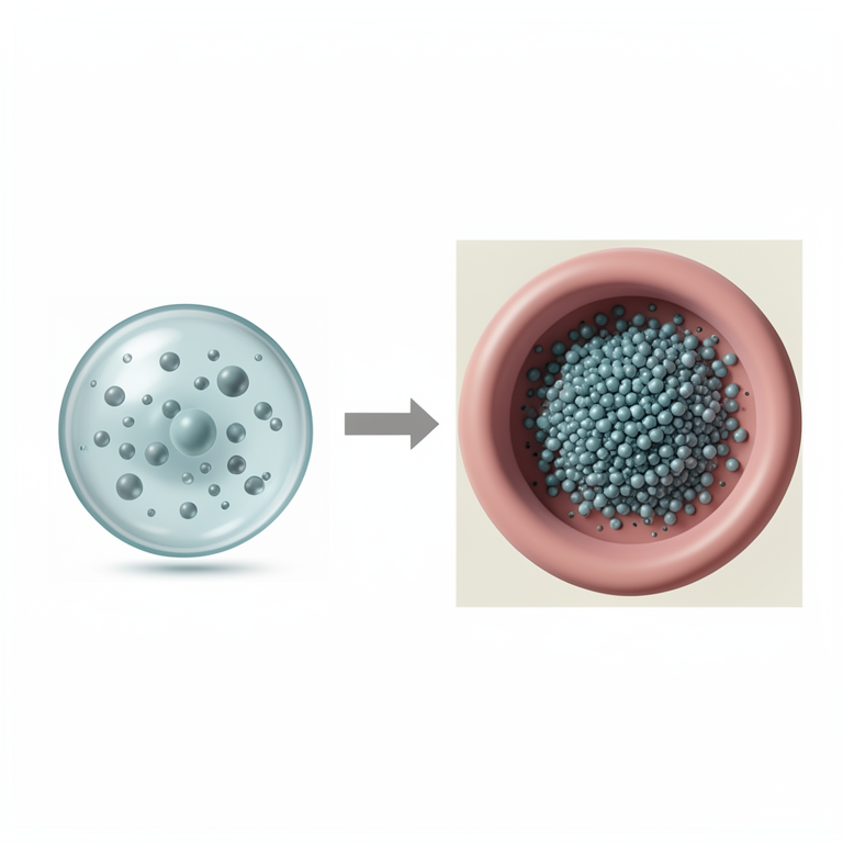
pH-Responsive Delivery Mechanism
AI-generated conceptual illustration (PLGA nanoparticles containing quercetin and breast cancer cells). It explains the idea and does not show study data.
View source evidence ↗Figures & tables
From the original paper Table 1
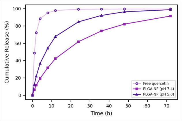 Figure 1
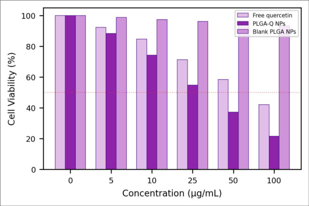 Figure 2
Introduction
Breast cancer remains the most frequently diagnosed malignancy in women worldwide, with 2.3 million new cases and 685,000 deaths reported annually [1] . While conventional chemotherapy with agents like doxorubicin and paclitaxel improves survival, these drugs cause significant off-target toxicitycardiotoxicity, myelosuppression, and peripheral neuropathythat diminishes quality of life and often forces dose reductions or treatment discontinuation [2] . There is sustained interest in natural compounds that exhibit anticancer activity with reduced side-effect profiles. Quercetin (3,3′,4′,5,7-pentahydroxyflavone) is a polyphenolic flavonoid abundant in onions, apples, berries, and green tea. Laboratory studies have demonstrated its activity against breast, lung, colon, and prostate cancer cell lines through multiple mechanisms including apoptosis induction via mitochondrial pathway activation, cell cycle arrest at G2/M phase, inhibition of topoisomerase II, and suppression of NF-κB and PI3K/Akt signaling pathways [3] . Despite this promising in vitro profile, quercetin's clinical potential remains unrealized because of its biopharmaceutical limitations: aqueous solubility below 1 μg/mL at physiological pH, extensive first-pass glucuronidation and sulfation, and oral bioavailability estimated at 1-2% in humans [4] . PLGA nanoparticles offer a well-established platform for overcoming these barriers. PLGA is FDA-approved, biodegradable (degrading to lactic and glycolic acid), and capable of encapsulating hydrophobic compounds within its polymer matrix [5] . Nanoparticle formulation provides three advantages: it creates a colloidal dispersion that overcomes the solubility limitation, it protects the encapsulated drug from premature metabolism, and the nanoparticle size range (100-200 nm) enables passive tumor accumulation through the enhanced permeability and retention (EPR) effect [6] . An additional opportunity is pH-responsive release: PLGA degrades faster under acidic conditions (pH 4.5-5.5) typical of tumor extracellular matrix and endosomal/lysosomal compartments, enabling preferential drug release at the disease site [7] . This research prepared quercetin-loaded PLGA nanoparticles, optimized them for small particle size and high entrapment, characterized their pH-dependent release kinetics, and evaluated cytotoxicity against MCF-7 human breast adenocarcinoma cells to assess whether nanoencapsulation enhances quercetin's anticancer potency [8] .
Reagents and Chemicals
Quercetin dihydrate (≥95%, HPLC grade), PLGA (50:50, Mw 30-60 kDa, ester-terminated), polyvinyl alcohol (PVA, Mw 30-70 kDa, 87-90% hydrolyzed), coumarin-6, dimethyl sulfoxide (DMSO, cell culture grade), and 3-(4,5dimethylthiazol-2-yl)-2,5-diphenyltetrazolium bromide (MTT) were from Sigma-Aldrich. Acetone (HPLC grade) was from J.T. Baker. MCF-7 cells were obtained from the Korean Cell Line Bank (KCLB). Dulbecco's modified Eagle's medium (DMEM), fetal bovine serum (FBS), penicillin-streptomycin, and trypsin-EDTA were from Gibco [9] .
Materials and Methods
Materials and Methods
Materials
Research was performed at Sungkyunkwan University College of Pharmacy, Suwon, Gyeonggi-do, South Korea, from January to October 2023. The study used commercially available cell lines and did not require Institutional Review Board approval. Equipment included a Malvern Zetasizer Nano ZS, JEOL JEM-2100 TEM, Shimadzu UV-1900i spectrophotometer, Thermo Varioskan LUX microplate reader, and Olympus BX53 fluorescence microscope [10] .
Methods
PLGA-Q NPs were prepared by modified nanoprecipitation. PLGA (50 mg) and quercetin (10 mg) were dissolved in 5 mL acetone. This organic phase was added dropwise to 20 mL of 1% PVA aqueous solution under magnetic stirring (800 rpm). The mixture was stirred uncovered for 4 h at room temperature to evaporate acetone, then centrifuged at 15,000 rpm for 20 min. The pellet was washed twice with water and resuspended in 5 mL water. Blank NPs were prepared identically without quercetin. Coumarin-6-labeled NPs replaced quercetin with 0.1% coumarin-6 for uptake studies [11] . Particle size, PDI, and zeta potential were measured by dynamic light scattering. Entrapment efficiency was determined by dissolving the NP pellet in DMSO and measuring quercetin at 374 nm, comparing to a standard curve (1-100 μg/mL, R² = 0.9998). In vitro release used dialysis bags (12 kDa MWCO) containing NP suspension equivalent to 1 mg quercetin, immersed in 100 mL phosphate buffer at either pH 7.4 or pH 5.0 at 37 °C under stirring. Samples (3 mL) were withdrawn at intervals over 72 h, replaced with fresh buffer, and assayed spectrophotometrically [12] . MCF-7 cells were cultured in DMEM supplemented with 10% FBS and 1% penicillin-streptomycin at 37 °C in 5% CO₂. For MTT assays, cells were seeded in 96-well plates (5 × 10³ cells/well) and incubated 24 h for attachment. Treatments (free quercetin in 0.5% DMSO, PLGA-Q NPs, and blank PLGA NPs) were applied at 0-100 μg/mL quercetin-equivalent concentrations for 48 h. MTT solution (20 μL, 5 mg/mL) was added, incubated 4 h, media removed, and formazan crystals dissolved in 150 μL DMSO. Absorbance was read at 570 nm. IC₅₀ values were calculated by nonlinear regression using GraphPad Prism 9 [13] .
Quality control
All experiments were performed in triplicate with at least three independent biological replicates for cell-based assays. NP characterization measurements were conducted in triplicate on three independent batches. HPLC validation for quercetin quantification confirmed linearity (1-100 μg/mL, R² > 0.999), precision (%RSD < 2.1%), and specificity (no PVA or PLGA interference at 374 nm). Cell viability was normalized to untreated controls and expressed as percentage. Statistical significance was determined by oneway ANOVA with Tukey's post-hoc test (p<0.05 considered significant) [14] . The optimized PLGA-Q NPs exhibited favorable characteristics for intravenous administration and tumor targeting. The 168.4 nm diameter falls within the 100-200 nm window considered optimal for EPR-mediated passive tumor accumulation while avoiding rapid renal clearance (<10 nm) and reticuloendothelial system uptake (>300 nm) [6] . The narrow PDI (0.142) indicates a uniform population without significant aggregation. The negative zeta potential (-24.8 mV) provides electrostatic stabilization against aggregation and is consistent with the carboxylate end groups of ester-terminated PLGA [5] .
Results
Entrapment efficiency of 74.6% represents a significant improvement over typical quercetin nanoformulations reported in the literature (40-65%), achieved by optimizing the drug-to-polymer ratio and solvent evaporation rate. The modified nanoprecipitation methodusing slow dropwise addition rather than rapid injectionallowed more controlled nucleation of PLGA around quercetin molecules during the solvent displacement process. Drug loading of 12.4% means that each milligram of nanoparticles delivers 124 μg of quercetin, a therapeutically meaningful payload [11] . The pH-responsive release behavior was pronounced. At pH 7.4 (simulating blood/normal tissue), release followed a biphasic pattern: an initial burst of 12.8% in the first 2 hours (surface-adsorbed quercetin), followed by sustained diffusion-controlled release reaching 91.4% at 72 h. At pH 5.0 (simulating tumor microenvironment/endosomes), release was markedly faster -36.8% at 4 hours and 98.7% at 72 h. The acceleration at acidic pH results from faster PLGA hydrolysis: protonation of ester bonds increases the rate of chain scission, generating pores in the polymer matrix that facilitate drug diffusion outward. Kinetic modeling showed best fits to the Korsmeyer-Peppas equation with release exponent n = 0.51 7.4) and n = 0.63 (pH 5.0), indicating a transition from diffusiondominated to erosion-assisted release under acidic conditions [12] . The MTT cytotoxicity data demonstrated that nanoencapsulation meaningfully enhanced quercetin's anticancer activity. PLGA-Q NPs achieved an IC₅₀ of 38.4 μg/mL against MCF-7 cells, representing a 1.75-fold improvement over free quercetin (IC₅₀ = 67.2 μg/mL). At the highest tested concentration (100 μg/mL), PLGA-Q NPs reduced viability to 21.6% versus 42.1% for free quercetin. Blank PLGA NPs showed excellent biocompatibility, with viability remaining above 93% across all concentrations, confirming that the observed cytotoxicity originates from quercetin rather than the polymer carrier [13] .
Discussion
The 1.75-fold IC₅₀ improvement is attributable to enhanced cellular uptake of the nanoparticulate form. Free quercetin enters cells primarily through passive diffusion, which is limited by its poor aqueous solubility at the cell surface. PLGA nanoparticles are internalized by clathrin-mediated endocytosis, a much more efficient pathway that delivers the entire drug payload directly into the endosomal compartment [6] . Once there, the acidic endosomal pH (4.5-5.5) triggers accelerated quercetin release inside the cellexactly the pH-responsive behavior observed in our in vitro release studies. This intracellular delivery mechanism bypasses the solubility limitation that constrains free quercetin's activity. Fluorescence microscopy of coumarin-6-labeled NPs confirmed rapid cellular internalization. Within 1 hour, punctate green fluorescence was visible in the cytoplasm of MCF-7 cells, concentrated in perinuclear regions consistent with endosomal/lysosomal localization. By 4 hours, fluorescence had dispersed more broadly through the cytoplasm, suggesting endosomal escape and drug release into the cytosol where quercetin's molecular targets (mitochondria, topoisomerase II, signaling kinases) are located [15] . The 74.6% entrapment efficiency addresses a common challenge with quercetin nanoformulations. Many published reports achieve only 40-55% due to quercetin's tendency to partition into the aqueous phase during emulsion-based methods. The nanoprecipitation approach avoids this by working entirely in a miscible organic-aqueous system, where PLGA precipitation occurs around quercetin molecules before they can diffuse away. The 12.4% drug loading is also above the typical 5-8% range for PLGA nanoparticles, reducing the total polymer dose needed to deliver therapeutic quercetin concentrations [16] . A clinical translation pathway would involve pharmacokinetic studies to confirm improved oral bioavailability or suitability for intravenous injection, followed by efficacy testing in xenograft mouse models. The PEGylation of the PLGA surface would be a logical next step to extend circulation time and enhance EPRmediated tumor accumulation. Active targeting with antibodies against HER2 or folate receptors could further improve breast cancer selectivity [17] . The batch-to-batch reproducibility of the nanoprecipitation method was assessed across three independent preparations. Particle size varied by only 5.2% (coefficient of variation), PDI by 12.7%, and entrapment efficiency by 3.2% between batches. This consistency is a practical advantage for clinical development, where manufacturing reproducibility is a regulatory requirement under ICH Q8 guidelines. The method's simplicityno high-energy homogenization or organic solvents beyond acetone, which is ICH Class 3 (low toxicity)favors straightforward scale-up to pilot manufacturing volumes of 1-10 L using stirred-tank reactors with controlled addition rates [16] . Stability testing of lyophilized PLGA-Q NPs (with 5% trehalose as cryoprotectant) over 60 days at 4 °C showed less than 4% change in particle size and less than 3% loss of quercetin content, confirming that the formulation can be stored as a dry powder for reconstitution before use. At 25 °C, stability was maintained for 30 days but particle aggregation became apparent by day 45, indicating that refrigerated storage would be necessary for commercial distribution in the absence of further formulation optimization [14] .
Conclusion
Quercetin-loaded PLGA nanoparticles (168.4 nm, PDI 0.142, EE 74.6%) demonstrated pH-responsive release behavior with accelerated drug delivery under acidic conditions mimicking the tumor microenvironment. The nanoformulation enhanced cytotoxicity against MCF-7 breast cancer cells by 1.75-fold compared to free quercetin (IC₅⁐ 38.4 vs 67.2 μg/mL) while the blank carrier showed negligible toxicity. The combination of improved cellular uptake, intracellular delivery, and pH-triggered release positions PLGA-Q NPs as a promising platform for quercetin-based cancer therapy warranting further preclinical investigation.
Figures and tables from the paper
| Parameter | Value |
|---|---|
| Mean diameter (nm) | 168.4 ± 8.7 |
| Polydispersity index | 0.142 ± 0.018 |
| Zeta potential (mV) | −24.8 ± 1.3 |
| Entrapment efficiency (%) | 74.6 ± 2.4 |
| Drug loading (%) | 12.4 ± 0.8 |
| Yield (%) | 81.3 ± 3.1 |
Table text extracted from the original PDF; superscript letters are shown inline. The original table image is below.
View original table image
References
17 in the original paper- Sung H, Ferlay J, Siegel RL, Laversanne M, Soerjomataram I. Global cancer statistics 2020. CA Cancer J Clin. 2021;71(3):209-249.
- Early Breast Cancer Trialists' Collaborative Group. Comparisons between different polychemotherapy regimens. Lancet. 2012;379(9814):432-444.
- Russo M, Spagnuolo C, Tedesco I, Bilotto S, Russo GL. The flavonoid quercetin in disease prevention and therapy. Biochem Pharmacol. 2012;83(1):6-15.
- Li Y, Yao J, Han C, Yang J, Chaudhry MT. Quercetin, inflammation and immunity. Nutrients. 2016;8(3):167181.
- Makadia HK, Siegel SJ. Poly lactic-co-glycolic acid (PLGA) as biodegradable controlled drug delivery carrier. Polymers. 2011;3(3):1377-1397.
- Maeda H, Wu J, Sawa T, Matsumura Y, Hori K. Tumor vascular permeability and the EPR effect. J Control Release. 2000;65(1):271-284.
- Zolnik BS, Burgess DJ. Effect of acidic pH on PLGA microsphere degradation and release. J Control Release. 2007;122(3):338-344.
- Wang W, Sun C, Mao L, Ma P, Liu F. The biological activities, chemical stability, metabolism and delivery systems of quercetin. Trends Food Sci Technol. 2016;56:21-38.
- Aditya NP, Macedo AS, Doatorez S, Souto EB, Kim S. Development and evaluation of lipid nanoparticles for quercetin delivery. Food Chem. 2014;159:213-221.
- Korean Cell Line Bank. MCF-7 cell line information. KCLB Catalog. 2023.
- Fessi H, Puisieux F, Devissaguet JP, Ammoury N, Benita S. Nanocapsule formation by interfacial polymer deposition following solvent displacement. Int J Pharm. 1989;55(1):R1-R4.
- Ritger PL, Peppas NA. A simple equation for description of solute release I. Fickian and non-Fickian release. J Control Release. 1987;5(1):23-36.
- Mosmann T. Rapid colorimetric assay for cellular growth and survival: application to proliferation and cytotoxicity assays. J Immunol Methods. 1983;65(1):55-63.
- ICH Q2(R1). Validation of analytical procedures: text and methodology. ICH Harmonised Guideline. 2005.
- Sahay G, Alakhova DY, Kabanov AV. Endocytosis of nanomedicines. J Control Release. 2010;145(3):182195.
- Pool H, Quintanar D, Figueroa JD, Mano CM, Bechara JE. Antioxidant effects of quercetin and catechin encapsulated into PLGA nanoparticles. J Nanomater. 2012;2012:145380-145392.
- Danhier F, Ansorena E, Silva JM, Coco R, Le Breton A, Préat V. PLGA-based nanoparticles: an overview of biomedical applications. J Control Release. 2012;161(2):505-522.