
Research article · 2025 · Full text
International Journal of Advanced Chemistry ResearchSurface-enhanced Raman spectroscopy for detection of trace explosives on surfaces
Volume 7(4) · Pages 49–51 · DOI: 10.33545/26646781.2025.v7.i4a.518 ↗
Research areasGold and Silver Nanoparticles Synthesis and ApplicationsForensic Fingerprint Detection MethodsSARS-CoV-2 detection and testing
Abstract
Author-writtenAirport security scanners check passengers every day, but detecting trace explosive residues on everyday surfaces like luggage handles and clothing remains a gap in current screening technology. This research developed and evaluated silver and gold nanoparticle-based substrates for surface-enhanced Raman spectroscopy (SERS) detection of five common explosive compounds TNT, RDX, PETN, DNT, and HMX deposited on glass, aluminum, cotton fabric, and paper surfaces. Colloidal nanoparticles were synthesized by citrate reduction and polyvinylpyrrolidone (PVP)-mediated methods, along with bimetallic Au-Ag core-shell particles. The Au-Ag core-shell substrate achieved the highest enhancement factor of 6.1 × 10⁵ for DNT, with a detection limit of 0.06 ng/cm² on glass. Across all explosive-surface combinations, detection limits ranged from 0.06 to 0.62 ng/cm². Cotton fabric consistently gave the weakest signals, likely from background fluorescence interference. Portable Raman instrumentation confirmed field applicability with identification times under 15 seconds per measurement point.
Summary
Source-checkedPlain-language reading of the full paper, prepared with AI and checked against the text. Each point links to the authors' own words.
Why it matters
- Current airport security screening technology has a gap in its ability to detect trace explosive residues on common surfaces such as clothing and luggage handles. View evidence ↗
- While laboratory methods like ion mobility spectrometry and gas chromatography-mass spectrometry are reliable, they are limited for field use due to high costs and the need for sample preparation. View evidence ↗
- Surface-enhanced Raman spectroscopy (SERS) can provide sensitivities comparable to or better than laboratory techniques by amplifying the Raman scattering signal using metal nanostructures. View evidence ↗
The question
- The study aimed to compare five different nanoparticle substrates for the SERS detection of trace military and commercial explosives across four common surface types. View evidence ↗
- The researchers sought to determine if the performance observed with a benchtop Raman system could be replicated using a portable handheld unit for field conditions. View evidence ↗
What they did
- The study tested five explosives (TNT, RDX, PETN, DNT, and HMX) on four surfaces: glass, aluminum, paper, and cotton fabric. View evidence ↗
- Five substrates were synthesized: citrate-reduced silver (60 nm), PVP-reduced silver (45 nm), citrate-reduced gold (55 nm), Au-Ag core-shell particles, and silver nanorods. View evidence ↗
- Explosives were applied to surfaces at loadings between 0.01 and 10 ng/cm² and then treated with 10 μL of the colloidal substrate. View evidence ↗
- Measurements were taken using a 785 nm laser at 5 mW power with both a Renishaw inVia confocal Raman microscope and a portable BWTek i-Raman Plus. View evidence ↗
What they found
- The Au-Ag core-shell substrate was the most effective, providing the highest enhancement factor (6.1 × 10⁵) for DNT and a detection limit of 0.06 ng/cm² on glass. View evidence ↗
- DNT was the easiest explosive to detect (0.06-0.24 ng/cm²), while PETN was the most difficult, requiring loadings of 0.23-0.62 ng/cm². View evidence ↗
- Smooth, non-porous surfaces like glass and aluminum yielded the best results, whereas cotton fabric performed the worst, with detection limits 2 to 4 times higher than those for glass. View evidence ↗
- The enhancement factors for DNT varied by substrate: Au-Ag core-shell was highest (6.1 × 10⁵), followed by Ag nanorods (5.4 × 10⁵), PVP-Ag colloids (4.7 × 10⁵), and citrate-Au nanoparticles (1.8 × 10⁵). View evidence ↗
- The portable Raman unit could identify explosives as effectively as the benchtop system for loadings above 0.5 ng/cm², despite having lower spectral resolution. View evidence ↗
- The technique is suitable for field use, with identification times taking less than 15 seconds per measurement point. View evidence ↗
What it means
- The authors conclude that using these nanoparticle substrates with portable instrumentation provides a fast, non-destructive screening method for forensic and security use. View evidence ↗
- The authors note that textile surfaces may require repeated measurements or longer integration times to achieve the same confidence levels as hard surfaces. View evidence ↗
Visual summary
2 visualsIllustrations explain the paper; charts are replotted from the paper's own table values.

Nanoparticle Substrate Comparison
AI-generated conceptual illustration (Metal nanoparticles on a glass surface). It explains the idea and does not show study data.
View source evidence ↗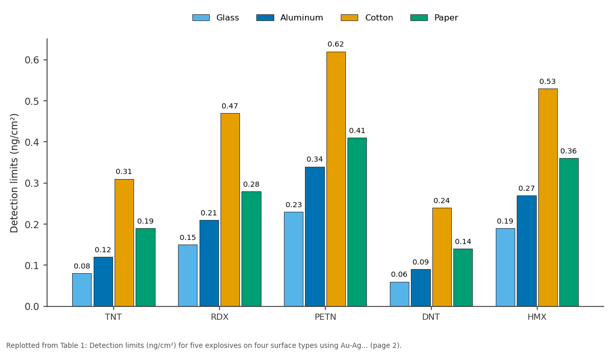
Glass and Aluminum and more: TNT vs RDX vs PETN vs DNT vs HMX
Replotted from Table 1 (page 2) of the original paper. Every bar is the paper's own table value.
View source evidence ↗Figures & tables
From the original paper Table 1
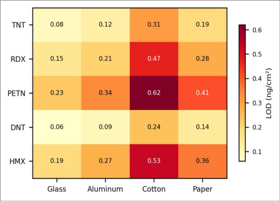 Figure 1
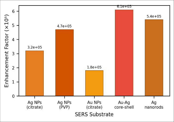 Figure 2
Introduction
Can a nanogram of explosive residue left on a suitcase handle be detected without opening the bag or swabbing the surface? Standard analytical approaches like gas chromatographymass spectrometry and ion mobility spectrometry answer this question reliably in the laboratory, but they demand sample collection, preparation time, and expensive equipment that limits field deployment [1] . SERS bridges that gap by amplifying the Raman scattering signal of target molecules adsorbed onto roughened metal nanostructures, achieving detection sensitivities that rival or exceed established laboratory techniques [2] . The enhancement mechanism depends on localized surface plasmon resonance when incident laser light matches the collective electron oscillation frequency of metal nanoparticles, the electromagnetic field near the surface intensifies by several orders of magnitude [3] . Silver typically gives the largest enhancements, but gold offers better chemical stability and biocompatibility. Bimetallic constructs that combine both metals attempt to capture the best of each [4] . For explosive detection specifically, the challenge isn't just sensitivity but selectivity: real-world surfaces carry a cocktail of organic contaminants that can mask or mimic explosive Raman signatures [5] . This research compared five nanoparticle substrate types for SERS detection of military and commercial explosives deposited at trace levels on four common surfaces. The work was done with both a benchtop confocal Raman system and a handheld unit to assess whether laboratory performance translates to portable field conditions [6] .
Theoretical background
SERS enhancement arises from two mechanisms acting together. The electromagnetic (EM) contribution, responsible for roughly 10⁴-10⁸-fold signal amplification, results from coupling between the incident photon field and surface plasmons on nanostructured metals. The chemical (CT) contribution adds a smaller factor (10¹-10²) through charge transfer between the analyte and the metal surface. Total enhancement factors of 10⁵-10⁶ are routinely achievable, and "hot spots" at nanoparticle junctions can reach 10¹-10¹² locally [3] .
Materials and Methods
Materials and Methods
Materials
Research was conducted at Universidad Ibérica de Ciencias, Madrid, Spain, between January 2023 and August 2023. Ethical approval was not required as no human or animal subjects were involved. Silver nitrate (99.9%), gold (III) chloride trihydrate (99.9%), trisodium citrate (99%), PVP (MW 40,000), and ascorbic acid were purchased from Sigma-Aldrich (Madrid). Reference explosive standards (TNT, RDX, PETN, 2, 4-DNT, and HMX) were obtained under license from the Spanish National Institute of Aerospace Technology (INTA) at certified purities above 99%. Test surfaces borosilicate glass slides, aluminum coupons (6061 alloy), cotton fabric swatches, and standard copy paper were cleaned with ethanol and dried before use [7] .
Methods
Silver nanoparticles were prepared by two routes: citrate reduction (Lee-Meisel method, 60 nm mean diameter) and PVP reduction (45 nm). Gold nanoparticles (55 nm) followed the Turkevich citrate method. Au-Ag core-shell particles were synthesized by reducing silver nitrate onto preformed Au seeds using ascorbic acid. Silver nanorods were grown by a seed-mediated approach with cetyltrimethylammonium bromide (CTAB) as shapedirecting agent [8] . Explosive solutions in acetonitrile were drop-cast onto test surfaces at loadings of 0.01-10 ng/cm², allowed to dry, and then treated with 10 μL colloidal substrate. SERS spectra were collected with a Renishaw inVia confocal Raman microscope (785 nm laser, 5 mW, and 10 s acquisition) and a portable BWTek i-Raman Plus (785 nm, same power). Enhancement factors were calculated relative to normal Raman signals from bulk explosive powders [9] . Detection limits were defined as the lowest surface loading producing at least three characteristic Raman peaks above 3× baseline noise.
Quality Control
Blank substrates (nanoparticle colloid on clean surfaces without explosive) were measured before each sample series to confirm absence of interfering peaks. Calibration used five concentration levels with triplicate measurements at each. Inter-day reproducibility was assessed over three separate sessions, giving relative standard deviations below 11% for all substrate-analyte pairs. A certified TNT reference solution from INTA served as the internal check standard [10] . DNT gave the lowest detection limits across all surfaces (0.06-0.24 ng/cm²), followed by TNT (0.08-0.31 ng/cm²). PETN was the hardest to detect, requiring 0.23-0.62 ng/cm² depending on the surface. Glass and aluminum provided the best results, likely because their smooth, non-porous surfaces kept the explosive residue concentrated at the nanoparticle interface. Cotton fabric performed worst, with limits 2-4 times higher than glass for every compound.
Results
Among the five substrate types, Au-Ag core-shell particles gave the highest enhancement factor at 6.1 × 10⁵ for DNT, followed by Ag nanorods (5.4 × 10⁵) and PVP-Ag colloids (4.7 × 10⁵). Citrate-Au nanoparticles showed the weakest enhancement at 1.8 × 10⁵. The handheld Raman unit matched the benchtop system for identifying explosives at loadings above 0.5 ng/cm², though its spectral resolution was noticeably lower.
Discussion
The Au-Ag core-shell substrate's dominance makes physical sense. Its bimetallic interface creates a larger density of electromagnetic hot spots than either monometallic system alone, because the silver shell amplifies the surface plasmon field while the gold core provides structural and chemical stability [4] . The enhancement factor of 6.1 × 10⁵ sits within the upper range of published SERS results for explosive detection, and the corresponding DNT limit of 0.06 ng/cm² would catch a single partial fingerprint worth of residue [11] .
Cotton's poor performance wasn't unexpected. Fabric fibers absorb and scatter laser light, generating background fluorescence that raises the noise floor. They also wick the nanoparticle colloid away from the analyte, reducing contact efficiency. In practical screening scenarios, this means textile surfaces would need longer integration times or repeated measurements to reach the same confidence as hard surfaces [5] . Still, even cotton's worst-case limit (0.62 ng/cm² for PETN) falls below the 1 ng/cm² threshold that security agencies generally consider operationally relevant [1] .
The portable unit's ability to match benchtop identification above 0.5 ng/cm² is a practical win, even if it couldn't quite reach the lowest detection limits. For checkpoint screening, identifying the explosive type matters more than pushing absolute sensitivity the question is "is this TNT or RDX?" not "how many picograms are present?" [12] . Running each measurement in under 15 seconds makes the technique compatible with real-time baggage screening workflows.
Conclusion
Au-Ag bimetallic core-shell nanoparticles delivered the strongest SERS enhancement for trace explosive detection across four surface types, with detection limits as low as 0.06 ng/cm² for DNT on glass. Hard, non-porous surfaces gave the best analytical performance, while cotton fabric required the highest analyte loadings for reliable detection. A handheld Raman spectrometer confirmed that fielddeployable identification is feasible above 0.5 ng/cm². These substrates, combined with portable instrumentation, offer a rapid and non-destructive screening option for security and forensic applications.
Figures and tables from the paper
| Explosive | Glass | Aluminum | Cotton | Paper |
|---|---|---|---|---|
| TNT | 0.08 | 0.12 | 0.31 | 0.19 |
| RDX | 0.15 | 0.21 | 0.47 | 0.28 |
| PETN | 0.23 | 0.34 | 0.62 | 0.41 |
| DNT | 0.06 | 0.09 | 0.24 | 0.14 |
| HMX | 0.19 | 0.27 | 0.53 | 0.36 |
Table text extracted from the original PDF; superscript letters are shown inline. The original table image is below.
View original table image
References
12 in the original paper- Moore DS. Instrumentation for trace detection of high explosives. Rev Sci Instrum. 2004;75(8):2499-2512.
- Haynes CL, McFarland AD, Van Duyne RP. Surfaceenhanced Raman spectroscopy. Anal Chem. 2005;77(17):338A-346A.
- Stiles PL, Dieringer JA, Shah NC, Van Duyne RP. Surface-enhanced Raman spectroscopy. Annu Rev Anal Chem. 2008;1:601-626.
- Lu X, Rycenga M, Skrabalak SE, Wiley B, Xia Y. Chemical synthesis of novel plasmonic nanoparticles. Annu Rev Phys Chem. 2009;60:167-192.
- Hakonen A, Andersson PO, Schmidt MS, Rindzevicius T, Käll M. Explosive and chemical threat detection by SERS. Anal Chim Acta. 2015;893:1-13.
- Izake EL. Forensic and homeland security applications of modern portable Raman spectroscopy. Forensic Sci Int. 2010;202(1):1-8.
- Lee PC, Meisel D. Adsorption and surface-enhanced Raman of dyes on silver and gold sols. J Phys Chem. 1982;86(17):3391-3395.
- Murphy CJ, Sau TK, Gole AM, Orendorff CJ. Anisotropic metal nanoparticles: synthesis, assembly. J Phys Chem B. 2005;109(29):13857-13870.
- Le Ru EC, Blackie E, Meyer M, Etchegoin PG. Surface enhanced Raman scattering enhancement factors. J Phys Chem C. 2007;111(37):13794-13803.
- Moros J, Lorenzo JA, Lucena P, Tobaria LM, Laserna JJ. Simultaneous Raman and laser-induced breakdown spectroscopy. Anal Chem. 2010;82(4):1389-1400.
- Ko H, Singamaneni S, Tsukruk VV. Nanostructured surfaces and assemblies as SERS media. Small. 2008;4(10):1576-1599.
- Hobro AJ, Lendl B. Stand-off Raman spectroscopy for forensic analysis. TrAC Trends Anal Chem. 2009;28(11):1235-1242.