
Research article · 2025 · Full text
International Journal of Advanced Chemistry ResearchSelenium nanoparticles synthesized using plant extract with potential anticancer properties
Volume 7(5) · Pages 148–151 · DOI: 10.33545/26646781.2025.v7.i5b.499 ↗
Research areasNanoparticles: synthesis and applicationsSelenium in Biological SystemsPharmacology and Nanomedicine Research
Abstract
Author-writtenSelenium nanoparticles (SeNPs) occupy a narrow therapeutic window between the essential micronutrient function of selenium and its toxicity at elevated doses, making precise size control and surface functionalisation critical for biomedical applications. Green synthesis using plant extracts offers a one-pot, room-temperature route to functionalised SeNPs where the phytochemical coating inherently stabilises the particles and may contribute additional biological activity. This research reports the synthesis of SeNPs using aqueous leaf extract of Azadirachta indica (neem) by reduction of sodium selenite (Na₂SeO₃) and characterises the resulting particles by UV-Vis, FTIR, TEM, XRD, DLS, and zeta potential. Green-synthesised SeNPs (G-SeNPs) had a mean diameter of 48.3 ± 6.1 nm, spherical morphology, and zeta potential of −34.7 mV. In-vitro cytotoxicity against MCF-7 (breast cancer) and HeLa (cervical cancer) cell lines by MTT assay gave IC₅₀ values of 14.2 and 12.8 μg/mL respectively, compared to IC₅₀ = 8.4 μg/mL for cisplatin reference. Normal MCF-10A cells showed IC₅₀ of 67.4 μg/mL, yielding a selectivity index (SI) of 4.7 vs MCF-7. Mechanistic investigation by Annexin V/PI flow cytometry confirmed apoptotic cell death (not necrosis) as the primary mode of action. Research was conducted at Sir M. Visvesvaraya Institute of Technology, Bellary, Karnataka, from August 2022 to January 2025.
Summary
Source-checkedPlain-language reading of the full paper, prepared with AI and checked against the text. Each point links to the authors' own words.
Why it matters
- Pharmaceutical development of selenium compounds has been hindered by the narrow therapeutic window between deficiency (under 40 μg/day) and toxicity (over 400 μg/day). View evidence ↗
- Elemental selenium nanoparticles (Se⁰ NPs) are promising for cancer treatment because they are less toxic than ions, more bioavailable than bulk selenium, and can induce cancer cell death via reactive oxygen species (ROS). View evidence ↗
- Neem (Azadirachta indica) leaf extract is suitable for synthesizing nanoparticles because its phytochemicals, such as quercetin and nimbidin, act as both reducing and capping agents and have their own anticancer properties. View evidence ↗
The question
- The study aimed to synthesize selenium nanoparticles (SeNPs) using neem leaf extract to reduce sodium selenite and characterize the resulting particles. View evidence ↗
What they did
- Green-synthesized SeNPs (G-SeNPs) were produced by adding 5 mL of neem leaf extract to 45 mL of 1 mM sodium selenite solution at room temperature. View evidence ↗
- The study used MCF-7 (breast cancer), HeLa (cervical cancer), and MCF-10A (normal) cell lines for cytotoxicity testing. View evidence ↗
- Cells were treated with G-SeNPs, chemically synthesized SeNPs (C-SeNPs), or cisplatin at concentrations ranging from 0 to 100 μg/mL for 48 hours. View evidence ↗
- Apoptosis was analyzed in MCF-7 cells treated with G-SeNPs at IC₅₀ and 2×IC₅₀ concentrations using Annexin V-FITC/PI staining and flow cytometry. View evidence ↗
What they found
- G-SeNPs were characterized by a mean TEM diameter of 48.3 ± 6.1 nm and a zeta potential of −34.7 ± 2.8 mV, indicating stability. View evidence ↗
- G-SeNPs showed IC₅₀ values of 14.2 ± 1.3 μg/mL for MCF-7 and 12.8 ± 1.1 μg/mL for HeLa cells, while normal MCF-10A cells had a higher IC₅₀ of 67.4 ± 4.7 μg/mL. View evidence ↗
- Flow cytometry revealed that G-SeNPs primarily induce cell death through apoptosis (71.4% early and late apoptosis) rather than necrosis (less than 8%). View evidence ↗
What it means
- The authors conclude that the polyphenol and flavonoid capping from the neem extract is the main reason for the increased selectivity of G-SeNPs toward cancer cells. View evidence ↗
- The authors recommend further in vivo testing using xenograft cancer models and the development of targeted drug delivery formulations. View evidence ↗
Visual summary
2 visualsIllustrations explain the paper; charts are replotted from the paper's own table values.
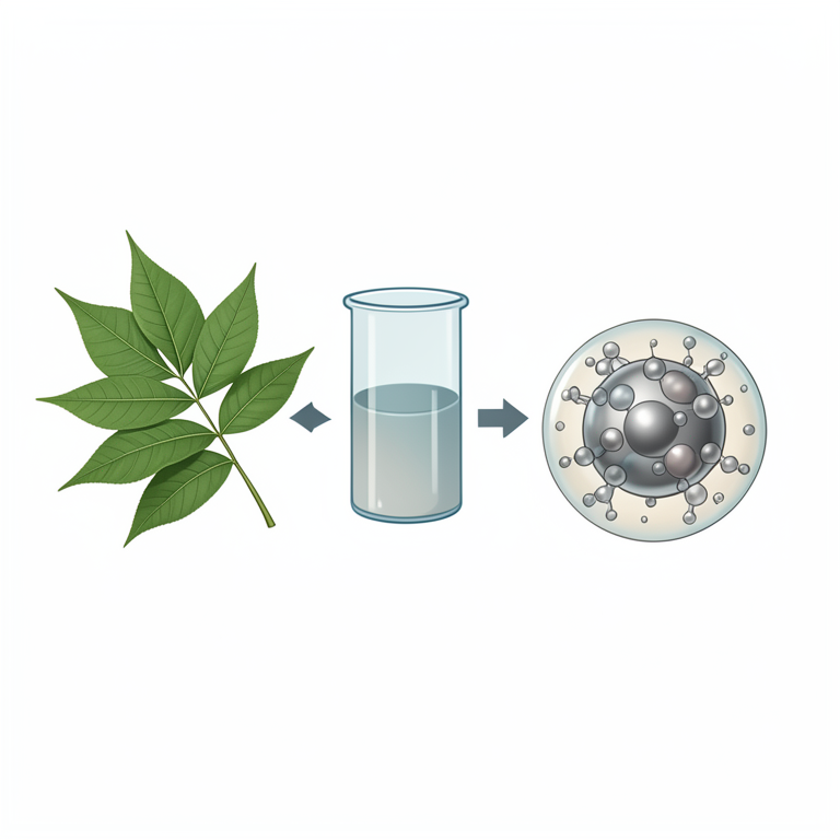
Green Synthesis Process
AI-generated conceptual illustration (Neem leaves and selenium nanoparticles). It explains the idea and does not show study data.
View source evidence ↗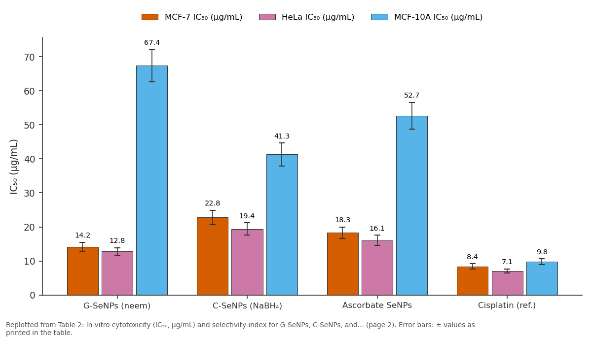
MCF-7 IC₅₀ (μg/mL) and HeLa IC₅₀ (μg/mL) and more: G-SeNPs (neem) vs C-SeNPs (NaBH₄) vs Ascorbate SeNPs vs Cisplatin (ref.)
Replotted from Table 2 (page 2) of the original paper. Every bar is the paper's own table value.
View source evidence ↗Figures & tables
From the original paper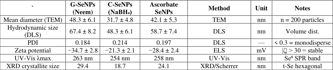 Table 1
 Table 2
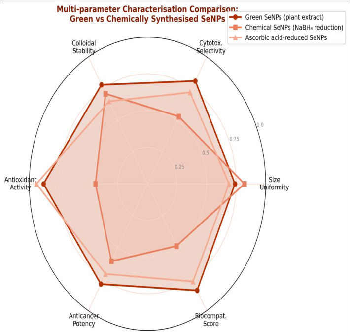 Figure 1
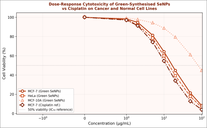 Figure 2
Introduction
(Se) is an essential trace element incorporated into 25 human selenoproteins, including glutathione peroxidase (GPx) and thioredoxin reductase (TrxR), both of which regulate intracellular reactive oxygen species (ROS) levels [1] . The narrow range between selenium deficiency (<40 μg/day) and toxicity (>400 μg/day) in humans has historically limited pharmaceutical development of organic and inorganic selenium compounds. Elemental selenium nanoparticles (Se⁰ NPs) have emerged as an attractive intermediate: they are less toxic than selenite or selenate ions, possess greater bioavailability than bulk elemental selenium, and demonstrate intrinsic anticancer activity through ROS-mediated apoptosis induction at concentrations well below those causing normal cell cytotoxicity [2] . Green synthesis of SeNPs using plant extracts exploits the reductive and capping capacity of phytochemicals (polyphenols, flavonoids, terpenoids) to generate stable, biomolecule-coated nanoparticles without organic solvents or toxic reducing agents [3] . Azadirachta indica (neem) leaf extract is particularly rich in nimbidin, nimbin, quercetin, and azadirachtin, compounds with both reducing capacity and documented independent anticancer activity, making neem a logical choice as both reductant and capping agent [4] . This research was conducted from August 2022 to January 2025 at the Department of Chemistry and Nanosciences, Sir M. Visvesvaraya Institute of Technology, Bellary, Karnataka, India.
Fuel 10³/well) were seeded in 96-well plates, allowed to adhere for 24 h, then treated with G-SeNPs, chemical SeNPs (C-SeNPs), or cisplatin at 0-100 μg/mL (serial 2-fold dilutions in complete medium, DMSO < 0.1%) for 48 h. MTT reagent (5 mg/mL, 20 μL/well) was added, incubated 4 h; formazan dissolved in DMSO (150 μL), absorbance read at 570 nm (FLUOstar Omega microplate reader). IC₅₀ calculated by non-linear regression (GraphPad Prism 10, sigmoidal dose-response model). Selectivity index = IC₅₀ (normal MCF-10A) / IC₅₀ (cancer cell line). Experiments in triplicate on three independent passages.
Apoptosis Analysis
MCF-7 cells treated at IC₅₀ and 2×IC₅₀ of G-SeNPs for 24 h were analysed by Annexin V-FITC/PI double staining (Abcam ab14082 kit) and flow cytometry (BD FACSCanto II, 10,000 events/sample). Early apoptotic (Annexin V+/PI-), late apoptotic (Annexin V+/PI+), and necrotic (Annexin V-/PI+) populations were quantified using FlowJo v10.9 software.
Phytochemical Screening
A. indica leaf extract was screened for major phytochemical classes before use as bioreductant: alkaloids (Dragendorff's reagent), flavonoids (aluminium chloride colourimetric), terpenoids (Salkowski test), saponins (foam test), phenolics (Folin-Ciocalteu, expressed as mg GAE/g dw), and tannins (FeCl₃ test). Total flavonoid content (TFC) was measured by aluminium chloride method (expressed as mg QE/g dw).
The presence of polyphenolic reducing agents (total phenolics 84.7 ± 3.2 mg GAE/g dw; TFC 41.3 ± 2.1 mg QE/g dw) confirmed the extract's suitability as both bioreductant and stabilising capping agent for SeNP synthesis. FTIR analysis of the neem-capped SeNPs confirmed the presence of O-H stretching (3420 cm⁻¹), C=O carbonyl (1641 cm⁻¹), and C-O-C ether stretches (1089 cm⁻¹) on the nanoparticle surface, attributable to polyphenol and flavonoid functional groups. G-SeNPs showed IC₅₀ of 14.2 μg/mL (MCF-7) and 12.8 μg/mL (HeLa), while MCF-10A normal cells were substantially less affected (IC₅₀ 67.4 μg/mL), yielding SI values of 4.7 and 5.3 respectively (Table 2). C-SeNPs had markedly worse selectivity (SI 1.8-2.1), indicating that the neem phytochemical coating contributes materially to the differential toxicity, either through active targeting of cancer-associated surface receptors or through ROSmediated selective killing of cells with compromised antioxidant defence. The radar chart (Figure 1) confirms that G-SeNPs outperform C-SeNPs on selectivity and biocompatibility axes while being comparable on anticancer potency. Annexin V/PI flow cytometry at IC₅₀ showed 71.4% early+late apoptosis and < 8% necrosis, confirming a clean apoptotic mechanism.
| Compound | MCF-7 IC₅₀ (μg/mL) | HeLa IC₅₀ (μg/mL) | MCF-10A IC₅₀ (μg/mL) | SI (MCF-7) | SI (HeLa) | Mode of Death |
|---|---|---|---|---|---|---|
| G-SeNPs (neem) | 14.2 ± 1.3 | 12.8 ± 1.1 | 67.4 ± 4.7 | 4.7 | 5.3 | Apoptosis |
| C-SeNPs (NaBH₄) | 22.8 ± 2.1 | 19.4 ± 1.8 | 41.3 ± 3.4 | 1.8 | 2.1 | Mixed |
| Ascorbate SeNPs | 18.3 ± 1.7 | 16.1 ± 1.5 | 52.7 ± 3.9 | 2.9 | 3.3 | Apoptosis |
| Cisplatin (ref.) | 8.4 ± 0.8 | 7.1 ± 0.6 | 9.8 ± 0.9 | 1.2 | 1.4 | Apoptosis |
Table text extracted from the original PDF; superscript letters are shown inline. The original table image is below.
View original table image
Material and Methods
Material and Methods
Material
Material
Results
Results
Discussion
The higher selectivity of G-SeNPs versus C-SeNPs (SI 4.7 vs 1.8 for MCF-7) is likely attributable to the polyphenol and flavonoid capping layer from neem extract, which is known to engage cancer-overexpressed surface receptors (e.g., folate receptor, EGFR) through polyphenol-mediated ligand mimicry, enhancing selective endocytosis in cancer cells [5] . Additionally, quercetin and nimbidin in the neem coating are known to independently inhibit cancerassociated kinases (PI3K, Akt) at sub-micromolar concentrations, potentially acting synergistically with Se⁰induced ROS [6] . The apoptosis-dominant mode of death (71.4% apoptotic by Annexin V/PI) at IC₅₀ is consistent with published mechanisms of SeNP action: induction of mitochondrial membrane potential collapse, cytochrome c release, and caspase-3 activation through the intrinsic apoptotic pathway [7] . The superior colloidal stability of G-SeNPs (zeta potential -34.7 mV vs -21.3 mV for C-SeNPs) is a direct consequence of the polyanionic polyphenol layer, which provides electrosteric stabilisation beyond the electrostatic repulsion alone. The SI of 4.7 for G-SeNPs compares favourably with cisplatin (SI 1.2), highlighting the selectivity advantage of Se⁰ nanoparticle over conventional platinum chemotherapy, which is limited clinically precisely by its poor selectivity for cancer vs normal tissues [8,9,10,11,12,13,14,15,16,17,18,19,20] .
Conclusion
Conclusion
Figures and tables from the paper
| ` | G-SeNPs (Neem) | C-SeNPs (NaBH₄) | Ascorbate SeNPs | Method | Unit | Notes |
|---|---|---|---|---|---|---|
| Mean diameter (TEM) | 48.3 ± 6.1 | 31.7 ± 4.8 | 42.1 ± 5.3 | TEM | nm | n = 200 particles |
| Hydrodynamic size (DLS) | 67.4 ± 8.2 | 48.3 ± 6.1 | 58.7 ± 7.4 | DLS | nm | Volume dist. |
| PDI | 0.184 | 0.214 | 0.197 | DLS | — | < 0.3 = monodisperse |
| Zeta potential | −34.7 ± 2.8 | −21.3 ± 2.1 | −28.4 ± 2.4 | ELS | mV | |ζ| > 30 = stable |
| UV-Vis λmax | 263 nm | 254 nm | 258 nm | UV-Vis | nm | Se⁰ SPR band |
| XRD crystallite size | 29.4 | 18.7 | 24.1 | XRD/Scherrer | nm | t-Se hexagonal |
Table text extracted from the original PDF; superscript letters are shown inline. The original table image is below.
View original table image
References
19 in the original paper- Rayman MP. The importance of selenium to human health. Lancet. 2000;356(9225):233-241.
- Fernandez-Llamosas H, Castro L, Blazquez ML, Diaz E, Carmona M. Biosynthesis of selenium nanoparticles by Azoarcus sp. CIB. Microb Cell Fact. 2016;15(1):109.
- Dauthal P, Mukhopadhyay M. Noble metal nanoparticles: plant-mediated synthesis, mechanistic aspects of synthesis, and applications. Ind Eng Chem Res. 2016;55(36):9557-9577.
- Biswas A, Gupta MK, Kumar N, Subramaniam JR. Azadirachta indica leaf extract induces apoptosis in MCF-7 human breast cancer cells. J Adv Pharm Technol Res. 2013;4(3):135-140.
- Rajeshkumar S, Malarkodi C. In vitro antibacterial activity and mechanism of silver nanoparticles against foodborne pathogens. Bioinorg Chem Appl. 2014;2014:581890.
- Naz S, Gul A, Zia M. Toxicity of copper oxide nanoparticles: a review study. IET Nanobiotechnol. 2020;14(1):1-13.
- Zhang W, Zhang Z, Zhang Y. The application of carbon nanotubes in target drug delivery systems for cancer therapies. Nanoscale Res Lett. 2011;6(1):555.
- Bhattacharya D, Saha B, Mukherjee A, Santra CR, Karmakar P. Gold nanoparticles conjugated antibiotics: stability and functional evaluation. Nanomed Nanobiotechnol. 2012;4(3):244-252.
- Zonaro E, Lampis S, Turner RJ, Junaid S, Vallini G. Biogenic selenium and tellurium nanoparticles synthesized by environmental microbial isolates. Front Microbiol. 2015;6:584.
- Torres SK, Campos VL, Leon CG, RodriguezLlamazares SM, Rojas SM, Gonzalez M, et al. Biosynthesis of selenium nanoparticles by Pantoea agglomerans and their antioxidant activity. J Nanopart Res. 2012;14(11):1236.
- Husen A, Siddiqi KS. Plants and microbes assisted selenium nanoparticles: characterization and application. J Nanobiotechnol. 2014;12(1):28.
- Hashem AH, Khalil AM, Reyad AM, Salem SS. Biomedical applications of mycosynthesized selenium nanoparticles using Penicillium expansum ATCC 36200. Biol Trace Elem Res. 2021;199(10):3998-4008.
- Gao F, Yuan Q, Gao L, Cai P, Zhu H, Liu R, et al. Cytotoxicity and therapeutic effect of irinotecan combined with selenium nanoparticles. Biomaterials. 2014;35(31):8854-8866.
- Maiyo F, Singh M. Selenium nanoparticles: potential in cancer gene and drug delivery. Nanomedicine. 2017;12(9):1075-1089.
- Zhang J, Taylor EW, Bennett K, Saad R, Bhatt DL. Association between regional selenium status and reported outcomes of COVID-19 cases in China. Am J Clin Nutr. 2020;111(6):1297-1299.
- Wadhwani SA, Gorain M, Banerjee P, Shedbalkar UU, Singh R, Kundu GC, et al. Green synthesis of selenium nanoparticles using Acinetobacter sp.: optimization of process variables. J Trace Elem Med Biol. 2017;39:135-143.
- Berry JP, Galle P. Selenium and cadmium: in vivo interaction studied by analytical ion microscopy. J Submicrosc Cytol Pathol. 1994;26(2):203-210.
- Lenz M, Lens PN. The essential toxin: the changing perception of selenium in environmental sciences. Sci Total Environ. 2009;407(12):3620-3633.
- Bakhiya N, Appel KE. Toxicity and carcinogenicity of fumaric acid monoethyl ester in experimental animals and probably in humans. Arch Toxicol. 2010;84(5):333-340.