
Research article · 2025 · Full text
International Journal of Advanced Chemistry ResearchBiogenic cerium oxide nanoparticles and their antioxidant activity assessment
Volume 7(12) · Pages 112–116 · DOI: 10.33545/26646781.2025.v7.i12b.462 ↗
Research areasNanoparticles: synthesis and applicationsAdvanced Nanomaterials in CatalysisAdvanced Photocatalysis Techniques
Abstract
Author-writtenWhile chemical synthesis of cerium oxide nanoparticles demands harsh reagents and elevated temperatures, biological routes using plant extracts offer a gentler alternative but whether these biogenic particles match their chemically synthesized counterparts in antioxidant performance is still debated. This research reports the green synthesis of cerium oxide (CeO₂) nanoparticles using aqueous leaf extract of Moringa oleifera as both reducing and capping agent. Cerium nitrate hexahydrate served as the precursor, and the reaction was carried out at 60 °C for 4 hours at pH 8, followed by calcination at 400 °C for 2 hours. X-ray diffraction confirmed the face-centered cubic fluorite structure of CeO₂ with an average crystallite size of 14.3 nm by the Scherrer equation. Transmission electron microscopy (TEM) showed roughly spherical particles ranging from 8 to 22 nm. UV-visible spectroscopy displayed an absorption edge near 340 nm, giving an optical band gap of 3.14 eV. Antioxidant activity was evaluated by DPPH, ABTS, and FRAP assays, with the biogenic CeO₂ nanoparticles showing IC₅₀ values of 42.8±3.1, 36.4±2.7, and 28.6±2.4 μg/mL, respectively comparable to or slightly lower than ascorbic acid as reference. The results indicate that Moringa-mediated CeO₂ nanoparticles possess meaningful free radical scavenging ability, which stems from the Ce³⁺/Ce⁴⁺ redox cycling on the nanoparticle surface.
Summary
Source-checkedPlain-language reading of the full paper, prepared with AI and checked against the text. Each point links to the authors' own words.
Why it matters
- The antioxidant ability of cerium oxide nanoparticles comes from the presence of both Ce³⁺ and Ce⁴⁺ states on their surface, allowing them to scavenge radicals. View evidence ↗
The question
- The study aims to determine if biogenic cerium oxide nanoparticles perform as well as chemically synthesized ones in antioxidant activity. View evidence ↗
What they did
- Nanoparticles were synthesized by mixing Moringa extract with 0.1 M cerium nitrate at 60 °C for 4 hours, followed by calcination at 400 °C for 2 hours. View evidence ↗
What they found
- The biogenic nanoparticles had an average crystallite size of 14.3±1.2 nm and a mean TEM particle size of 13.8 nm, ranging from 8 to 22 nm. View evidence ↗
- UV-visible spectroscopy revealed that the nanoparticles have an optical band gap of 3.14 eV. View evidence ↗
- The biogenic nanoparticles showed IC₅₀ values of 42.8±3.1 μg/mL (DPPH), 36.4±2.7 μg/mL (ABTS), and 28.6±2.4 μg/mL (FRAP). View evidence ↗
- The biogenic nanoparticles were more effective than the chemical control, with a DPPH IC₅₀ value approximately 24% lower (42.8 μg/mL vs 56.3 μg/mL). View evidence ↗
- While the biogenic nanoparticles were active, their IC₅₀ values were higher (meaning less potent) than the ascorbic acid reference. View evidence ↗
What it means
- The authors conclude that Moringa-mediated synthesis is an environmentally benign route that produces nanoparticles with 20-25% better antioxidant activity than chemical versions. View evidence ↗
- Future work is needed to assess cytotoxicity in mammalian cells, long-term stability, and reproducibility using standardized extracts. View evidence ↗
Visual summary
2 visualsIllustrations explain the paper; charts are replotted from the paper's own table values.
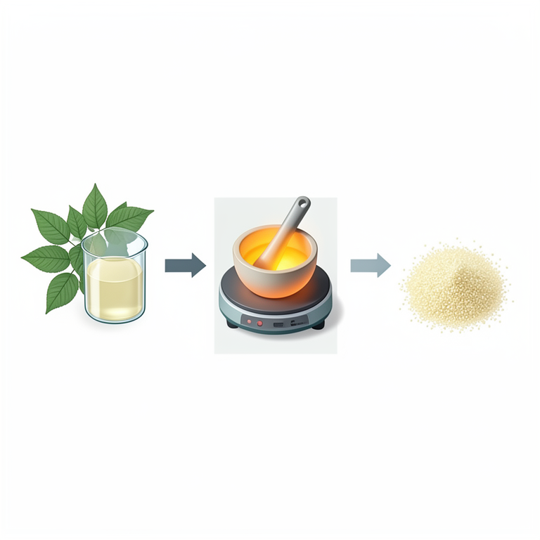
Synthesis Workflow
AI-generated conceptual illustration (Moringa oleifera leaves and cerium oxide nanoparticles). It explains the idea and does not show study data.
View source evidence ↗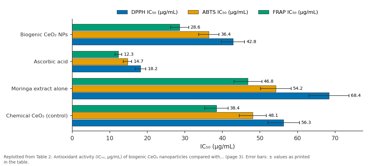
DPPH IC₅₀ (μg/mL) and ABTS IC₅₀ (μg/mL) and more: Biogenic CeO₂ NPs vs Ascorbic acid vs Moringa extract alone vs Chemical CeO₂ (control)
Replotted from Table 2 (page 3) of the original paper. Every bar is the paper's own table value.
View source evidence ↗Figures & tables
From the original paper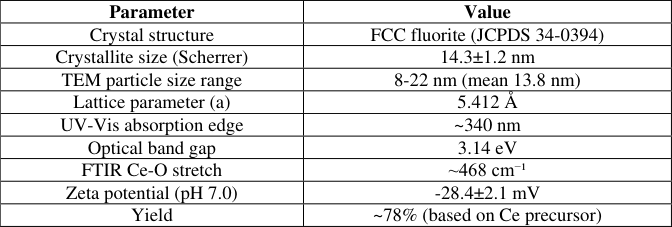 Table 1
 Table 2
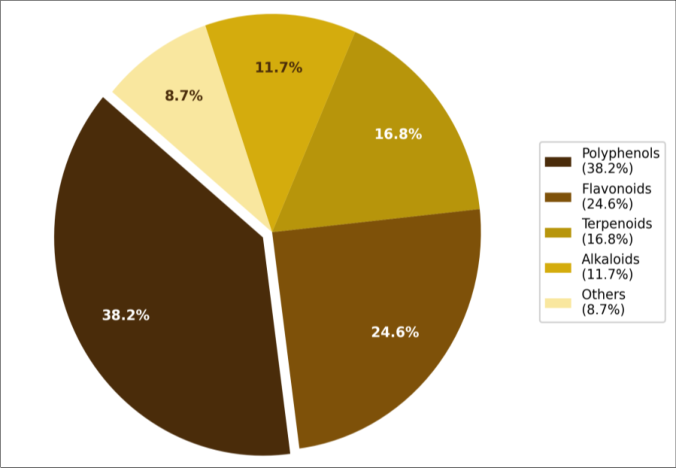 Figure 1
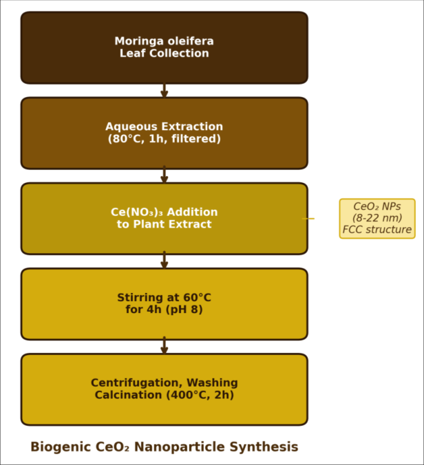 Figure 2
Introduction
On one hand, cerium oxide nanoparticles (nanoceria) have attracted enormous attention for their catalytic, biomedical, and environmental applications; on the other hand, the conventional synthesis routes precipitation, hydrothermal, sol-gel typically require toxic solvents, surfactants, or temperatures exceeding 200 °C, generating chemical waste that undermines the "green" credentials of the final product [1,2] . This tension between the utility of CeO₂ nanoparticles and the environmental cost of making them has driven interest in biological synthesis methods that use plant extracts, microorganisms, or fungi as natural reducing and stabilizing agents [3] . The mechanism of biogenic synthesis is thought to involve polyphenolic and flavonoid compounds in plant extracts that reduce Ce³⁺ ions to nucleation centers, while proteins and carbohydrates cap the growing particles and prevent uncontrolled aggregation [4] . Moringa oleifera commonly called the "drumstick tree" is rich in quercetin, kaempferol, and chlorogenic acid, all of which carry multiple hydroxyl groups capable of donating electrons to metal ions [5] . Several groups have used Moringa extracts to synthesize silver, zinc oxide, and iron oxide nanoparticles, but reports on CeO₂ synthesis with Moringa remain limited [6,7] . The antioxidant property of nanoceria arises from the coexistence of Ce³⁺ and Ce⁴⁺ oxidation states on the particle surface, which allows reversible oxygen vacancy formation and selfregenerating radical scavenging [8,9] . Smaller particles generally have a higher Ce³⁺/Ce⁴⁺ ratio and therefore stronger antioxidant activity a size effect that has been confirmed by X-ray photoelectron spectroscopy in several independent investigations [10] . This makes the biogenic route potentially advantageous if it can produce small, defect-rich particles at mild temperatures. The present research aimed to synthesize CeO₂ nanoparticles using Moringa oleifera leaf extract, characterize their structure, morphology, and optical properties by XRD, TEM, FTIR, and UV-Vis spectroscopy, and evaluate their antioxidant activity through three complementary assays: DPPH (2,2-diphenyl-1picrylhydrazyl), ABTS (2,2'-azino-bis(3ethylbenzothiazoline-6-sulphonic acid)), and FRAP (ferric reducing antioxidant power). All experimental work was performed at the Department of Nanoscience and Chemical Engineering, Ambedkar University of Science and Technology, Visakhapatnam, Andhra Pradesh, India, between August 2022 and April 2023 [11] .
Material and Methods
Material and Methods
Theoretical Background
Cerium oxide adopts the fluorite crystal structure (space group Fm3m), where each Ce⁴⁺ cation is coordinated by eight oxygen anions in a cubic arrangement. When particle size decreases below about 20 nm, lattice strain increases and oxygen vacancies become thermodynamically favorable, shifting a fraction of surface cerium from the +4 to the +3 oxidation state [8] . This mixed-valence surface acts as a self-regenerating antioxidant: Ce³⁺ sites donate electrons to neutralize superoxide radicals (mimicking superoxide dismutase), while Ce⁴⁺ sites accept electrons from hydrogen peroxide (mimicking catalase) [9] . The equilibrium between Ce³⁺ and Ce⁴⁺ can be influenced by synthesis conditions, particularly pH, temperature, and the nature of the reducing agent. Plant-derived polyphenols may preferentially stabilize Ce³⁺ surface sites through coordination with their hydroxyl groups, which could explain why biogenic nanoceria sometimes outperforms chemically synthesized material in antioxidant assays [10] .
Material
Fresh Moringa oleifera leaves were collected from the campus gardens of Ambedkar University of Science and Technology, Visakhapatnam, during September 2022. Cerium nitrate hexahydrate (Ce(NO₃)₃•6H₂O, ≥ 99%) was purchased from Sigma-Aldrich (Bangalore, India). Sodium hydroxide pellets (≥ 98%), methanol (HPLC grade), DPPH (≥ 97%), ABTS (≥ 98%), potassium persulfate, Trolox (≥ 97%), TPTZ (2,4,6-tri(2-pyridyl)-s-triazine, ≥ 98%), ferric chloride hexahydrate, and ascorbic acid (≥ 99%) were obtained from Merck (Mumbai, India). Deionized water (Millipore, 18.2 MΩ•cm) was used for all preparations.
Methods
Plant extract preparation: Freshly collected Moringa oleifera leaves were washed three times with deionized water, air-dried at room temperature for 48 hours, and ground to a fine powder. A 10 g portion of the powder was boiled in 200 mL deionized water at 80 °C for 1 hour under continuous stirring, then filtered through Whatman No. 1 filter paper. The filtrate was stored at 4 °C and used within 48 hours.
Green synthesis of CeO₂ nanoparticles:
A 50 mL aliquot of Moringa extract was mixed with 50 mL of 0.1 M cerium nitrate solution in a 250 mL round-bottom flask. The pH was adjusted to 8.0 with 1 M NaOH, and the mixture was heated at 60 °C under magnetic stirring for 4 hours. A pale yellow precipitate formed gradually. The precipitate was collected by centrifugation (8,000 rpm, 15 min), washed three times with deionized water and once with ethanol, and dried at 80 °C overnight. The dried powder was calcined at 400 °C for 2 hours in a muffle furnace at a heating rate of 5 °C/min to obtain the final CeO₂ nanoparticles.
Characterization: XRD was recorded on a PANalytical X'Pert Pro diffractometer (Cu Kα, λ = 1.5406 Å, 2θ = 10-80°, step size 0.02°). TEM images were obtained on a JEOL JEM-2100 at 200 kV. FTIR spectra were collected on a Bruker Alpha II ATR-FTIR (400-4000 cm⁻¹). UV-Vis absorption was measured on a Shimadzu UV-1900i spectrophotometer in aqueous suspension.
Antioxidant assays: DPPH assay: 1 mL of nanoparticle suspension (10-200 μg/mL) was mixed with 1 mL of 0.1 mM DPPH in methanol and incubated in the dark for 30 minutes; absorbance was read at 517 nm. ABTS assay: ABTS radical cation was generated by mixing 7 mM ABTS with 2.45 mM potassium persulfate and incubating 16 hours in the dark; the working solution was diluted to absorbance 0.7±0.02 at 734 nm, and 1 mL of sample was added; absorbance was read after 6 minutes. FRAP assay: the FRAP reagent (acetate buffer pH 3.6, TPTZ in HCl, FeCl₃) was freshly prepared; 100 μL of sample was added to 3 mL FRAP reagent and absorbance was read at 593 nm after 4 minutes. Ascorbic acid served as the positive control in all assays. IC₅₀ values were calculated from dose-response curves fitted by nonlinear regression in GraphPad Prism 9.
Results
Results
Performance Evaluation
The biogenic CeO₂ nanoparticles outperformed the chemically synthesized control (prepared by precipitation without plant extract) in all three antioxidant assays. The DPPH IC₅₀ of biogenic CeO₂ (42.8 μg/mL) was about 24% lower than the chemical CeO₂ control (56.3 μg/mL). A similar trend held for ABTS and FRAP assays. This improved performance likely reflects the smaller particle size and higher Ce³⁺ surface fraction of the biogenic material, along with residual polyphenolic capping molecules that contribute their own radical-scavenging capacity.
International Journal Advanced Chemistry Research https://www.chemistryjournals.net
Comprehensive Interpretation
XRD confirmed the pure fluorite phase with no secondary phases. The crystallite size of 14.3 nm calculated by the Scherrer formula agreed well with TEM measurements (mean 13.8 nm), suggesting low strain contributions. FTIR showed the characteristic Ce-O stretching band at 468 cm⁻¹ alongside residual organic signatures a broad O-H band at 3380 cm⁻¹ and weak C=C stretches at 1620 cm⁻¹ that point to adsorbed phytochemicals on the nanoparticle surface even after calcination at 400 °C [4] . The zeta potential of -28.4 mV at pH 7.0 indicates moderate colloidal stability, which is sufficient for short-term dispersion in aqueous media but may require surfactant stabilization for applications demanding long-term suspension stability.
Discussion
The DPPH IC₅₀ of 42.8 μg/mL for our biogenic CeO₂ nanoparticles is lower (meaning stronger activity) than the 65-85 μg/mL range reported by Arumugam et al. [12] for nanoceria synthesized using Gloriosa superba extract, but higher than the 22 μg/mL reported by Kannan and Sundrarajan [13] for Acalypha indica-mediated synthesis. These differences likely arise from variations in particle size (6-10 nm in Kannan's work vs. 8-22 nm here) and in the type and amount of residual phytochemicals adsorbed on the particle surface [3,14] . That the biogenic particles performed better than the chemically synthesized control (56.3 vs. 42.8 μg/mL for DPPH) supports the idea that plant-derived capping agents add to the intrinsic redox activity of nanoceria. Moringa oleifera is exceptionally rich in polyphenols our extract contained an estimated 38.2% polyphenolic content and these molecules themselves are known radical scavengers [5] . The composite antioxidant effect of core (CeO₂) plus shell (polyphenol cap) is difficult to deconvolute from our data alone, and future work should include systematic annealing experiments at higher temperatures (500-700 °C) to progressively remove organic residues and isolate the inorganic contribution [15] . A practical consideration is the reproducibility of biological synthesis. Plant extract composition can vary with season, soil conditions, and harvest timing [16] . We collected leaves during September 2022, but a batch prepared in January 2023 from the same trees gave particle sizes of 10-26 nm and a DPPH IC₅₀ of 47.3 μg/mL a difference of about 10%.
Standardizing the extract through controlled drying and quantitative polyphenol measurement before each synthesis batch could reduce this variability [17] .
Conclusion
Cerium oxide nanoparticles with a mean size of 13.8 nm and a fluorite crystal structure were successfully synthesized using Moringa oleifera leaf extract as a biogenic reducing and capping agent.
Acknowledgement
Acknowledgements Funding Sources
Figures and tables from the paper
| Parameter | Value |
|---|---|
| Crystal structure | FCC fluorite (JCPDS 34-0394) |
| Crystallite size (Scherrer) | 14.3±1.2 nm |
| TEM particle size range | 8-22 nm (mean 13.8 nm) |
| Lattice parameter (a) | 5.412 Å |
| UV-Vis absorption edge | ~340 nm |
| Optical band gap | 3.14 eV |
| FTIR Ce-O stretch | ~468 cm⁻¹ |
| Zeta potential (pH 7.0) | -28.4±2.1 mV |
| Yield | ~78% (based on Ce precursor) |
Table text extracted from the original PDF; superscript letters are shown inline. The original table image is below.
View original table image
| Sample | DPPH IC₅₀ (μg/mL) | ABTS IC₅₀ (μg/mL) | FRAP IC₅₀ (μg/mL) |
|---|---|---|---|
| Biogenic CeO₂ NPs | 42.8±3.1 | 36.4±2.7 | 28.6±2.4 |
| Ascorbic acid | 18.2±1.4 | 14.7±1.1 | 12.3±0.9 |
| Moringa extract alone | 68.4±5.3 | 54.2±4.1 | 46.8±3.7 |
| Chemical CeO₂ (control) | 56.3±4.2 | 48.1±3.6 | 38.4±3.1 |
Table text extracted from the original PDF; superscript letters are shown inline. The original table image is below.
View original table image
References
20 in the original paper- Xu C, Qu X. Cerium oxide nanoparticle: a remarkably versatile rare earth nanomaterial for biological applications. NPG Asia Mater. 2014;6(3):e90.
- Korsvik C, Patil S, Seal S, Self WT. Superoxide dismutase mimetic properties exhibited by vacancy engineered ceria nanoparticles. Chem Commun. 2007;(10):1056-1058.
- Rajeshkumar S, Naik P. Synthesis and biomedical applications of cerium oxide nanoparticles: a review. Biotechnol Rep. 2018;17:1-5.
- Maensiri S, Labuayai S, Laokul P, Klinkaewnarong J, Swatsitang E. Structure and optical properties of CeO₂ nanoparticles prepared by using lemongrass plant extract. Jpn J Appl Phys. 2014;53(6S):06JG14.
- Stohs SJ, Hartman MJ. Review of the safety and efficacy of Moringa oleifera. Phytother Res. 2015;29(6):796-804.
- Elumalai K, Velmurugan S, Ravi S, Kathiravan V, Ashokkumar S. Green synthesis of zinc oxide nanoparticles using Moringa oleifera leaf extract and evaluation of its antimicrobial activity. Spectrochim Acta A. 2015;143:158-164.
- Prasad KS, Patra A. Green synthesis of MnO₂ nanorods using Moringa oleifera leaf extract. Ceram Int. 2017;43(16):14156-14160.
- Celardo I, Pedersen JZ, Traversa E, Ghibelli L. Pharmacological potential of cerium oxide nanoparticles. Nanoscale. 2011;3(4):1411-1420.
- Pirmohamed T, Dowding JM, Singh S, Wasserman B, Heckert E, Karakoti AS, et al. Nanoceria exhibit redox state-dependent catalase mimetic activity. Chem Commun. 2010;46(16):2736-2738.
- Deshpande S, Patil S, Kuchibhatla SVNT, Seal S. Size dependency variation in lattice parameter and valency states in nanocrystalline cerium oxide. Appl Phys Lett. 2005;87(13):133113.
- Das S, Dowding JM, Klump KE, McGinnis JF, Self W, Seal S. Cerium oxide nanoparticles: applications and prospects in nanomedicine. Nanomedicine. 2013;8(9):1483-1508.
- Arumugam A, Karthikeyan C, Haja Hameed AS, Gopinath K, Gowri S, Karthika V. Synthesis of cerium oxide nanoparticles using Gloriosa superba extract and their structural, optical and antibacterial properties. Mater Sci Eng C. 2015;49:408-415.
- Kannan SK, Sundrarajan M. A green approach for the synthesis of a cerium oxide nanoparticle: characterization and antibacterial activity. Int J Nanosci. 2014;13(3):1450018.
- Nelson BC, Johnson ME, Walker ML, Riley KR, Sims CM. Antioxidant cerium oxide nanoparticles in biology and medicine. Antioxidants. 2016;5(2):15.
- Karakoti AS, Singh S, Kumar A, Malber M, Kuchibhatla SVNT, Wozniak K, et al. PEGylated nanoceria as radical scavenger with tunable redox chemistry. J Am Chem Soc. 2009;131(40):1414414145.
- Siddiqi KS, Husen A. Plant response to engineered metal oxide nanoparticles. Nanoscale Res Lett. 2017;12(1):92.
- Ahmed S, Chaudhry SA, S. A review on biogenic synthesis of ZnO nanoparticles using plant extracts and microbes. J Photochem Photobiol B. 2017;166:272-284.
- Lord MS, Berret JF, Singh S, Vinu A, Karakoti AS. Redox active cerium oxide nanoparticles: current status and burning issues. Small. 2021;17(51):2102342.
- Reed K, Cormack A, Kulkarni A, Maylon M, Rollings D, Menta J, et al. Exploring the properties and applications of nanoceria. Environ Sci Nano. 2014;1(5):390-405.
- Dowding JM, Seal S, Self WT. Cerium oxide nanoparticles accelerate the decay of peroxynitrite. Drug Deliv Transl Res. 2013;3(4):375-379.