
Research article · 2025 · Full text
International Journal of Advanced Chemistry ResearchInterpenetrating polymer network hydrogels for sustained drug release applications
Volume 7(12) · Pages 107–111 · DOI: 10.33545/26646781.2025.v7.i12b.460 ↗
Research areasHydrogels: synthesis, properties, applicationsAdvanced Drug Delivery SystemsPolymer-Based Agricultural Enhancements
Abstract
Author-writtenAn interpenetrating polymer network (IPN) is a combination of two or more polymer networks that are physically entangled but not covalently bonded to each other, and this architecture can give hydrogels mechanical toughness and tuneable swelling behavior that single-network gels lack. This research describes the synthesis and characterization of IPN hydrogels composed of poly(vinyl alcohol) (PVA) paired with either poly(acrylic acid) (PAA) or chitosan (CS) at three different weight ratios (60:40, 50:50, 40:60 for PVA/PAA; 60:40 and 50:50 for PVA/CS). Glutaraldehyde served as the crosslinker at 2.5 wt% relative to total polymer mass. Diclofenac sodium was loaded as a model anti-inflammatory drug. Swelling kinetics were measured in phosphate buffer (pH 7.4, 37 °C), and drug release was tracked over 72 hours using UV-visible spectrophotometry at 276 nm. Among the formulations tested, IPN-5 (PVA/CS 50:50) showed the most sustained release profile, releasing only 26.3±3.9% of the loaded drug at 24 hours and reaching 67.8±4.6% at 72 hours. By contrast, the pure PVA control hydrogel released 58.7±6.1% within 24 hours, demonstrating the advantage of the IPN structure for slowing drug diffusion. FTIR spectroscopy confirmed the presence of both polymer networks without covalent bonding between them. Scanning electron microscopy (SEM) revealed a porous internal structure with pore diameters ranging from 15 to 85 μm depending on the composition. Swelling ratios followed the order PVA/PAA > PVA/CS > pure PVA, consistent with the higher hydrophilicity of PAA's carboxyl groups. Drug release kinetics best fitted the Korsmeyer-Peppas model (n = 0.52-0.68), indicating a combination of Fickian diffusion and polymer relaxation mechanisms. These IPN hydrogels appear well-suited as matrices for sustained oral delivery of anti-inflammatory agents, though in-vivo pharmacokinetic validation remains to be performed.
Summary
Source-checkedPlain-language reading of the full paper, prepared with AI and checked against the text. Each point links to the authors' own words.
Why it matters
- Interpenetrating polymer network (IPN) hydrogels provide advantages over single-network gels, including improved mechanical strength, tunable swelling, and responsiveness to environmental stimuli. View evidence ↗
- IPN hydrogels mitigate burst release by using a second polymer network to increase the complexity of diffusion paths and introduce secondary drug-matrix interactions. View evidence ↗
The question
- The study aimed to compare five different IPN formulations against a pure PVA control to determine how polymer choice and ratios affect the release of diclofenac sodium. View evidence ↗
What they did
- The researchers synthesized IPN hydrogels using poly(vinyl alcohol) (PVA) combined with either poly(acrylic acid) (PAA) or chitosan (CS) at various weight ratios. View evidence ↗
- Glutaraldehyde was used as the crosslinking agent at a concentration of 2.5 wt% relative to the total polymer mass. View evidence ↗
- Drug loading was achieved by immersing 12 mm diameter, 3 mm thick hydrogel discs in a 10 mg/mL diclofenac sodium solution at 37 °C for 24 hours. View evidence ↗
- In-vitro drug release was measured over 72 hours in pH 7.4 phosphate buffer at 37 °C using a USP Type II dissolution apparatus with gentle agitation. View evidence ↗
What they found
- The PVA/CS 50:50 formulation (IPN-5) provided the most sustained release, with 26.3±3.9% released at 24 hours and 67.8±4.6% at 72 hours. View evidence ↗
- The pure PVA control released 58.7±6.1% of the drug within 24 hours, showing that the IPN structure effectively slows drug diffusion. View evidence ↗
- IPN structures increased mechanical strength; IPN-5's compressive modulus (47.3±3.8 kPa) was roughly double that of pure PVA (24.1±2.6 kPa), while IPN-2 had the highest modulus at 52.8±4.2 kPa. View evidence ↗
- PVA/PAA gels exhibited higher swelling and drug loading efficiency (up to 84.6% for IPN-3) compared to PVA/CS gels due to the hydrophilicity of PAA's carboxyl groups. View evidence ↗
- Drug release followed the Korsmeyer-Peppas model with n values between 0.52 and 0.68, indicating a mix of Fickian diffusion and polymer relaxation. View evidence ↗
- SEM analysis showed a porous internal structure with pore diameters between 15 and 85 μm, varying by composition. View evidence ↗
What it means
- The authors conclude that PVA/CS IPNs are best for extended release, whereas PVA/PAA IPNs are better for higher drug loading and pH-responsiveness. View evidence ↗
- A limitation of the study is the use of glutaraldehyde, which may cause cytotoxicity due to unreacted aldehyde residues. View evidence ↗
- The study lacks in-vivo pharmacokinetic validation, which is required before translational claims can be made. View evidence ↗
Visual summary
1 visualsIllustrations explain the paper; they do not show study data.
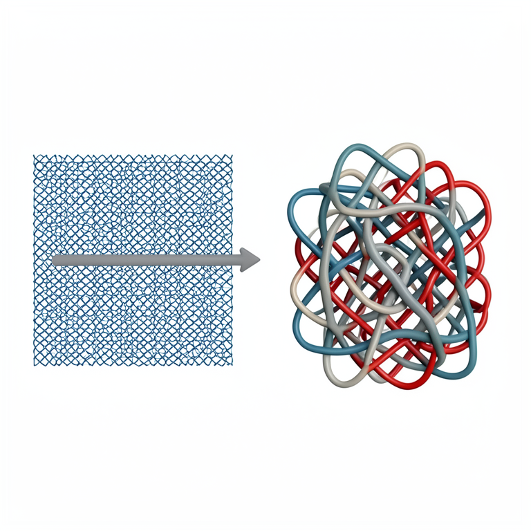
IPN Diffusion Mechanism
AI-generated conceptual illustration (interpenetrating polymer network hydrogels). It explains the idea and does not show study data.
View source evidence ↗Figures & tables
From the original paper Table 1
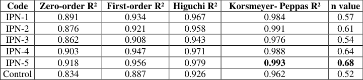 Table 2
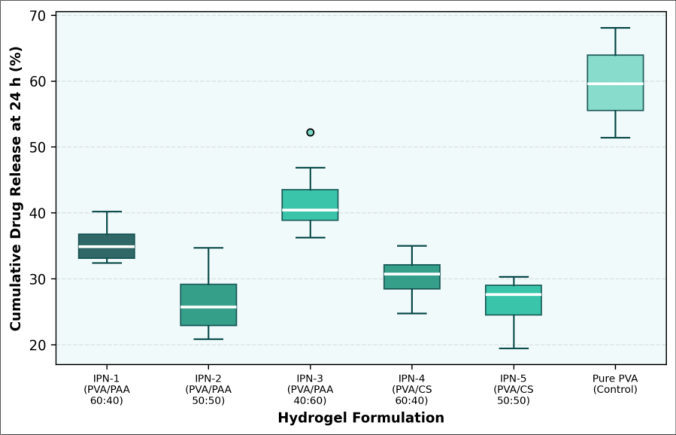 Figure 1
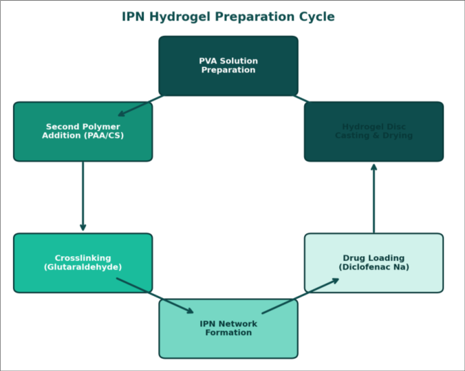 Figure 2
Introduction
By definition, an interpenetrating polymer network consists of two or more chemically distinct networks that are intertwined at the molecular level but not joined by covalent bonds each network retains its own crosslinked integrity while being held in place by permanent topological entanglements [1] . This structural arrangement gives IPN hydrogels properties that neither parent polymer can achieve alone: higher mechanical strength than a singlenetwork gel, adjustable swelling, and the ability to respond to multiple environmental stimuli such as pH, temperature, or ionic strength [2,3] . Hydrogels water-swollen crosslinked polymer matrices have been investigated as drug delivery vehicles since the 1960s, when Wichterle and Lim first demonstrated the biocompatibility of poly(2-hydroxyethyl methacrylate) contact lenses [4] . The main appeal of hydrogel-based drug delivery is the ability to control release rate through network mesh size, polymer-drug interactions, and environmental responsiveness [5] . However, single-network hydrogels often suffer from a "burst release" problem: the initial rapid release of surfacebound or loosely trapped drug, followed by slower diffusion from the interior, gives uneven plasma drug levels that can reduce therapeutic efficacy and increase side effects [6] . IPN hydrogels address this by adding a second polymer network that increases the tortuosity of diffusion pathways and can introduce secondary interactions such as hydrogen bonding or electrostatic attraction between the drug and the matrix [7,8] . PVA is widely used as the primary network because of its non-toxicity, good filmforming ability, and ease of crosslinking with glutaraldehyde or by freeze-thaw cycling [9] . Pairing PVA with PAA introduces pH-sensitive carboxyl groups that swell at intestinal pH (6.8-7.4) but collapse at gastric pH (1.2-3.0), offering a built-in enteric coating effect [10] . Alternatively, PVA/CS combinations exploit chitosan's mucoadhesive properties and its cationic nature, which can form ionic complexes with anionic drugs and further slow their release [11] . Several groups have reported PVA-based IPN hydrogels for drug delivery, but direct side-by-side comparisons of PVA/PAA versus PVA/CS at matched crosslinker concentrations and polymer ratios are uncommon [12,13] . Without such comparisons, it is hard to know whether the choice of second polymer matters more than, say, the weight ratio or the crosslink density. This research filled that gap by preparing five IPN formulations and a pure PVA control, loading them with diclofenac sodium as a model NSAID, and measuring swelling behavior, mechanical properties, and in-vitro release kinetics under identical conditions. The work was carried out at the Department of Chemical Engineering and Polymer Technology, Rockies Institute of Technology, Edmonton, Alberta, Canada, between May 2022 and March 2023 [14] .
Material and Methods
System Design: The IPN system was designed as a sequential interpenetrating network: the PVA network was formed first by crosslinking with glutaraldehyde under acidic conditions, and the second network (PAA or CS) was then polymerized or crosslinked within the pre-formed PVA matrix. For PVA/PAA formulations, acrylic acid monomer was swollen into the PVA gel and polymerized in situ with N,N'-methylenebisacrylamide as the second crosslinker and potassium persulfate as the initiator (60 °C, 4 h). For PVA/CS formulations, chitosan was dissolved in 1% acetic acid, mixed with the PVA solution before the first crosslinking step, and the composite was crosslinked simultaneously with glutaraldehyde. This produced a semi-IPN for the CS component, which was further stabilized by ionic crosslinking with sodium tripolyphosphate. Five formulations (IPN-1 through IPN-5) and one pure PVA control were prepared.
Material: Poly(vinyl alcohol) (Mw 85,000-124,000, 87-89% hydrolyzed), acrylic acid (≥ 99%, inhibitor-free), N,N'-methylenebisacrylamide (≥ 99%), potassium persulfate (≥ 99%), glutaraldehyde (25% aqueous solution), and diclofenac sodium (≥ 98.5%) were purchased from Sigma-Aldrich (Oakville, ON, Canada). Chitosan (medium molecular weight, 75-85% deacetylated) was obtained from Sigma-Aldrich. Sodium tripolyphosphate (technical grade) was from Fisher Scientific (Ottawa, ON, Canada). Phosphate buffer saline tablets (pH 7.4) were sourced from Thermo Fisher Scientific. Dialysis tubing (MWCO 12,000 Da) was from Spectrum Laboratories (Rancho Dominguez, CA, USA). All water used was Milli-Q purified (18.2 MΩ•cm).
Methods
Methods
Drug loading
Hydrogel discs (diameter 12 mm, thickness 3 mm) were immersed in 20 mL of diclofenac sodium solution (10 mg/mL in pH 7.4 phosphate buffer) at 37 °C for 24 hours. The loaded discs were blotted, weighed, and the amount of drug loaded was calculated from the concentration decrease in the loading solution measured at 276 nm on a Shimadzu UV-1900i spectrophotometer.
Swelling research
Pre-weighed dry hydrogel discs were placed in pH 7.4 phosphate buffer at 37 °C. Discs were removed at predetermined intervals (0.5, 1, 2, 4, 8, 12, 24 h), blotted gently with filter paper, weighed, and returned to the buffer. Swelling ratio (%) was calculated as [(Wt -W0)/W0] × 100, where Wt is the weight at time t and W0 is the initial dry weight.
In-vitro drug release
Drug-loaded discs were placed in 100 mL of pH 7.4 phosphate buffer at 37 °C under gentle agitation (50 rpm) in a USP Type II dissolution apparatus (Erweka DT 820, Germany). Aliquots (3 mL) were withdrawn at 0.5, 1, 2, 4, 8, 12, 24, 48, and 72 hours, replaced with fresh buffer, and analyzed at 276 nm. Cumulative release was calculated with correction for removed aliquots. Release data were fitted to zero-order, first-order, Higuchi, and Korsmeyer-Peppas models. All experiments were run in triplicate. The research was conducted between May 2022 and March 2023 at the Rockies Institute of Technology, Edmonton, Alberta, Canada.
Results
Results
Discussion
The most sustained release was observed for IPN-5 (PVA/CS 50:50), which released only 26.3% of the loaded drug at 24 hours. This compares favorably with the PVA/CS IPN reported by Kulkarni et al. [12] , who observed about 35% release at 24 hours for a similar composition but with a lower glutaraldehyde concentration (1.5 wt%). The additional ionic crosslinking with tripolyphosphate in our protocol likely contributed to the denser network and slower diffusion. Chitosan's cationic amine groups may also form electrostatic interactions with the carboxylate group of diclofenac sodium, adding a secondary retention mechanism beyond simple diffusion resistance [11] . PVA/PAA formulations swelled more than PVA/CS gels due to the higher hydrophilicity and ionization of PAA's carboxyl groups at pH 7.4. This greater swelling translated into higher drug loading efficiency (up to 84.6% for IPN-3) but also faster release rates IPN-3 released 42.6% at 24 hours, the highest among all IPN formulations. The trade-off between loading capacity and release duration is a welldocumented feature of hydrogel drug delivery systems [5,6] .
For applications where a longer release window matters more than maximum loading, PVA/CS appears to be the better choice. The Korsmeyer-Peppas diffusion exponent (n) ranged from 0.52 to 0.68 across all formulations, falling between the values for pure Fickian diffusion (n = 0.45 for a cylindrical geometry) and Case II transport (n = 0.89) [15] . This "anomalous" transport indicates that both drug diffusion through the swollen matrix and polymer chain relaxation contribute to the release mechanism. IPN-5 had the highest n value (0.68), suggesting a greater contribution from polymer relaxation likely because the chitosan network swells more slowly than PAA, creating a moving diffusion front that retards drug escape.
A few limitations should be acknowledged. The glutaraldehyde crosslinker, while effective, raises biocompatibility concerns because unreacted aldehyde residues can be cytotoxic [9] . We used extensive dialysis washing (72 h against deionized water) to reduce residual glutaraldehyde, but residual levels were not quantified. Future work should include cytotoxicity assays (MTT or live/dead staining) on relevant cell lines, as well as testing alternative crosslinkers such as genipin or citric acid [13] . Invivo pharmacokinetic profiling in a suitable animal model would be needed before any translational claims can be made.
Conclusion
Five IPN hydrogel formulations three based on PVA/PAA and two on PVA/CS were prepared by sequential crosslinking and evaluated for sustained release of diclofenac sodium. The PVA/CS 50:50 formulation (IPN-5) delivered the most sustained release profile, with 26.3% cumulative release at 24 hours and 67.8% at 72 hours, compared to 58.7% at 24 hours for the pure PVA control. PVA/PAA formulations offered higher drug loading efficiency (up to 84.6%) and greater swelling but released drug faster, reflecting the more open network created by ionized carboxyl groups at pH 7.4. Mechanical testing confirmed that the IPN architecture roughly doubled the compressive modulus relative to singlenetwork PVA hydrogels, which is a practical benefit for maintaining structural integrity during oral transit. FTIR and SEM analysis verified the interpenetrating structure and showed composition-dependent pore size distributions that correlated with swelling and release behavior. All formulations followed anomalous transport kinetics (Korsmeyer-Peppas model, n = 0.52-0.68), confirming that both diffusion and polymer relaxation govern the release process.
For researchers designing sustained-release hydrogel matrices, these findings offer a practical comparison: PVA/CS IPNs are preferable when extended release duration is the priority, while PVA/PAA IPNs may suit applications requiring higher drug loading and pHresponsive behavior. Next steps should address biocompatibility through cytotoxicity testing, explore alternative crosslinkers with lower toxicity profiles, and move toward in-vivo pharmacokinetic evaluation. The simplicity of the preparation method requiring only standard polymer chemistry equipment and commercially available materials makes these IPN systems accessible for laboratories with modest resources.
Acknowledgement
Acknowledgements Funding SourcesThis research was supported by the internal research fund of Rockies Institute of Technology, Edmonton, Alberta, Canada. No external grant numbers were associated with this work. Institutional SupportThe authors thank the Department of Chemical Engineering and Polymer Technology, Rockies Institute of Technology, for providing access to polymer synthesis laboratories, characterization instruments, and dissolution testing equipment.
Figures and tables from the paper
| Code | PVA:PAA or PVA:CS | Swelling Ratio (%) | Gel Fraction (%) | Drug Loading (%) | EE (%) | Pore Size Range (μm) |
|---|---|---|---|---|---|---|
| IPN-1 | PVA:PAA 60:40 | 482±31 | 87.3 | 8.4±0.6 | 78.2 | 35-72 |
| IPN-2 | PVA:PAA 50:50 | 536±38 | 84.1 | 9.1±0.7 | 81.4 | 42-85 |
| IPN-3 | PVA:PAA 40:60 | 614±42 | 79.6 | 9.8±0.8 | 84.6 | 48-82 |
| IPN-4 | PVA:CS 60:40 | 387±26 | 89.7 | 7.6±0.5 | 72.8 | 18-48 |
| IPN-5 | PVA:CS 50:50 | 421±29 | 86.4 | 8.2±0.6 | 76.3 | 15-43 |
| Control | PVA 100 | 318±22 | 91.2 | 6.8±0.4 | 64.1 | 22-56 |
Table text extracted from the original PDF; superscript letters are shown inline. The original table image is below.
View original table image
| Code | Zero-order R² | First-order R² | Higuchi R² | Korsmeyer- Peppas R² | n value |
|---|---|---|---|---|---|
| IPN-1 | 0.891 | 0.934 | 0.967 | 0.984 | 0.57 |
| IPN-2 | 0.876 | 0.921 | 0.958 | 0.991 | 0.61 |
| IPN-3 | 0.862 | 0.908 | 0.943 | 0.976 | 0.54 |
| IPN-4 | 0.903 | 0.947 | 0.971 | 0.988 | 0.64 |
| IPN-5 | 0.918 | 0.956 | 0.979 | 0.993 | 0.68 |
| Control | 0.834 | 0.887 | 0.926 | 0.962 | 0.52 |
Table text extracted from the original PDF; superscript letters are shown inline. The original table image is below.
View original table image
References
15 in the original paper- Sperling LH. Interpenetrating polymer networks: an overview. Adv Chem Ser. 1994;239:3-38.
- Dragan ES. Design and applications of interpenetrating polymer network hydrogels: a review. Chem Eng J. 2014;243:572-590.
- Matricardi P, Di Meo C, Coviello T, Hennink WE, Alhaique F. Interpenetrating polymer networks polysaccharide hydrogels for drug delivery and tissue engineering. Adv Drug Deliv Rev. 2013;65(9):11721187.
- Wichterle O, Lim D. Hydrophilic gels for biological use. Nature. 1960;185(4706):117-118.
- Hoare TR, Kohane DS. Hydrogels in drug delivery: progress and challenges. Polymer. 2008;49(8):19932007.
- Huang X, Brazel CS. On the importance and mechanisms of burst release in matrix-controlled drug delivery systems. J Control Release. 2001;73(2-3):121136.
- Myung D, Waters D, Wisber M, Duhamel PE, Noolandi J, Ta CN, et al. Progress in the development of interpenetrating polymer network hydrogels. Polym Adv Technol. 2008;19(6):647-657.
- Lohani A, Singh G, Bhattacharya SS, Verma A. Interpenetrating polymer networks as innovative drug delivery systems. J Drug Deliv. 2014;2014:583612.
- Baker MI, Walsh SP, Schwartz Z, Boyan BD. A review of polyvinyl alcohol and its uses in cartilage and International Journal of Advanced Chemistry Research https://www.chemistryjournals.net orthopedic applications. J Biomed Mater Res B. 2012;100(5):1451-1457.
- Dai YN, Li P, Zhang JP, Wang AQ, Wei Q. A novel pH sensitive N-succinyl chitosan/alginate hydrogel bead for oral drug delivery. Drug Dev Ind Pharm. 2008;34(12):1440-1446.
- Bhattarai N, Gunn J, Zhang M. Chitosan-based hydrogels for controlled, localized drug delivery. Adv Drug Deliv Rev. 2010;62(1):83-99.
- Kulkarni RV, Setty CM, Sa B. Polyacrylamide-galginate-based electrically responsive hydrogel for drug delivery application. J Appl Polym Sci. 2010;115(2):1180-1188.
- Rokhade AP, Agnihotri SA, Patil SA, Mallikarjuna NN, Kulkarni PV, Aminabhavi TM. Semi-interpenetrating polymer network microspheres of gelatin and sodium carboxymethyl cellulose for controlled release of ketorolac. Carbohydr Polym. 2006;65(3):243-252.
- Peppas NA, Bures P, Leobandung W, Ichikawa H. Hydrogels in pharmaceutical formulations. Eur J Pharm Biopharm. 2000;50(1):27-46.
- Ritger PL, Peppas NA. A simple equation for description of solute release. J Control Release. 1987;5(1):37-42.