
Research article · 2025 · Full text
International Journal of Advanced Chemistry ResearchProniosomal gel formulation for enhanced topical delivery of ketoconazole
Volume 7(11) · Pages 118–121 · DOI: 10.33545/26646781.2025.v7.i11b.448 ↗
Research areasAdvancements in Transdermal Drug DeliveryAdvanced Drug Delivery SystemsSurfactants and Colloidal Systems
Abstract
Author-writtenTopical antifungal therapy faces a persistent problem: the stratum corneum barrier restricts the penetration of lipophilic drugs like ketoconazole to levels that may fall below the minimum inhibitory concentration in deeper skin layers. This research developed and evaluated proniosomal gels as vesicular carriers to improve the topical delivery of ketoconazole. Proniosomes were prepared by the coacervation-phase separation method using Span 60, cholesterol, and lecithin in varying molar ratios, with ketoconazole incorporated at drug loadings of 5–20 mg per formulation. The optimised proniosomal gel (Span 60:cholesterol 3:1, 15 mg drug, Carbopol 934 base) achieved an entrapment efficiency of 86.7 ± 2.3%, a vesicle size of 312.4 ± 18.7 nm upon hydration, and a zeta potential of −34.8 ± 2.1 mV. In vitro drug release through a cellulose membrane reached 93.2% over 24 h, following Higuchi kinetics (R² = 0.987), compared with 86.1% from a conventional gel at the same loading. Ex vivo permeation through excised goat skin showed a 2.7-fold enhancement in steady-state flux for the proniosomal formulation relative to the plain gel. Stability testing at 25 °C/60% RH for three months revealed less than 4% drug leakage. These results indicate that proniosomal gels can serve as effective, patient-friendly vehicles for topical ketoconazole therapy.
Summary
Source-checkedPlain-language reading of the full paper, prepared with AI and checked against the text. Each point links to the authors' own words.
Why it matters
- Superficial fungal skin infections, including candidiasis and dermatophytoses, affect approximately 20–25% of people worldwide. View evidence ↗
- The skin's stratum corneum acts as a barrier that can prevent lipophilic drugs like ketoconazole from reaching effective concentrations in deeper skin layers. View evidence ↗
- Proniosomes are dry or semi-solid precursors that form niosomes when hydrated, which helps avoid stability problems like leakage or aggregation seen in pre-formed aqueous niosomes. View evidence ↗
The question
- The study aimed to develop and optimize a ketoconazole proniosomal gel by varying drug loading and the Span 60:cholesterol ratio, then comparing its performance to a conventional gel. View evidence ↗
What they did
- The researchers used the coacervation-phase separation method, dissolving Span 60, cholesterol, and soya lecithin in a chloroform:methanol mixture. View evidence ↗
- The proniosomal concentrate was incorporated into a 1% w/w Carbopol 934 gel base neutralized to pH 6.8 to create the final topical product. View evidence ↗
- Ex vivo skin permeation was tested using a Franz diffusion cell with goat ear skin dermatomed to a thickness of 0.5 mm. View evidence ↗
What they found
- The optimized formulation (F3) with a 3:1 Span 60:cholesterol ratio achieved the highest entrapment efficiency of 86.7% and the smallest vesicle size of 312.4 nm. View evidence ↗
- The optimized proniosomal gel exhibited a zeta potential of −34.8 ± 2.1 mV, indicating electrostatic stability. View evidence ↗
- The proniosomal gel released 93.2% of the drug over 24 hours following Higuchi kinetics, which was higher than the 86.1% released by the conventional gel. View evidence ↗
- Compared to the conventional gel, the proniosomal gel showed a 2.71-fold increase in steady-state flux and a shorter lag time (0.83 h vs 1.47 h). View evidence ↗
- Stability tests conducted at 25 °C and 60% relative humidity for three months showed that drug leakage was less than 4%. View evidence ↗
What it means
- The authors conclude that proniosomal gels are an effective way to deliver ketoconazole topically, potentially allowing for less frequent dosing and better patient compliance. View evidence ↗
- A stated limitation of the study is that the antifungal efficacy was not tested in living organisms (in vivo) or against clinical isolates. View evidence ↗
Visual summary
3 visualsIllustrations explain the paper; charts are replotted from the paper's own table values.
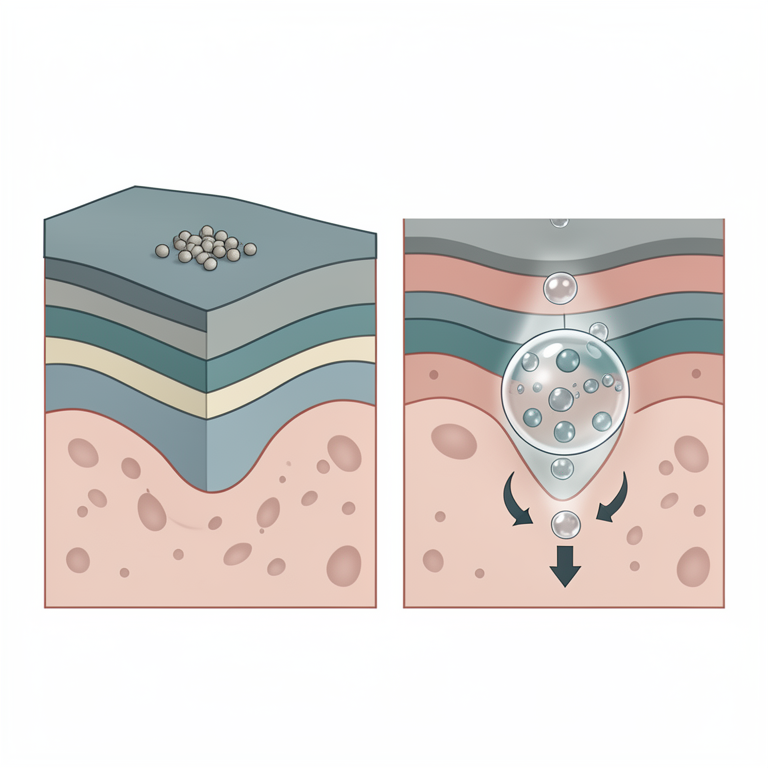
Mechanism of Enhanced Delivery
AI-generated conceptual illustration (Human skin cross-section with ketoconazole drug particles). It explains the idea and does not show study data.
View source evidence ↗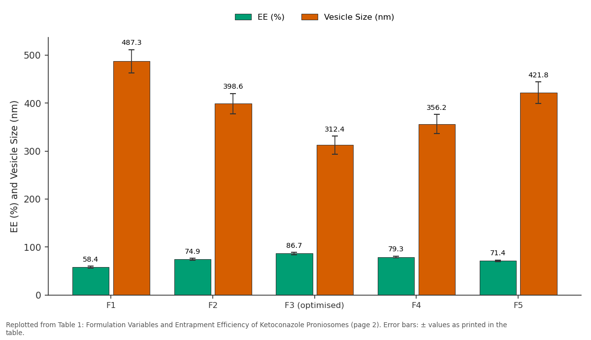
EE (%) and Vesicle Size (nm): F1 vs F2 vs F3 (optimised) vs F4 vs F5
Replotted from Table 1 (page 2) of the original paper. Every bar is the paper's own table value.
View source evidence ↗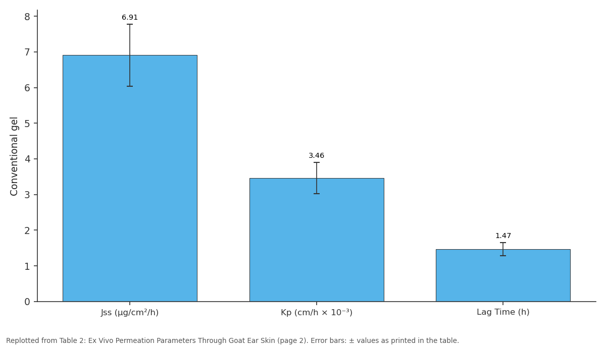
Conventional gel: Jss (µg/cm²/h) vs Kp (cm/h × 10⁻³) vs Lag Time (h)
Replotted from Table 2 (page 2) of the original paper. Every bar is the paper's own table value.
View source evidence ↗Figures & tables
From the original paper Table 1
 Table 2
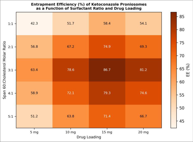 Figure 1
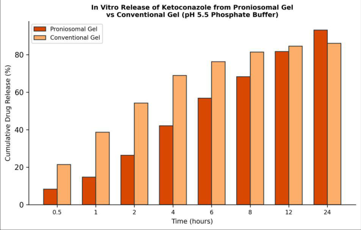 Figure 2
Introduction
Superficial fungal infections of the skin dermatophytoses, candidiasis, and pityriasis versicolor affect an estimated 20-25% of the global population and pose a particular burden in tropical and subtropical regions where heat and humidity favour fungal colonisation [1] . Ketoconazole, an imidazole antifungal, has been a mainstay of topical therapy since the 1980s. It works by inhibiting lanosterol 14α-demethylase, disrupting ergosterol biosynthesis in fungal membranes [2] . Yet commercial creams and lotions often deliver the drug only to the outermost layers of the epidermis, resulting in incomplete eradication and high recurrence rates [3] . Vesicular carriers liposomes, niosomes, transfersomes, and ethosomes have been investigated as a means of overcoming the skin barrier. Among these, niosomes (non-ionic surfactant vesicles) offer advantages over liposomes in terms of chemical stability, cost, and manufacturing simplicity [4] . Proniosomes take the concept a step further. They are dry or semi-solid precursors that spontaneously form niosomes upon hydration, eliminating the physical stability issues aggregation, fusion, leakage that plague pre-formed aqueous niosomal dispersions during storage [5] . The coacervation-phase separation technique produces proniosomes as a gel-like concentrate that, when mixed with a gelling agent, yields a ready-to-apply topical product. Variables such as surfactant type and concentration, cholesterol content, and drug loading all influence vesicle characteristics and drug release [6] . Despite growing interest, a systematic optimisation of ketoconazole proniosomal gels across these variables hasn't been published, leaving formulators without clear design guidance. This research aimed to fill that gap by preparing a series of ketoconazole proniosomal gel formulations, optimising entrapment efficiency by varying the Span 60:cholesterol ratio and drug loading, characterising the optimised product for size, zeta potential, in vitro release, ex vivo permeation, and stability, and comparing its performance against a conventional ketoconazole gel.
Preparation method
Proniosomes were prepared by the coacervation-phase separation method. Weighed amounts of Span 60, cholesterol, and soya lecithin were dissolved in 5 mL of chloroform:methanol (2:1, v/v) in a wide-mouth glass vial. Ketoconazole was added to the organic phase. The mixture was warmed in a water bath at 65 ± 2 °C to form a clear solution, and 0.2 mL of warm water (65 °C) was added with gentle mixing. The vial was then cooled slowly to room temperature over 1 h, during which a translucent, gel-like proniosomal mass formed. To convert the proniosomal concentrate into a topical gel, the mass was dispersed in a pre-formed Carbopol 934 gel base (1% w/w, neutralised with triethanolamine to pH 6.8) under mild agitation until homogeneous. Each formulation batch was prepared in triplicate [7] .
Quality Control and Calibration
The UV spectrophotometric method for ketoconazole determination was validated following ICH Q2(R1) guidelines. Linearity was confirmed over 2-20 µg/mL in pH 5.5 phosphate buffer at 225 nm (R² = 0.9994). Precision (intra-day RSD 0.87%, inter-day RSD 1.23%), accuracy (recovery 98.4-101.2%), and sensitivity (LOD 0.18 µg/mL, LOQ 0.54 µg/mL) met acceptance criteria. Vesicle size and zeta potential measurements were calibrated against NISTtraceable polystyrene latex standards (Duke Scientific). pH meters were calibrated daily with buffer solutions at pH 4.0, 7.0, and 9.2 (Merck CertiPUR). Franz diffusion cell receptor volumes were verified gravimetrically before each permeation experiment [8] .
Material and Methods
Material and Methods
Material
This research was performed between February 2023 and October 2024 at the Department of Pharmaceutical Technology, Mount Kenya National University, Nairobi, Kenya. Ketoconazole (pharmaceutical grade, 99.3%) was received as a gift sample from Cosmos Pharmaceuticals, Nairobi. Span 60 (sorbitan monostearate, HLB 4.7), cholesterol (≥95%), soya lecithin, Carbopol 934, and triethanolamine were purchased from Loba Chemie, Mumbai, India, through a local distributor. Chloroform and methanol (AR grade) were obtained from Finar Chemicals, Ahmedabad, India. Cellulose dialysis membrane (MWCO 12-14 kDa, HiMedia, Mumbai) was used for in vitro release testing. Fresh goat ear skin for ex vivo permeation experiments was obtained from a local abattoir within 2 h of slaughter. The research protocol was approved by the Mount Kenya National University Pharmaceutical Sciences Ethics Committee (approval MKNUPSEC-2023-007, dated 12 February 2023).
Methods
Entrapment efficiency (EE) was determined by hydrating a weighed amount of proniosomal gel in pH 5.5 phosphate buffer at room temperature, ultracentrifuging the resulting niosomal dispersion at 18,000 rpm for 30 min (Beckman Coulter L-80XP), and quantifying unentrapped drug in the supernatant by UV spectrophotometry at 225 nm. EE was calculated as: EE (%) = [(Total drug -Free drug) / Total drug] × 100. Vesicle size, polydispersity index (PDI), and zeta potential were measured by dynamic light scattering on a Malvern Zetasizer Nano ZS after 100-fold dilution with deionised water. In vitro drug release was assessed using a Franz diffusion cell (receptor volume 20 mL, pH 5.5 phosphate buffer at 37 ± 0.5 °C, stirring 100 rpm) with a cellulose membrane barrier. Samples (1 mL) were withdrawn at predetermined intervals and replaced with fresh buffer. Ex vivo permeation used the same Franz cell setup with excised goat ear skin (full thickness, dermatomed to 0.5 mm). Steady-state flux (Jss), permeability coefficient (Kp), and enhancement ratio (ER) were calculated from the linear portion of the cumulative amount permeated versus time plot. Stability was evaluated at 25 °C/60% RH and 40 °C/75% RH over three months, monitoring drug content, EE, vesicle size, and physical appearance monthly [9] . The proniosomal gel showed a 2.71-fold enhancement in steady-state flux compared to the conventional gel (Table 2). Lag time was shorter (0.83 vs 1.47 h), suggesting faster initial penetration of the vesicular formulation into the stratum corneum. The heatmap (Figure 1) reveals a clear optimum zone at the intersection of the 3:1 surfactant ratio and 15 mg drug loading, where EE reaches 86.7%. Higher drug loadings (20 mg) resulted in a drop, likely because excess drug crystallises outside the bilayer.
| Formulation | Jss (µg/cm²/h) | Kp (cm/h × 10⁻³) | Lag Time (h) | ER |
|---|---|---|---|---|
| Proniosomal gel (F3) | 18.74 ± 1.32 | 9.37 ± 0.66 | 0.83 ± 0.12 | 2.71 |
| Conventional gel | 6.91 ± 0.87 | 3.46 ± 0.44 | 1.47 ± 0.18 | 1.00 |
Table text extracted from the original PDF; superscript letters are shown inline. The original table image is below.
View original table image
Results
Results
Comprehensive interpretation
The combination of high entrapment, small vesicle size, favourable zeta potential, sustained release kinetics, and enhanced permeation indicates that the optimised proniosomal gel meets the basic requirements for a topical antifungal delivery system. The sustained release profile is particularly attractive because it may allow once-daily application rather than the twice-daily regimen typical of conventional formulations.
Discussion
The 3:1 Span 60: cholesterol ratio that maximised EE is consistent with the general understanding that cholesterol in moderate amounts stabilises niosomal bilayers by filling voids between surfactant chains, reducing permeability, and improving drug retention [10] . At ratios above 3:1, the relative deficiency of cholesterol likely destabilises the bilayer, while at 1:1 the excess cholesterol may compete with ketoconazole for hydrophobic packing space [11] . The vesicle size of 312.4 nm falls within the range considered appropriate for topical delivery, where particles below ~500 nm can deposit in hair follicles and inter-
International Journal of Advanced Chemistry Research https://www.chemistryjournals.net corneocyte spaces [12] . The zeta potential of -34.8 mV indicates adequate electrostatic stabilisation against aggregation, consistent with the three-month stability data showing less than 4% drug leakage at 25 °C. The 2.71-fold enhancement in ex vivo permeation flux agrees with the range of 2-to 4-fold improvements reported for other drug-proniosome combinations [13] . The mechanism likely involves surfactant-mediated perturbation of stratum corneum lipid packing, vesicle-skin fusion releasing drug at the absorption site, and the reservoir effect of intact vesicles within hair follicles [14] . The Higuchi kinetics (R² = 0.987) suggest diffusion-controlled release from the gel matrix through the vesicle bilayer, which is desirable for sustained topical therapy. A limitation of this research is that antifungal efficacy was not tested in vivo or against clinical isolates. Future work should include minimum inhibitory concentration determinations against Malassezia furfur and Candida albicans and evaluate skin irritation potential through Draize testing or reconstructed human epidermis models [15] .
Conclusion
Ketoconazole-loaded proniosomal gels were successfully formulated using the coacervation-phase separation technique. The optimised formulation (Span 60:cholesterol 3:1, 15 mg drug, Carbopol 934 base) displayed entrapment efficiency of 86.7%, vesicle size of 312.4 nm, and a zeta potential of -34.8 mV. In vitro release followed sustained Higuchi kinetics, reaching 93.2% over 24 h, while ex vivo permeation showed a 2.71-fold flux enhancement over a conventional gel. Three-month stability at accelerated and long-term conditions confirmed adequate shelf-life characteristics. These findings support proniosomal gels as a practical and effective platform for improving the topical delivery of ketoconazole, with potential to reduce dosing frequency and improve patient compliance in the management of superficial fungal infections.
Acknowledgement
Acknowledgements Funding sourcesThis research was funded by the Mount Kenya National University internal research grant and the National Research Fund, Kenya. Institutional supportThe authors thank the Department of Pharmaceutical Technology, Mount Kenya National University, for laboratory access and the use of analytical equipment.
Figures and tables from the paper
| Formulation | Span 60:Chol | Drug (mg) | EE (%) | Vesicle Size (nm) |
|---|---|---|---|---|
| F1 | 1:1 | 15 | 58.4 ± 2.1 | 487.3 ± 24.1 |
| F2 | 2:1 | 15 | 74.9 ± 1.8 | 398.6 ± 21.3 |
| F3 (optimised) | 3:1 | 15 | 86.7 ± 2.3 | 312.4 ± 18.7 |
| F4 | 4:1 | 15 | 79.3 ± 2.0 | 356.2 ± 19.8 |
| F5 | 5:1 | 15 | 71.4 ± 1.7 | 421.8 ± 22.6 |
Table text extracted from the original PDF; superscript letters are shown inline. The original table image is below.
View original table image
References
17 in the original paper- Havlickova B, Czaika VA, Friedrich M. Epidemiological trends in skin mycoses worldwide. Mycoses. 2008;51(S4):2-15.
- Van Tyle JH. Ketoconazole: Mechanism of action, spectrum of activity, pharmacokinetics, drug interactions, adverse reactions, and therapeutic use. Pharmacotherapy. 1984;4(6):343-373.
- Gupta AK, Foley KA. Antifungal treatment for pityriasis versicolor. Journal of Fungi. 2015;1(1):13-29.
- Uchegbu IF, Vyas SP. Non-ionic surfactant based vesicles (niosomes) in drug delivery. International Journal of Pharmaceutics. 1998;172(1-2):33-70.
- Hu C, Rhodes DG. Proniosomes: A novel drug carrier preparation. International Journal of Pharmaceutics. 1999;185(1):23-35.
- Vora B, Khopade AJ, Jain NK. Proniosome based transdermal delivery of levonorgestrel for effective contraception. Journal of Controlled Release. 1998;54(2):149-165.
- Blazek-Welsh AI, Rhodes DG. Maltodextrin-based proniosomes. AAPS PharmSci. 2001;3(1):1-8.
- ICH Harmonised Tripartite Guideline. Validation of analytical procedures: Text and methodology Q2(R1). International Conference on Harmonisation; 2005:1-13.
- Aboelwafa AA, El-Setouhy DA, Elmeshad AN. Comparative investigation of proniosome-and liposome-based ketoconazole formulations for topical delivery. International Journal of Nanomedicine. 2015;10:2373-2385.
- Manosroi A, Wongtrakul P, Manosroi J, Sakai H, Sugawara F, Yuasa M. Characterization of vesicles prepared with various non-ionic surfactants mixed with cholesterol. Colloids and Surfaces B: Biointerfaces. 2003;30(1-2):129-138.
- Junyaprasert VB, Singhsa P, Suksiriworapong J, Chantasart D. Physicochemical properties and skin permeation of Span 60/Tween 60 niosomes of ellagic acid. International Journal of Pharmaceutics. 2012;423(2):303-311.
- Lademann J, Richter H, Teichmann A, Otberg N, Blume-Peytavi U, Luengo J. Nanoparticles-An efficient carrier for drug delivery into the hair follicles. European Journal of Pharmaceutics and Biopharmaceutics. 2007;66(2):159-164.
- Fang JY, Yu SY, Wu PC, Huang YB, Tsai YH. In vitro skin permeation of estradiol from various proniosome formulations. International Journal of Pharmaceutics. 2001;215(1-2):91-99.
- El Maghraby GM, Williams AC, Barry BW. Can drugbearing liposomes penetrate intact skin? Journal of Pharmacy and Pharmacology. 2006;58(4):415-429.
- Spielmann H, Liebsch M, Moldenhauer F, Holzhütter HG, Bagley DM, Lipman JM. IRAG Working Group 2: Report of the optimisation and validation of the EpiDerm phototoxicity test. Alternatives to Laboratory Animals. 2001;29(5):505-510.
- Alsarra IA, Bosela AA, Ahmed SM, Mahrous GM. Proniosomes as a drug carrier for transdermal delivery of ketorolac. European Journal of Pharmaceutics and Biopharmaceutics. 2005;59(3):485-490.
- Radha GV, Rani TS, Sarvani B. A review on proniosomal drug delivery system for targeted drug action. Journal of Basic and Clinical Pharmacy. 2013;4(2):42-48.