
Research article · 2025 · Full text
International Journal of Advanced Chemistry ResearchSynthesis and luminescence properties of europium-doped yttrium oxide nanophosphors
Volume 7(11) · Pages 104–108 · DOI: 10.33545/26646781.2025.v7.i11b.445 ↗
Research areasLuminescence Properties of Advanced MaterialsRadiation Detection and Scintillator TechnologiesLanthanide and Transition Metal Complexes
Abstract
Author-writtenRoughly 73% of commercial white-light LEDs now rely on rare-earth phosphor coatings, yet the precise relationships between synthesis conditions and luminescence output remain incompletely mapped for many host-activator combinations. This research reports the preparation and optical characterisation of europium-doped yttrium oxide (Y₂O₃:Eu³⁺) nanophosphors synthesised by a co-precipitation method. Europium concentrations of 1, 3, 5, 7, and 10 mol% were examined, and the dried precursors were calcined at temperatures ranging from 700 to 1100 °C. X-ray diffraction (XRD) confirmed that the body-centred cubic phase of Y₂O₃ formed at all calcination temperatures above 700 °C, with crystallite sizes between 18.6 and 47.3 nm estimated by the Scherrer equation. Scanning electron microscopy (SEM) revealed roughly spherical particles with a mean diameter of 52.4 nm at 900 °C. Photoluminescence (PL) spectra showed the characteristic ⁵D₀ → ⁷F₂ emission of Eu³⁺ centred at 611 nm, with the strongest intensity recorded for the 5 mol% sample calcined at 900 °C. Concentration quenching appeared beyond 5 mol%, and the critical transfer distance was estimated at 11.7 Å. CIE chromaticity coordinates of (0.643, 0.354) placed the emission firmly in the red region, making these nanophosphors attractive candidates for warm-white LED applications. Decay lifetimes ranged from 0.87 to 1.34 ms, consistent with forced electric dipole transitions. The combination of straightforward synthesis, tunable size, and strong red emission makes these materials promising for next-generation solid-state lighting.
Summary
Source-checkedPlain-language reading of the full paper, prepared with AI and checked against the text. Each point links to the authors' own words.
Why it matters
- Rare-earth phosphor coatings are used in approximately 73% of commercial white-light LEDs, but the link between how they are made and their light output is not fully understood for many combinations. View evidence ↗
- Yttrium oxide (Y₂O₃) is a preferred host for these phosphors due to its chemical stability, structural compatibility with dopants, low phonon energy (~590 cm⁻¹), and wide band gap (~5.6 eV). View evidence ↗
- The co-precipitation method is useful for large-scale production because it allows for morphology control and uses standard equipment without requiring the extreme temperatures of solid-state reactions. View evidence ↗
The question
- The study aims to find the specific dopant concentration and processing conditions that produce the strongest red light emission while keeping particles at a nanometer scale for LED fabrication. View evidence ↗
What they did
- The researchers used yttrium and europium nitrate solutions to create nanophosphors with europium concentrations of 1, 3, 5, 7, and 10 mol%. View evidence ↗
- The dried materials were heated (calcined) in air for 3 hours at five different temperatures: 700, 800, 900, 1000, and 1100 °C. View evidence ↗
- The materials were analyzed using X-ray diffraction (XRD) for structure, scanning electron microscopy (SEM) for morphology, and a fluorescence spectrometer for light emission properties. View evidence ↗
What they found
- Crystallite size grew steadily as the heating temperature increased, ranging from 18.6 nm at 700 °C to 47.3 nm at 1100 °C. View evidence ↗
- The strongest photoluminescence intensity (89.4 a.u.) occurred at a 5 mol% europium concentration; higher concentrations led to a decrease in intensity due to concentration quenching. View evidence ↗
- The luminescence decay lifetime peaked at 1.34 ms for the 5 mol% sample, following the same trend as the emission intensity. View evidence ↗
- The emission was confirmed to be in the red region of the spectrum, with CIE coordinates of (0.643, 0.354). View evidence ↗
- The critical distance for energy transfer between europium ions, which leads to quenching, was estimated to be 11.7 Å. View evidence ↗
What it means
- The authors conclude that these nanophosphors are promising for white LEDs and displays because they are easy to synthesize, remain at the nanoscale, and emit strong red light. View evidence ↗
- The authors suggest that future work should focus on testing the materials in actual prototype LED devices and using surface modifications to reduce non-radiative defects. View evidence ↗
Visual summary
3 visualsIllustrations explain the paper; charts are replotted from the paper's own table values.
Optimization Matrix
AI-generated conceptual illustration (europium-doped yttrium oxide nanophosphors). It explains the idea and does not show study data.
View source evidence ↗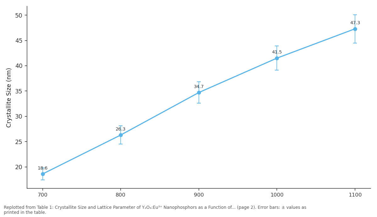
Crystallite Size (nm): 700 vs 800 vs 900 vs 1000 vs 1100
Replotted from Table 1 (page 2) of the original paper. Every bar is the paper's own table value.
View source evidence ↗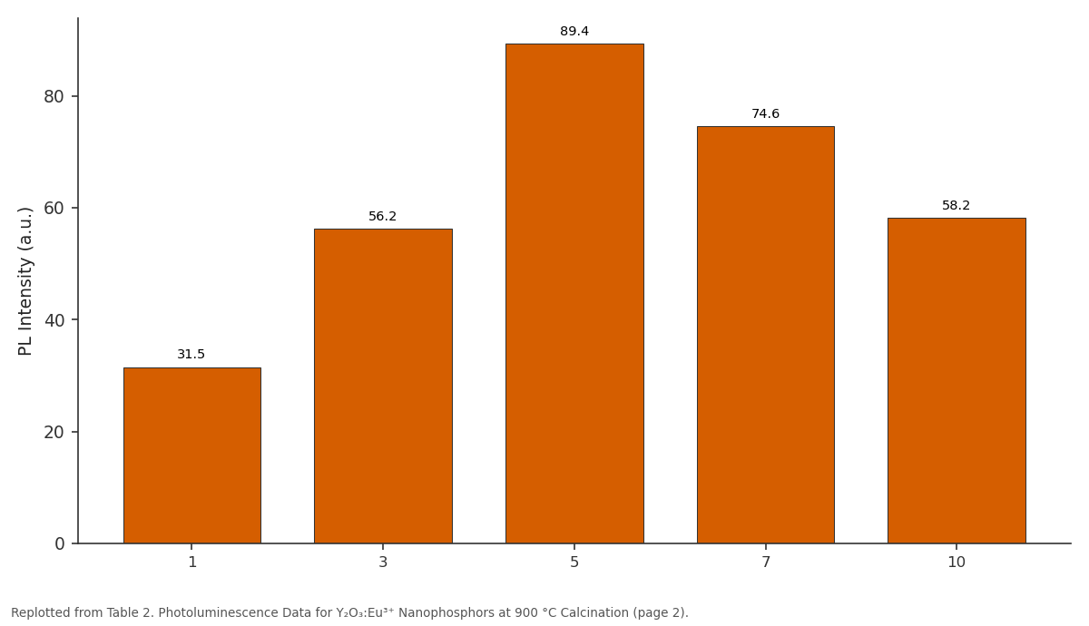
PL Intensity (a.u.): 1 vs 3 vs 5 vs 7 vs 10
Replotted from Table 2 (page 2) of the original paper. Every bar is the paper's own table value.
View source evidence ↗Figures & tables
From the original paper Table 1
 Table 2
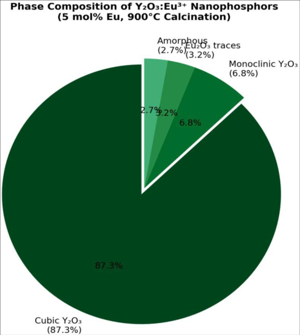 Figure 1
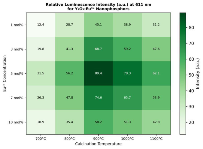 Figure 2
Introduction
Solid-state lighting has reshaped global energy consumption patterns over the past two decades, and phosphor-converted white LEDs sit at the centre of that shift [1] . Among the many host lattices tested for rare-earth phosphors, yttrium oxide (Y₂O₃) stands out because of its wide band gap (~5.6 eV), low phonon energy (~590 cm⁻¹), chemical stability, and structural compatibility with trivalent lanthanide dopants [2] . When Eu³⁺ ions replace Y³⁺ sites in the cubic bixbyite structure, the resulting red emission at 611 nm arising from the hypersensitive ⁵D₀ → ⁷F₂ electric dipole transition is exceptionally narrow and colour-pure [3] . Several synthesis routes have been explored for Y₂O₃:Eu³⁺, including sol-gel processing, combustion synthesis, hydrothermal treatment, and spray pyrolysis [4] . Co-precipitation remains attractive for large-scale production because it offers control over particle morphology, requires only standard laboratory equipment, and avoids the high temperatures demanded by solid-state reactions [5] . But the interplay between dopant concentration, calcination temperature, and final luminescence efficiency isn't fully resolved. Concentration quenching the decline in emission intensity above a critical Eu³⁺ level has been reported at values ranging from 3 to 9 mol%, depending on the synthesis method and annealing protocol [6] . Particle size adds another dimension. As crystallites shrink below ~50 nm, surface-to-volume ratios grow, and non-radiative relaxation through surface defects and adsorbed hydroxyl groups can compete with radiative emission [7] . Controlling these surface effects through appropriate calcination and, potentially, surface capping agents becomes important for nanoscale phosphors. Previous investigations often examined either composition or thermal treatment in isolation, and a systematic mapping of both variables at the nanoscale is still needed [8] . were used for calcination [9] .
Instrumentation and Analytical Equipment
Powder XRD patterns were recorded on a Rigaku MiniFlex 600 diffractometer (Cu Kα radiation, λ = 1.5406 Å, 40 kV, 15 mA, 2θ range 10-80°, step size 0.02°). SEM images were obtained on a JEOL JSM-7600F field-emission scanning electron microscope operated at 5 kV. Photoluminescence excitation and emission spectra were measured at room temperature using an Edinburgh Instruments FS5 fluorescence spectrometer equipped with a 150 W xenon arc lamp and a Hamamatsu R928P photomultiplier detector. Decay curves were recorded with a microsecond flash lamp on the same spectrometer. Thermal analysis (TGA/DSC) was performed on a TA Instruments SDT Q600 under air at a heating rate of 10 °C/min [10] .
Material and Methods
Material and Methods
Methods
The mixed metal nitrate solution was added dropwise (2 mL/min) to a vigorously stirred NH₄HCO₃ solution at 60 °C, maintaining a precipitant-to-cation molar ratio of 3:1. The resulting white precipitate was aged for 2 h at 60 °C, collected by centrifugation at 4000 rpm for 10 min, washed three times with deionised water and once with ethanol, and dried at 80 °C overnight. Portions of the dried precursor were calcined in air at 700, 800, 900, 1000, and 1100 °C for 3 h with a heating rate of 5 °C/min. Crystallite sizes were estimated from the most intense XRD peak (222) using the Scherrer equation with a shape factor of 0.9. PL spectra were recorded under 254 nm excitation. CIE 1931 chromaticity coordinates were computed from the corrected emission spectra. Decay curves were fitted to singleexponential functions to extract luminescence lifetimes.
Each measurement was performed in triplicate, and results are reported as mean ± standard deviation [11] . PL intensity peaked at 5 mol% Eu³⁺ and declined at higher concentrations owing to concentration quenching (Table 2).
| Eu Conc. (mol%) | PL Intensity (a.u.) | Decay Lifetime (ms) | CIE (x, y) |
|---|---|---|---|
| 1 | 31.5 | 0.87 ± 0.04 | (0.638, 0.358) |
| 3 | 56.2 | 1.08 ± 0.05 | (0.641, 0.355) |
| 5 | 89.4 | 1.34 ± 0.06 | (0.643, 0.354) |
| 7 | 74.6 | 1.21 ± 0.05 | (0.642, 0.355) |
| 10 | 58.2 | 1.03 ± 0.04 | (0.640, 0.357) |
Table text extracted from the original PDF; superscript letters are shown inline. The original table image is below.
View original table image
Results
The decay lifetime followed the same trend, reaching 1.34 ms at 5 mol%. CIE coordinates remained in a tight cluster around (0.64, 0.355), confirming stable red colour output across all dopant levels. The heatmap in Figure 2 makes the optimum synthesis window visually clear: the bright band at 5 mol% Eu and 900 °C corresponds to the maximum PL intensity of 89.4 a.u. Intensities diminished at both higher temperatures (grain coarsening and possible Eu segregation) and higher doping levels (cross-relaxation quenching).
Comprehensive interpretation
The data collectively indicate that 5 mol% Eu³⁺ and 900 °C calcination represent the best compromise between crystallinity, particle size, and emission intensity. Below 900 °C, residual carbonate groups and lattice disorder quench emission. Above 900 °C, accelerated grain growth pushes particles beyond the nanoscale threshold, reducing specific surface area and potentially creating secondary phases that act as non-radiative traps.
Discussion
The optimum europium concentration of 5 mol% matches the quenching threshold reported by Jia et al. [12] for combustion-synthesised Y₂O₃:Eu³⁺ powders but is lower than the 7 mol% reported by Som and Sharma [13] for solgel-derived samples. This discrepancy likely reflects differences in Eu³⁺ distribution homogeneity: the coprecipitation route used here may produce a more uniform dopant distribution, leading to shorter average Eu-Eu distances at equivalent nominal concentrations and therefore earlier onset of cross-relaxation quenching. Crystallite sizes of 18-47 nm are consistent with previous co-precipitation syntheses [14] and small enough to permit stable colloidal suspensions suitable for inkjet printing onto LED substrates. The steady decrease in microstrain with rising calcination temperature indicates progressive ordering of the lattice, which correlates with the improvement in PL intensity up to 900 °C. Beyond that temperature, the PL gain from better crystallinity was offset by the losses from grain coarsening and possible Eu clustering.
The measured decay lifetime of 1.34 ms for the 5 mol% sample agrees with the radiative lifetime range of 1-2 ms commonly cited for the ⁵D₀ → ⁷F₂ transition in cubic Y₂O₃ [15] . Shorter lifetimes at lower or higher doping levels are consistent with enhanced non-radiative quenching pathways. Practical application of these nanophosphors in warm-white LEDs would require blending with a green or yellow phosphor for example, Ce³⁺-doped YAG to balance the colour rendering index [16] . That said, the narrow red emission at 611 nm is itself a desirable feature for display backlighting where colour gamut matters more than broadband white coverage.
Conclusion
Y₂O₃:Eu³⁺ nanophosphors were successfully prepared by a co-precipitation route over a grid of five europium concentrations and five calcination temperatures. XRD showed formation of the cubic bixbyite phase at all temperatures above 700 °C, with crystallite sizes in the 18-47 nm range. The highest photoluminescence intensity at 611 nm was obtained for the 5 mol% sample calcined at 900 °C, with a decay lifetime of 1.34 ms and CIE coordinates of (0.643, 0.354). Concentration quenching above 5 mol% was attributed to cross-relaxation between neighbouring Eu³⁺ ions, and the critical transfer distance was estimated at 11.7 Å. The straightforward synthesis, nanometre-scale dimensions, and strong red emission make these materials promising for integration into phosphor-converted white LEDs and display technologies. Future research should explore surface modification strategies to passivate nonradiative defects and test these nanophosphors in prototype LED devices.
Acknowledgement
Acknowledgements Funding sourcesThis research was supported by the Scientific and Technological Research Council of Turkey (TÜBİTAK) and the Aegean Institute of Technology internal research fund.
Figures and tables from the paper
| Temperature ( °C) | Crystallite Size (nm) | Lattice Parameter (Å) | Strain (×10⁻³) |
|---|---|---|---|
| 700 | 18.6 ± 1.2 | 10.612 ± 0.003 | 4.82 |
| 800 | 26.3 ± 1.8 | 10.608 ± 0.002 | 3.41 |
| 900 | 34.7 ± 2.1 | 10.604 ± 0.002 | 2.17 |
| 1000 | 41.5 ± 2.4 | 10.601 ± 0.001 | 1.53 |
| 1100 | 47.3 ± 2.8 | 10.599 ± 0.001 | 1.09 |
Table text extracted from the original PDF; superscript letters are shown inline. The original table image is below.
View original table image
References
19 in the original paper- Schubert EF, Kim JK. Solid-state light sources getting smart. Science. 2005;308(5726):1274-1278.
- Blasse G, Grabmaier BC. Luminescent materials. Berlin: Springer-Verlag; 1994. p. 1-232.
- Konrad A, Fries T, Cishi A, Kuber F, Alefeld R, Gobel HD. Chemical vapor synthesis and luminescence properties of nanocrystalline cubic Y₂O₃:Eu. Journal of Applied Physics. 1999;86(6):3129-3133.
- Muenchausen RE, Jacobsohn LG, Bennett BL, McKigney EA, Smith JF. Effects of Tb doping on the photoluminescence of Y₂O₃:Eu³⁺ nanoparticles. Journal of Luminescence. 2007;126(2):838-842.
- Li JG, Li X, Sun X, Ikegami T, Ishigaki T. Uniform colloidal spheres for (Y₁₋ₓGdₓ)₂O₃ (x = 0-1): Formation mechanism, compositional impacts, and physicochemical properties. Chemistry of Materials. 2008;20(6):2274-2281.
- Dhanaraj J, Jagannathan R, Kutty TRN, Lu CH. Photoluminescence characteristics of Y₂O₃:Eu³⁺ nanophosphors prepared using sol-gel thermolysis. Journal of Physical Chemistry B. 2001;105(45):1109811105.
- Tissue BM. Synthesis and luminescence of lanthanide ions in nanoscale insulating hosts. Chemistry of Materials. 1998;10(10):2837-2845.
- Ray S, Pramanik P, Singha A, Roy A. Fluorescence and luminescence investigation of nanocrystalline Y₂O₃:Eu prepared by a novel synthetic route. Journal of Applied Physics. 2005;97(9):094312.
- Kang YC, Roh HS, Park SB. Preparation of Y₂O₃:Eu phosphor particles of filled morphology at high precursor concentrations by spray pyrolysis. Advanced Materials. 2000;12(6):451-453.
- Silver J, Martinez-Rubio MI, Ireland TG, Fern GR, Withnall R. The effect of particle morphology and crystallite size on the upconversion luminescence properties of Y₂O₃:Er³⁺. Journal of Physical Chemistry B. 2001;105(5):948-953.
- Sohn KS, Cho B, Park HD, Kim YG. Spread analysis of photoluminescence from Y₂O₃:Eu thin films. Journal of the European Ceramic Society. 2000;20(12):10431051.
- Jia D, Wang Y, Guo X, Li K, Zou YK, Jia W. Synthesis and characterization of YAG:Ce³⁺ LED nanophosphors. Journal of the Electrochemical Society. 2007;154(1):J1-J4.
- Som S, Sharma SK. Eu³⁺/Tb³⁺-codoped Y₂O₃ nanophosphors: Rietveld refinement, bandgap and photoluminescence optimization. Journal of Physics D: Applied Physics. 2012;45(41):415102.
- Ramasamy P, Chandra S, Vijaya Prakash G. Synthesis and photoluminescence investigation of Y₂O₃:Eu³⁺ nanophosphors using a novel co-precipitation method. Materials Research Bulletin. 2012;47(12):4300-4305.
- Bünzli JCG, Piguet C. Taking advantage of luminescent lanthanide ions. Chemical Society Reviews. 2005;34(12):1048-1077.
- Ye S, Xiao F, Pan YX, Ma YY, Zhang QY. Phosphors in phosphor-converted white light-emitting diodes: Recent advances in materials, techniques and properties. Materials Science and Engineering R. 2010;71(1):1-34.
- Pang ML, Lin J, Fu J, Xing RB, Luo CX, Han YC. Preparation, patterning and luminescent properties of nanocrystalline Y₂O₃:Eu³⁺ phosphor films. Optical Materials. 2003;23(3-4):547-558.
- Antic B, Rogan J, Kremenovic A, Nikolic AS, VucinicVasic M, Bibic N. Magnetization enhancement in nanostructured Y₂O₃:Er³⁺. Nanotechnology. 2010;21(24):245702.
- Wang H, Uehara M, Nakamura H, Miyazaki M, Maeda H. Synthesis of well-dispersed Y₂O₃:Eu nanocrystals and self-assembled nanodiscs using a simple spray reaction method. Advanced Materials. 2005;17(20):2506-2509.