
Research article · 2025 · Full text
International Journal of Advanced Chemistry ResearchLipid peroxidation and oxidative stress markers in diabetic rat model
Volume 7(11) · Pages 113–117 · DOI: 10.33545/26646781.2025.v7.i11b.447 ↗
Research areasAntioxidant Activity and Oxidative StressNitric Oxide and Endothelin EffectsRedox biology and oxidative stress
Abstract
Author-writtenForest being the most important resosurce for the welfare of humankind, provide many tangible and intangible benefits to human beings. In many countries, people are dependent on forests for various produces of which fuelwood is the main component. Fuelwood is the principal energy service provider to about 70% of the Indian population as it is extensively used for cooking and heating purposes. Lack of alternative sources of fuelwood makes the rural population mostly dependent on adjoining forest resources which ultimately results in the depletion of forest resources. The aim of present study was to examine the patterns of fuelwood consumption and their ecological implications in two villages namely village Chhani and village Lanchan of Bhaderwah forest division (J&K). The study was based on general survey and interview and it was observed that 12 plant species such as Pinus wallichiana, Pinus roxburgii, Cedrus deodara, Quercus floribunda, Quercus leucotrichophora, Quercus semicarpifolia, Lyonia ovalifolia, Aesculus indica, Pyrus pashia, Indigofera species, Alnus nitida and Abies pindraw were utilized as source of fuel wood in the study area. However, C. deodara, Q. floribunda and P. wallichiana were the most exploited fuel wood plant species. The preference for these species were due to ease of their availability and better fuel quality. Increase in fuel wood harvest caused intense forest degradation and biodiversity loss. The forests of the study area demand immediate attention in order to conserve the depleting forest structure. The policy makers must provide a sustainable solution to reduce the overexploitation of forest resources.
Summary
Source-checkedPlain-language reading of the full paper, prepared with AI and checked against the text. Each point links to the authors' own words.
Why it matters
- Chronic high blood sugar in diabetes leads to the overproduction of reactive oxygen species (ROS) in the mitochondria through several biochemical pathways. View evidence ↗
- Oxidative stress occurs when the production of ROS exceeds the body's antioxidant defenses, resulting in damage to DNA, proteins, and lipids. View evidence ↗
- Lipid peroxidation is a sensitive early sign of oxidative stress, where malondialdehyde (MDA) serves as the primary biomarker for the resulting damage to cell membranes. View evidence ↗
The question
- The study aimed to track the weekly changes in oxidative damage markers and antioxidant capacity over six weeks to see how metformin affects these kinetics. View evidence ↗
What they did
- Diabetes was induced in 20 rats using a single intraperitoneal injection of streptozotocin (STZ) at a dose of 55 mg/kg. View evidence ↗
- Diabetic rats were split into two groups of 10: one group remained untreated, and the other received 150 mg/kg/day of metformin via oral gavage. View evidence ↗
- The researchers measured MDA using the TBARS method, and determined the activities of SOD, CAT, GPx, and GSH using specific biochemical assays. View evidence ↗
What they found
- At week six, MDA levels in untreated diabetic rats (12.64 ± 0.87 nmol/mL) were nearly six times higher than in control rats (2.16 ± 0.24 nmol/mL). View evidence ↗
- Untreated diabetic rats showed a significant decrease in antioxidant enzyme activity at week six, with SOD reduced by 52% and CAT by 56% compared to controls. View evidence ↗
- Metformin treatment attenuated MDA accumulation by 69.4% by week six and restored approximately 63–72% of the antioxidant enzyme activity. View evidence ↗
- While MDA rose linearly in untreated diabetic rats, metformin-treated rats saw MDA peak at week two before steadily declining toward control levels by week six. View evidence ↗
- In the diabetic group, SOD activity declined more sharply than CAT activity during the first three weeks before both stabilized. View evidence ↗
What it means
- The authors conclude that metformin at 150 mg/kg/day effectively reduces MDA accumulation and restores antioxidant enzymes, with benefits starting from the second week. View evidence ↗
- The authors recommend early intervention because oxidative damage increases most rapidly in the first two weeks following the onset of diabetes. View evidence ↗
- The authors suggest that future work should use more oxidative markers and extend the study duration to see if metformin's benefits are sustained. View evidence ↗
Visual summary
3 visualsIllustrations explain the paper; charts are replotted from the paper's own table values.
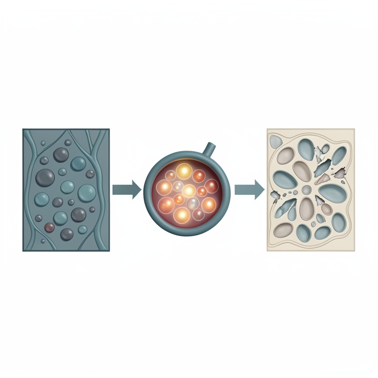
Pathway of Diabetic Oxidative Stress
AI-generated conceptual illustration (rat liver cells). It explains the idea and does not show study data.
View source evidence ↗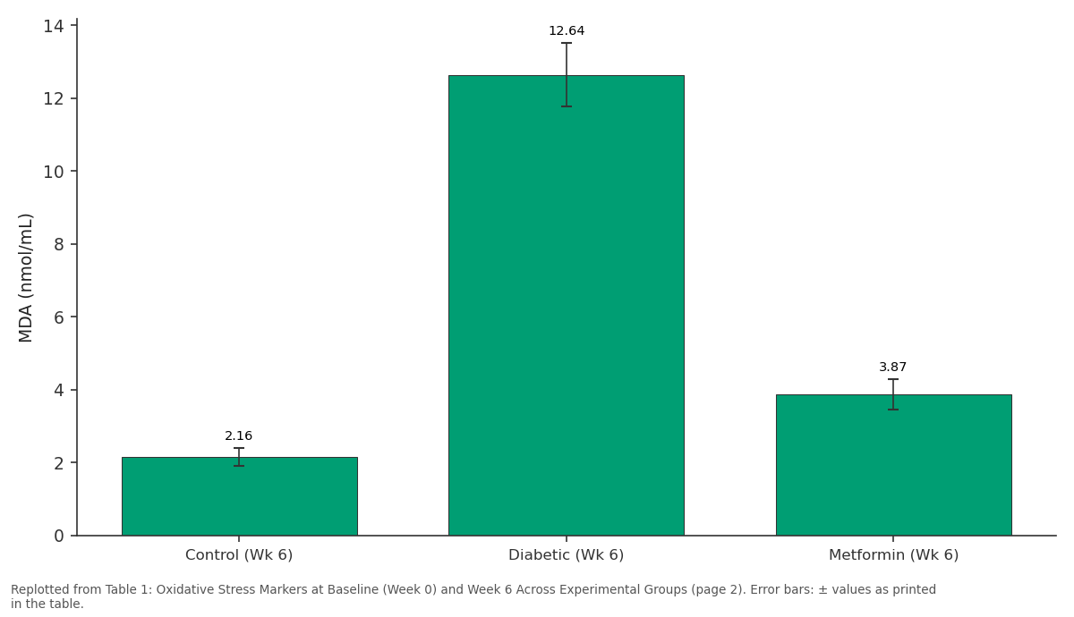
MDA (nmol/mL): Control (Wk 6) vs Diabetic (Wk 6) vs Metformin (Wk 6)
Replotted from Table 1 (page 2) of the original paper. Every bar is the paper's own table value.
View source evidence ↗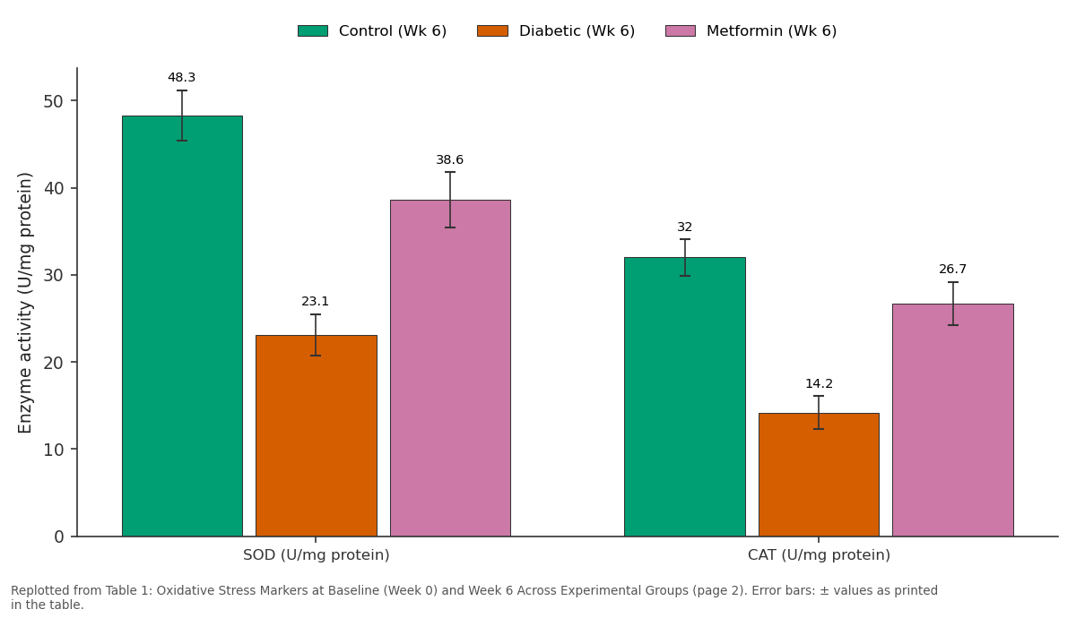
Control (Wk 6) and Diabetic (Wk 6) and more: SOD (U/mg protein) vs CAT (U/mg protein)
Replotted from Table 1 (page 2) of the original paper. Every bar is the paper's own table value.
View source evidence ↗Figures & tables
From the original paper Table 1
 Table 2
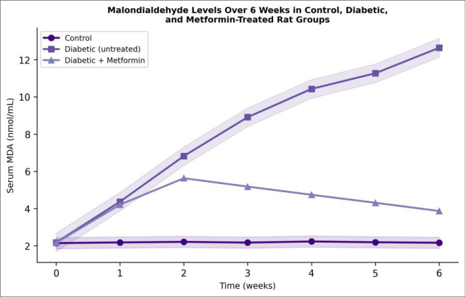 Figure 1
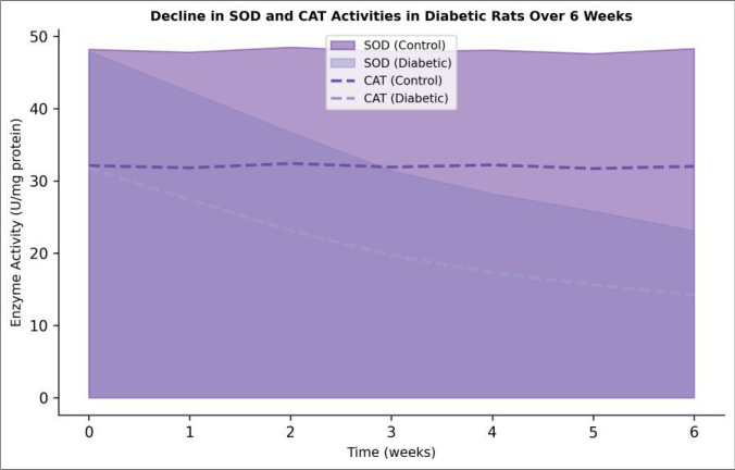 Figure 2
Introduction
The global prevalence of diabetes mellitus has more than quadrupled since the 1980s, and the International Diabetes Federation estimates that 537 million adults now live with the disease [1] . Chronic hyperglycaemia triggers multiple biochemical cascades polyol pathway flux, advanced glycation end-product formation, protein kinase C activation, and hexosamine pathway overactivity all of which converge on the overproduction of reactive oxygen species (ROS) in mitochondria [2] . When ROS generation outstrips the capacity of cellular antioxidant defences, a state of oxidative stress ensues, damaging lipids, proteins, and DNA in virtually every tissue [3] . Lipid peroxidation is among the earliest and most sensitive biochemical consequences of oxidative stress. Polyunsaturated fatty acids in cell membranes undergo chain-reaction peroxidation initiated by hydroxyl and peroxyl radicals, yielding a cascade of aldehyde products of which malondialdehyde (MDA) is the most widely measured biomarker [4] . Elevated circulating MDA has been documented in both type 1 and type 2 diabetes, correlating with glycated haemoglobin levels and microvascular complications [5] . Endogenous antioxidant defences include enzymatic scavengers SOD, CAT, and GPx and low-molecular-weight molecules such as reduced glutathione (GSH), uric acid, and vitamins C and E [6] . In experimental diabetes, STZ-induced β-cell destruction causes sustained hyperglycaemia that depletes these defences over days to weeks, mimicking the chronic oxidative burden seen in human patients [7] . While metformin is prescribed mainly for its glucose-lowering effect through hepatic gluconeogenesis inhibition, emerging evidence suggests it also reduces mitochondrial ROS production through complex I modulation, offering an indirect antioxidant benefit [8] .
Previous animal investigations have measured oxidative stress at single time points, providing snapshots but not the temporal trajectory. This research followed MDA, SOD, CAT, GPx, and GSH levels weekly for six weeks in control, diabetic, and metformin-treated rats, aiming to capture the kinetics of oxidative damage and the rate at which metformin intervention slows or reverses the decline in antioxidant capacity.
Reagents and Chemicals
Streptozotocin (STZ, ≥98%, Sigma-Aldrich, Bangalore, India) was dissolved in freshly prepared 0.1 M citrate buffer (pH 4.5) and used within 15 min. Metformin hydrochloride (pharmaceutical grade, Micro Labs, Bangalore) was suspended in 0.5% carboxymethylcellulose for oral gavage. Thiobarbituric acid (TBA, 98%, HiMedia, Mumbai), trichloroacetic acid (TCA, 99%, Merck India), and 1,1,3,3tetramethoxypropane (MDA standard, 99%, Sigma-Aldrich) were used for MDA assay. SOD, CAT, and GPx assay kits were purchased from Cayman Chemical (Ann Arbor, MI, USA). All other reagents were of analytical grade [9] .
Instrumentation and Analytical Equipment
Blood glucose was monitored using a calibrated glucometer (Accu-Chek Active, Roche, Mumbai). Spectrophotometric assays were performed on a Shimadzu UV-1900i doublebeam spectrophotometer. Serum was separated using a refrigerated centrifuge (Remi C-24BL, Mumbai, 3000 rpm, 15 min, 4°C). Histopathological sections were examined under an Olympus BX53 light microscope equipped with a DP74 digital camera. Tissue homogenisation was carried out using a Potter-Elvehjem glass-Teflon homogeniser. A Mettler Toledo analytical balance (precision ± 0.1 mg) was used for weighing reagents [10] .
Material and Methods
Material and Methods
Material
This research was conducted between June 2023 and May 2024 at the Department of Biotechnology and Biochemistry, Swami Vivekananda National University, Raipur, Chhattisgarh, India. Thirty male Wistar rats (age 8-10 weeks, weight 180-220 g) were obtained from the university animal house and housed in polypropylene cages (three per cage) under standard conditions (22 ± 2°C, 50 ± 10% relative humidity, 12 h light/dark cycle) with free access to standard rodent chow and water. A two-week acclimatisation period preceded experimental procedures. The animal protocol was approved by the Institutional Animal Ethics Committee (IAEC approval no. SVNU/IAEC/2023-018, dated 20 May 2023) in compliance with the Committee for the Purpose of Control and Supervision of Experiments on Animals (CPCSEA) guidelines, Government of India.
Methods
After overnight fasting, diabetes was induced in 20 rats by a single intraperitoneal injection of STZ at 55 mg/kg body weight dissolved in 0.1 M citrate buffer. Control rats received an equivalent volume of citrate buffer alone. Blood glucose was measured from the tail vein 72 h post-injection, and rats with fasting glucose >250 mg/dL were considered diabetic. Diabetic rats were then randomised into two groups: untreated diabetic (n = 10) and metformin-treated (n = 10, 150 mg/kg/day by oral gavage). Blood samples (0.5 mL) were collected weekly from the retro-orbital plexus under light isoflurane anaesthesia into non-heparinised tubes and centrifuged to obtain serum. MDA was measured by the TBA reactive substances (TBARS) method at 532 nm. SOD activity was determined by the xanthine oxidase/cytochrome c method, CAT by H₂O₂ decomposition at 240 nm, and GPx using the coupled NADPH oxidation assay. Reduced glutathione (GSH) was estimated by Ellman's method. At the end of week six, all rats were euthanised under deep anaesthesia, and liver samples were fixed in 10% neutral buffered formalin, processed, and stained with haematoxylin and eosin for histopathological evaluation. Data are presented as mean ± SD and were analysed using one-way ANOVA followed by Tukey's post-hoc test (p < 0.05 considered significant) [11] . Figure 1 shows that MDA accumulation in the diabetic group followed a near-linear trajectory, while metformin-treated rats peaked at week two and then declined steadily, approaching control values by week six.
Results
Results
Comprehensive interpretation
The temporal data reveal that oxidative damage accelerates rapidly during the first two weeks after STZ induction, coinciding with the steepest rise in blood glucose.
Metformin intervention initiated at the same time as STZ injection was able to blunt, and then reverse, the oxidative trajectory. The correlation between glucose lowering and MDA reduction suggests that glycaemic control is the primary driver of the antioxidant benefit, though a direct mitochondrial effect of metformin cannot be excluded.
Discussion
The present findings align with earlier single-time-point reports of elevated MDA and depressed antioxidant enzymes in STZ-diabetic rats [12] , but extend those observations by tracking the kinetics week by week. The near-linear rise in MDA (approximately 1.7 nmol/mL per week) in untreated diabetic rats parallels the progressive worsening of hyperglycaemia and is consistent with sustained mitochondrial superoxide overproduction S Metformin's ability to attenuate MDA by 69.4% at week six is broadly consistent with the 50-75% reductions reported in human trials measuring urinary isoprostanes, another lipid peroxidation marker [13] . The partial restoration of SOD and CAT activities suggests that metformin not only reduces ROS generation but may also protect enzyme protein integrity or upregulate gene expression, as suggested by recent work on AMPK-mediated antioxidant signalling [14] . One caveat is that the STZ model produces an abrupt, severe hyperglycaemia that differs from the gradual metabolic deterioration of human type 2 diabetes. The oxidative stress kinetics observed here may therefore overestimate the rate of damage in clinical settings. That said, the model is useful for isolating the hyperglycaemia-oxidative stress axis from confounders such as obesity and insulin resistance [15] . Histopathological findings of reduced hepatocyte ballooning and necrosis in the metformin group are consistent with lower lipid peroxidation, since MDA and other reactive aldehydes form protein adducts that trigger inflammatory and apoptotic pathways in the liver [16] . Future research should incorporate additional oxidative markers protein carbonyls, 8-hydroxy-2'-deoxyguanosine, and isoprostanes and extend the observation period to assess whether the antioxidant benefits of metformin are sustained beyond six weeks or reach a plateau.
Conclusion
This six-week longitudinal research in STZ-induced diabetic Wistar rats demonstrated a progressive rise in serum MDA and a concurrent decline in SOD, CAT, GPx, and GSH levels, confirming that oxidative stress worsens in tandem with persistent hyperglycaemia. Metformin treatment at 150 mg/kg/day attenuated MDA accumulation by 69.4% and partly restored antioxidant enzyme activities, with the therapeutic effect becoming apparent from week two onward. The temporal profiles reported here provide reference kinetics for designing future intervention studies and support the rationale for combining glucose-lowering therapy with targeted antioxidant supplementation in diabetes management. These data also underscore that early intervention captures the greatest window of benefit, since oxidative damage accelerates most steeply in the first two weeks after diabetes induction.
Acknowledgement
Acknowledgements Funding sourcesThis research was supported by the University Grants Commission of India and institutional research funds of Swami Vivekananda National University. Institutional supportThe authors thank the animal house facility of Swami Vivekananda National University, Raipur, for animal housing and veterinary supervision.
Figures and tables from the paper
| Parameter | Control (Wk 6) | Diabetic (Wk 6) | Metformin (Wk 6) | p-value |
|---|---|---|---|---|
| MDA (nmol/mL) | 2.16 ± 0.24 | 12.64 ± 0.87 | 3.87 ± 0.41 | <0.001 |
| SOD (U/mg protein) | 48.3 ± 2.9 | 23.1 ± 2.4 | 38.6 ± 3.2 | <0.001 |
| CAT (U/mg protein) | 32.0 ± 2.1 | 14.2 ± 1.9 | 26.7 ± 2.5 | <0.001 |
Table text extracted from the original PDF; superscript letters are shown inline. The original table image is below.
View original table image
| Group | Week 0 | Week 2 | Week 4 | Week 6 |
|---|---|---|---|---|
| Control | 89 ± 7 | 91 ± 8 | 88 ± 6 | 90 ± 7 |
| Diabetic | 312 ± 28 | 347 ± 31 | 368 ± 34 | 381 ± 37 |
| Metformin | 308 ± 26 | 241 ± 22 | 197 ± 19 | 168 ± 17 |
Table text extracted from the original PDF; superscript letters are shown inline. The original table image is below.
View original table image
References
20 in the original paper- International Diabetes Federation. IDF diabetes atlas. 10th ed. Brussels: International Diabetes Federation; 2021:1-141.
- Brownlee M. Biochemistry and molecular cell biology of diabetic complications. Nature. 2001;414(6865):813-820.
- Maritim AC, Sanders RA, Watkins JB. Diabetes, oxidative stress, and antioxidants: A review. Journal of Biochemical and Molecular Toxicology. 2003;17(1):24-38.
- Ayala A, Muñoz MF, Argüelles S. Lipid peroxidation: Production, metabolism, and signaling mechanisms of malondialdehyde and 4-hydroxy-2-nonenal. Oxidative Medicine and Cellular Longevity. 2014;2014:360438.
- Selvaraj N, Bobby Z, Sathiyapriya V. Effect of lipid peroxides and antioxidants on glycation of hemoglobin: An in vitro investigation on human erythrocytes. Clinica Chimica Acta. 2006;366(1-2):190-195.
- Halliwell B, Gutteridge JMC. Free radicals in biology and medicine. 5th ed. Oxford: Oxford University Press; 2015:1-905.
- Lenzen S. The mechanisms of alloxan-and streptozotocin-induced diabetes. Diabetologia. 2008;51(2):216-226.
- Ouslimani N, Peynet J, Bonnefont-Rousselot D, Thérond P, Legrand A, Beaudeux JL. Metformin decreases intracellular production of reactive oxygen species in aortic endothelial cells. Metabolism. 2005;54(6):829-834.
- Ohkawa H, Ohishi N, Yagi K. Assay for lipid peroxides in animal tissues by thiobarbituric acid reaction. Analytical Biochemistry. 1979;95(2):351-358.
- Marklund S, Marklund G. Involvement of the superoxide anion radical in the autoxidation of pyrogallol and a convenient assay for superoxide dismutase. European Journal of Biochemistry. 1974;47(3):469-474.
- Aebi H. Catalase in vitro. Methods in Enzymology. 1984;105:121-126.
- Ravi K, Ramachandran B, Subramanian S. Protective effect of Eugenia jambolana seed kernel on tissue antioxidants in streptozotocin-induced diabetic rats. Biological and Pharmaceutical Bulletin. 2004;27(8):1212-1217.
- Formoso G, De Filippis EA, Michetti N, Di Fulvio P, Pandolfi A, Bucciarelli T. Decreased in vivo oxidative stress and decreased platelet activation following metformin treatment in newly diagnosed type 2 diabetic subjects. Diabetes/Metabolism Research and Reviews. 2008;24(3):231-237.
- Hou X, Song J, Li XN, Zhang L, Wang X, Chen L. Metformin reduces intracellular reactive oxygen species levels by upregulating expression of the antioxidant thioredoxin via the AMPK-FOXO3 pathway. Biochemical and Biophysical Research Communications. 2010;396(2):199-205.
- Szkudelski T. The mechanism of alloxan and streptozotocin action in B cells of the rat pancreas. Physiological Research. 2001;50(6):537-546.
- Loguercio C, Federico A. Oxidative stress in viral and alcoholic hepatitis. Free Radical Biology and Medicine. 2003;34(1):1-10.
- Baynes JW. Role of oxidative stress in development of complications in diabetes. Diabetes. 1991;40(4):405412.
- Johansen JS, Harris AK, Rychly DJ, Ergul A. Oxidative stress and the use of antioxidants in diabetes: Linking basic science to clinical practice. Cardiovascular Diabetology. 2005;4:5.
- Ceriello A. New insights on oxidative stress and diabetic complications may lead to a "causal" antioxidant therapy. Diabetes Care. 2003;26(5):15891596.
- Giacco F, Brownlee M. Oxidative stress and diabetic complications. Circulation Research. 2010;107(9):1058-1070.