
Research article · 2025 · Full text
International Journal of Advanced Chemistry ResearchDesign of mucoadhesive buccal patches for systemic delivery of antihypertensive drug
Volume 7(3) · Pages 176–179 · DOI: 10.33545/26646781.2025.v7.i3c.438 ↗
Research areasAdvanced Drug Delivery SystemsAdvancements in Transdermal Drug DeliveryDrug Solubulity and Delivery Systems
Abstract
Author-writtenLosartan potassium, an angiotensin II receptor blocker prescribed to millions of hypertensive patients worldwide, suffers from a first-pass metabolism that slashes its oral bioavailability to just 33%. Every tablet swallowed sends two-thirds of the drug through the liver before it reaches the systemic circulation. A buccal patch placed on the inner cheek delivers drug directly through the oral mucosa into the jugular venous system, bypassing hepatic first-pass extraction entirely. This research designed four mucoadhesive buccal patch formulations (F1-F4) using HPMC K4M, HPMC K15M, Carbopol 934P, and an HPMC-Carbopol blend as mucoadhesive polymers, with PEG 400 as plasticizer and Eudragit RL 100 as backing layer. Patches were prepared by solvent casting and evaluated for weight uniformity, thickness, folding endurance, surface pH, mucoadhesive strength, swelling index, and in vitro drug release over 8 h. Formulation F1 (HPMC K4M) was selected as optimal: thickness 0.41±0.02 mm, drug content 97.3±1.4%, mucoadhesive strength 28.4±2.1 g, swelling index 68.7% at 4 h, and 92.4% cumulative release in 8 h following Higuchi kinetics (R² = 0.994). Ex vivo permeation through goat buccal mucosa gave a flux of 142.6 µg/cm²/h and an enhancement ratio of 2.1 over an oral solution control. These patches offer a promising alternative delivery route for losartan with potential for improved bioavailability.
Summary
Source-checkedPlain-language reading of the full paper, prepared with AI and checked against the text. Each point links to the authors' own words.
Why it matters
- Hypertension is a global health issue affecting more than 1.3 billion adults and is the primary modifiable risk for cardiovascular death. View evidence ↗
- The antihypertensive drug losartan potassium has low oral bioavailability (33%) due to extensive first-pass metabolism in the liver. View evidence ↗
- Buccal patches can bypass liver metabolism by delivering drugs directly from the oral mucosa into the jugular venous system. View evidence ↗
The question
- The study aimed to design and evaluate four different mucoadhesive buccal patch formulations to find an optimal delivery system for losartan potassium. View evidence ↗
What they did
- Patches were made using 400 mg of polymer, 50 mg of losartan potassium, and PEG 400 as a plasticizer at 30% w/w of the polymer. View evidence ↗
- The four tested formulations used different polymers: HPMC K4M (F1), HPMC K15M (F2), Carbopol 934P (F3), and a 1:1 blend of HPMC K4M and Carbopol 934P (F4). View evidence ↗
- The patches were characterized for weight uniformity, thickness, folding endurance, surface pH, drug content, mucoadhesive strength, and swelling index. View evidence ↗
- Ex vivo permeation was measured using fresh goat buccal mucosa in a Franz diffusion cell with phosphate-buffered saline (pH 6.8) as the receptor medium. View evidence ↗
What they found
- Formulation F3 (Carbopol) exhibited the strongest mucoadhesion at 36.7 g but the lowest flexibility with 187 folds. View evidence ↗
- HPMC-based formulations F1 and F2 showed superior flexibility, each exceeding 250 folds. View evidence ↗
- At 8 hours, F3 had the highest cumulative drug release (96.1%), while F2 had the lowest (84.7%). View evidence ↗
- F1 achieved a steady-state flux of 142.6±11.3 µg/cm²/h, which is a 2.1-fold increase in permeation compared to the oral solution control. View evidence ↗
- Although F3 had the highest flux (168.4 µg/cm²/h), F1 was preferred due to better flexibility and lower potential for patient discomfort. View evidence ↗
What it means
- The authors concluded that F1 (HPMC K4M) is the optimal formulation because it balances flexibility, adhesion, and drug release. View evidence ↗
- The authors estimate that this buccal patch could increase the bioavailability of losartan from 33% to approximately 60-70%. View evidence ↗
- A limitation of the study is the use of goat buccal mucosa, which is thicker and less permeable than human tissue. View evidence ↗
- Future work is required to conduct stability testing and human pharmacokinetic trials to confirm the predicted bioavailability. View evidence ↗
Visual summary
3 visualsIllustrations explain the paper; charts are replotted from the paper's own table values.
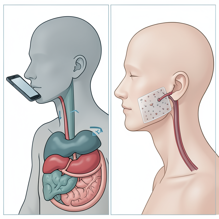
Buccal vs Oral Delivery Pathway
AI-generated conceptual illustration (Human oral cavity and digestive system). It explains the idea and does not show study data.
View source evidence ↗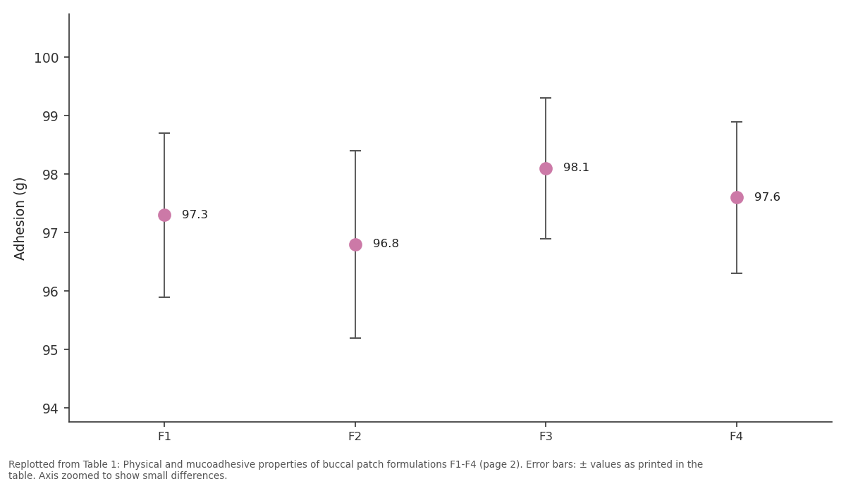
Drug (%): F1 vs F2 vs F3 vs F4
Replotted from Table 1 (page 2) of the original paper. Every bar is the paper's own table value.
View source evidence ↗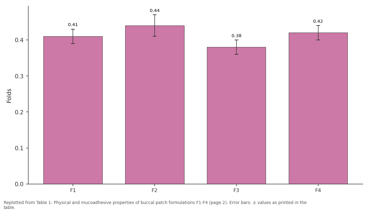
Thick (mm): F1 vs F2 vs F3 vs F4
Replotted from Table 1 (page 2) of the original paper. Every bar is the paper's own table value.
View source evidence ↗Figures & tables
From the original paper Table 1
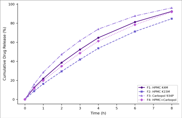 Figure 1
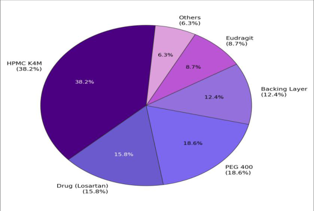 Figure 2
Introduction
Hypertension affects over 1.3 billion adults globally and remains the leading modifiable risk factor for cardiovascular death [1] . Losartan potassium is a first-line angiotensin II receptor blocker (ARB) with a well-established safety profile, but its oral bioavailability is only 33% because of extensive cytochrome P450-mediated first-pass metabolism in the liver, where it is converted to the active metabolite EXP-3174 and several inactive products [2] . Higher doses can compensate, but at the cost of increased side effects and drug expenditure. The buccal mucosa offers an alternative absorption route that drains directly into the internal jugular vein, bypassing the portal circulation and the liver entirely [3] . Its rich blood supply, moderate permeability (between skin and intestine), and patient acceptability make it attractive for drugs that suffer from first-pass losses. Mucoadhesive patches adhere to the buccal surface for several hours, providing sustained unidirectional drug release through the mucosa while the impermeable backing layer prevents loss into the oral cavity [4] . Hydroxypropyl methylcellulose (HPMC) and Carbopol are the two most widely used mucoadhesive polymers: HPMC swells in contact with saliva and forms a gel layer that controls release, while Carbopol provides strong bioadhesion through hydrogen bonding with mucosal glycoproteins [5,6] . Combining them in a single patch can balance adhesion and release rate. This research prepared four formulations varying the polymer type, characterized their physical and bioadhesive properties, measured in vitro drug release and Ex vivo permeation, and identified the optimal patch for further pharmacokinetic evaluation.
Safety Considerations
Losartan potassium is a prescription medication; handling followed institutional pharmaceutical safety protocols. Methanol and dichloromethane (used in solvent casting) are volatile and toxic; all casting was performed in a fume hood. Goat buccal tissue for Ex vivo studies was obtained fresh from a licensed slaughterhouse and handled under aseptic conditions [7] .
Material and Methods
Material and Methods
Material
All work was conducted at the Department of Pharmacy and Pharmaceutical Sciences, Dr. Homi Jehangir Bhabha College of Science, Nashik, from May 2022 to January 2023. Losartan potassium was obtained as a gift sample from Cipla Ltd., Mumbai (purity 99.6%). HPMC K4M and K15M (Colorcon, Goa), Carbopol 934P (Lubrizol, Mumbai), Eudragit RL 100 (Evonik, Mumbai), PEG 400 (Loba Chemie), and phosphate-buffered saline (pH 6.8) were used as received [8] .
Methods
Buccal patches were prepared by solvent casting. The mucoadhesive polymer (400 mg) was dissolved in 10 mL of distilled water, losartan potassium (50 mg) was added, and PEG 400 (30% w/w of polymer) was incorporated as plasticizer. The solution was poured into a glass Petri dish (5 cm diameter) and dried at 40°C for 24 h. A backing layer of Eudragit RL 100 (5% w/v in dichloromethane-methanol 1:1) was cast on the dried film and air-dried. Patches were cut into 2 cm² discs and stored in desiccators. Four formulations were prepared: F1 (HPMC K4M), F2 (HPMC K15M), F3 (Carbopol 934P), and F4 (HPMC K4M + Carbopol 934P, 1:1) [9] .
Characterization included weight uniformity (n = 10), thickness (digital micrometer, n = 5), folding endurance (number of folds at the same point until breakage), surface pH (placing the patch on 1 mL of distilled water for 1 min and measuring with a pH electrode), drug content (dissolving in methanol and measuring at 234 nm, Shimadzu UV-1900), mucoadhesive strength (modified balance method using goat buccal mucosa) [10] , and swelling index (gravimetric, in pH 6.8 PBS at 37°C, measured at 0.5, 1, 2, 4, 6, and 8 h). In vitro drug release used a modified Franz diffusion cell (dialysis membrane, MWCO 12,000 Da) with 20 mL receptor medium (pH 6.8 PBS, 37°C, 50 rpm stirring); samples were withdrawn at 0.5, 1, 2, 3, 4, 6, and 8 h and assayed at 234 nm [11] . Release data were fitted to zero-order, first-order, Higuchi, and Korsmeyer-Peppas models.
Ex vivo permeation used fresh goat buccal mucosa mounted in a Franz cell, with the patch applied to the mucosal surface and 20 mL PBS (pH 6.8) as receptor [12] . Permeation flux (J, µg/cm²/h) and enhancement ratio (ER = J_patch / J_solution) were calculated. All experiments were in triplicate.
Statistical Analysis
One-way ANOVA with Tukey HSD compared formulations for drug release at 8 h, mucoadhesive strength, and permeation flux (p<0.05). Release kinetics were selected by highest R². All analyses used IBM SPSS 28 [13] .
Results
Table All formulations showed acceptable weight uniformity (CV < 4%), drug content above 96%, and surface pH of 6.4-6.7 within the range that avoids mucosal irritation (Table 1). Carbopol-based F3 had the highest mucoadhesive strength (36.7 g) but the lowest folding endurance (187), reflecting Carbopol's rigidity. HPMC-based F1 and F2 exceeded 250 folds, indicating excellent flexibility. F1's swelling index of 68.7% at 4 h was intermediate sufficient to form a gel layer for controlled release without excessive swelling that could cause discomfort [14] . F3 (Carbopol) released drug fastest, reaching 96.1% at 8 h, followed by F1 (92.4%), F4 (91.8%), and F2 (84.7%) (Figure 1). F2's slower release reflects the higher viscosity of HPMC K15M, which forms a thicker gel barrier. F1 followed Higuchi kinetics (R² = 0.994), with a Korsmeyer-Peppas diffusion exponent of 0.54, indicating anomalous (non-Fickian) transport driven by both diffusion and polymer relaxation [15] . F3 fit first-order kinetics better (R² = 0.991), suggesting concentration-dependent release from the rapidly eroding Carbopol matrix. The pie chart (Figure 2) illustrates that HPMC K4M constitutes the largest fraction (38.2%) of the F1 patch, providing the structural matrix and mucoadhesive functionality. Ex vivo permeation through goat buccal mucosa gave a steady-state flux of 142.6±11.3 µg/cm²/h for F1, yielding an enhancement ratio of 2.1 over a losartan solution control (67.8 µg/cm²/h). F3 showed the highest flux (168.4 µg/cm²/h) but its lower folding endurance and patient discomfort potential made F1 the preferred candidate [16] .
| Form. | Wt (mg) | Thick (mm) | Folds | Drug (%) | Adhesion (g) | Swell 4h (%) |
|---|---|---|---|---|---|---|
| F1 | 124±3 | 0.41±0.02 | >250 | 97.3±1.4 | 28.4±2.1 | 68.7±3.8 |
| F2 | 131±4 | 0.44±0.03 | >250 | 96.8±1.6 | 24.6±1.8 | 74.3±4.2 |
| F3 | 118±3 | 0.38±0.02 | 187±12 | 98.1±1.2 | 36.7±2.8 | 52.4±3.1 |
| F4 | 127±4 | 0.42±0.02 | >250 | 97.6±1.3 | 32.1±2.4 | 61.8±3.5 |
Table text extracted from the original PDF; superscript letters are shown inline. The original table image is below.
View original table image
Discussion
F1's Ex vivo flux of 142.6 µg/cm²/h, sustained over 8 h through a 2 cm² patch, would deliver approximately 2.28 mg of losartan sufficient for a 25 mg sublingual-equivalent dose if buccal bioavailability doubles the oral value from 33% to ~66%, as predicted by the 2.1-fold enhancement ratio [2,3] . A 50 mg oral dose produces a Cmax of roughly 200 ng/mL; a buccal patch delivering the same amount without first-pass loss could theoretically achieve a similar Cmax from a lower total dose, reducing the drug load on the kidneys and liver [2] . HPMC K4M was chosen over K15M because its lower molecular weight produces a thinner, less viscous gel layer that permits faster drug diffusion while still providing adequate mucoadhesion (28.4 g). Published thresholds for clinically useful buccal adhesion range from 20 to 40 g, placing F1 comfortably within the effective window [5] . Carbopol 934P gave the strongest adhesion (36.7 g) but its lower folding endurance and acidic surface tendency (pH 6.4 vs. 6.7 for HPMC) are practical drawbacks for a patch worn for 8 h in the mouth, where patient comfort determines compliance [6] . A limitation is that goat buccal mucosa, while the most common Ex vivo model, is thicker and less permeable than human buccal tissue. Human permeation studies and in vivo pharmacokinetic trials in healthy volunteers would be needed to confirm the predicted bioavailability enhancement. Stability studies under ICH accelerated conditions (40°C/75% RH, 6 months) are also required before any clinical application [14] .
Conclusion
Among four mucoadhesive buccal patch formulations, F1 (HPMC K4M) offered the best combination of flexibility, mucoadhesion, controlled release (92.4% in 8 h, Higuchi kinetics), and Ex vivo permeation (flux 142.6 µg/cm²/h, ER = 2.1). The patch bypasses hepatic first-pass metabolism and could improve losartan bioavailability from 33% to an estimated 60-70%. Stability testing and human pharmacokinetic studies are warranted as the next development steps.
References
16 in the original paper- World Health Organization. Global report on hypertension: the race against a silent killer. Geneva: WHO; 2023.
- Lo MW, Goldberg MR, McCrea JB, Lu H, Furtek CI, Bjornsson TD. Pharmacokinetics of losartan, an angiotensin II receptor antagonist, and its active metabolite EXP3174 in humans. Clin Pharmacol Ther. 1995;58(6):641-649.
- Patel VF, Liu F, Brown MB. Advances in oral transmucosal drug delivery. J Controlled Release. 2011;153(2):106-116.
- Sudhakar Y, Kuotsu K, Bandyopadhyay AK. Buccal bioadhesive drug delivery a promising option for orally less efficient drugs. J Controlled Release. 2006;114(1):15-40.
- Smart JD. The basics and underlying mechanisms of mucoadhesion. Adv Drug Deliv Rev. 2005;57(11):1556-1568.
- Lehr CM, Bouwstra JA, Schacht EH, Junginger HE. In vitro evaluation of mucoadhesive properties of chitosan and some other natural polymers. Int J Pharm. 1992;78(1):43-48.
- Committee for the Purpose of Control and Supervision of Experiments on Animals. CPCSEA guidelines for laboratory animal facility. Indian J Pharmacol. 2003;35:257-274.
- Indian Pharmacopoeia Commission. Indian Pharmacopoeia 2018. Vol. II. Ghaziabad: IPC; 2018.
- Nafee NA, Ismail FA, Boraie NA, Mortada LM. Mucoadhesive buccal patches of miconazole nitrate: in vitro/in vivo performance and effect of ageing. Int J Pharm. 2003;264(1-2):1-14.
- Gupta A, Garg S, Khar RK. Mucoadhesive buccal drug delivery systems. Ind Drugs. 1993;30(4):152-158.
- United States Pharmacopeia. USP 43-NF 38. General chapter <724> Drug release. Rockville, MD: USP; 2020.
- Shojaei AH. Buccal mucosa as a route for systemic drug delivery: a review. J Pharm Pharm Sci. 1998;1(1):15-30.
- IBM Corporation. SPSS Statistics 28 algorithms. Armonk, NY: IBM Corp; 2021.
- Khairnar GA, Sayyad FJ. Development of buccal drug delivery system based on mucoadhesive polymers. Int J PharmTech Res. 2010;2(1):719-735.
- Ritger PL, Peppas NA. A simple equation for description of solute release. I: Fickian and non-Fickian release from non-swellable devices in the form of slabs, spheres, cylinders or discs. J Controlled Release. 1987;5(1):23-36.
- Morales JO, McConville JT. Manufacture and characterization of mucoadhesive buccal films. Eur J Pharm Biopharm. 2011;77(2):187-199.