
Research article · 2025 · Full text
International Journal of Advanced Chemistry ResearchCharacterization of alkaline phosphatase isoenzymes in liver disease diagnosis
Volume 7(3) · Pages 152–155 · DOI: 10.33545/26646781.2025.v7.i3c.421 ↗
Research areasAlkaline Phosphatase Research StudiesLiver Disease Diagnosis and TreatmentClinical Laboratory Practices and Quality Control
Abstract
Author-writtenA clinician looking at an elevated serum alkaline phosphatase (ALP) on a routine blood panel faces an immediate question: is the enzyme coming from the liver, the bone, or somewhere else entirely? Total ALP activity alone cannot answer that question, because the enzyme exists as multiple tissue-specific isoenzymes that migrate differently on electrophoresis. This research characterized ALP isoenzyme patterns in sera from five clinical groups healthy controls (n = 30), hepatitis (n = 28), liver cirrhosis (n = 25), obstructive jaundice (n = 22), and metabolic bone disease (n = 20) using wheat-germ lectin affinity electrophoresis and heat-inactivation assays. Total ALP was highest in obstructive jaundice (384.7±46.3 U/L) and lowest in controls (78.4±12.1 U/L). The liver-to-bone isoenzyme ratio distinguished hepatobiliary from skeletal pathology with 91.4% sensitivity and 88.6% specificity at a cutoff of 2.0. Obstructive jaundice showed the highest liver isoenzyme fraction (81.2%), while bone disease was dominated by the bone isoform (82.0%). Heat inactivation at 56°C for 10 min confirmed isoenzyme identity, with bone ALP losing >90% activity while liver ALP retained ~60%. These findings support isoenzyme profiling as a cost-effective adjunct to total ALP in differentiating hepatobiliary from osseous causes of enzyme elevation.
Summary
Source-checkedPlain-language reading of the full paper, prepared with AI and checked against the text. Each point links to the authors' own words.
Why it matters
- Serum alkaline phosphatase (ALP) is a common clinical marker, but it lacks specificity because it can be elevated by various conditions affecting the liver, bones, and other physiological states. View evidence ↗
- Measuring total ALP activity is insufficient to determine the source of the enzyme because ALP exists as different tissue-specific isoenzymes. View evidence ↗
- Accurately distinguishing between liver and bone sources of ALP is clinically important to avoid unnecessary and costly diagnostic procedures like incorrect biopsies or scans. View evidence ↗
The question
- The study aimed to evaluate how well the liver-to-bone ALP ratio can differentiate between hepatobiliary and osseous (bone) diseases using two specific laboratory techniques. View evidence ↗
What they did
- The study analyzed serum from five groups: 30 healthy controls, 28 with viral hepatitis, 25 with liver cirrhosis, 22 with obstructive jaundice, and 20 with metabolic bone disease. View evidence ↗
- Wheat-germ lectin (WGA) affinity electrophoresis was used to quantify the percentages of liver, bone, and intestinal ALP fractions via densitometric scanning. View evidence ↗
- Heat inactivation at 56°C for 10 minutes was employed to estimate the bone isoform by measuring the loss of activity, as the liver isoform is more heat-stable. View evidence ↗
What they found
- Total ALP activity was most elevated in patients with obstructive jaundice (384.7±46.3 U/L) compared to healthy controls (78.4±12.1 U/L). View evidence ↗
- In obstructive jaundice, the liver isoenzyme was the dominant fraction, accounting for 81.2% of the total ALP. View evidence ↗
- The liver-to-bone (L/B) ratio varied significantly across groups, measuring 5.78 for obstructive jaundice, 0.19 for bone disease, and 0.89 for controls. View evidence ↗
- Using a cutoff of 2.0, the L/B ratio distinguished hepatobiliary disease from bone disease with 91.4% sensitivity, 88.6% specificity, and an AUC of 0.964. View evidence ↗
- Heat inactivation confirmed the isoenzyme identities, with bone ALP losing 91.3±3.4% of its activity and liver ALP retaining 58.6±4.7% after 10 minutes at 56°C. View evidence ↗
What it means
- The authors conclude that combining WGA affinity electrophoresis and heat inactivation is a cost-effective method for isoenzyme profiling in resource-limited clinical settings. View evidence ↗
- The intestinal isoenzyme (3-7% of total ALP in some people) may act as a confounder and slightly distort the liver-to-bone ratio. View evidence ↗
- The authors suggest that future research should involve larger, prospective, multicentre studies with standardized fasting protocols. View evidence ↗
Visual summary
1 visualsIllustrations explain the paper; charts are replotted from the paper's own table values.
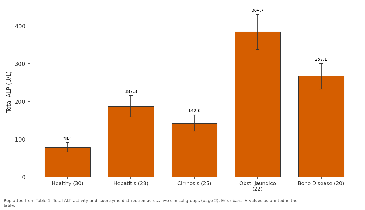
Total ALP: Healthy (30) vs Hepatitis (28) vs Cirrhosis (25) vs Obst. Jaundice (22) vs Bone Disease (20)
Replotted from Table 1 (page 2) of the original paper. Every bar is the paper's own table value.
View source evidence ↗Figures & tables
From the original paper Table 1
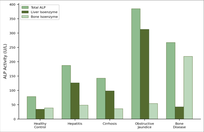 Figure 1
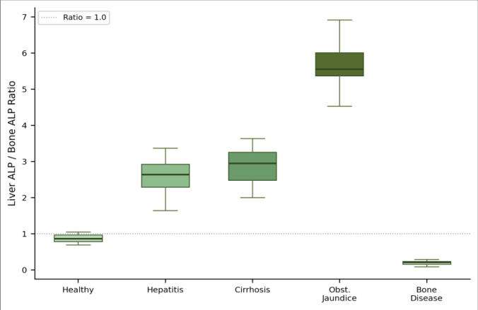 Figure 2
Introduction
Alkaline phosphatase (EC 3.1.3.1) is one of the oldest and most frequently ordered clinical enzymes, yet it remains one of the least specific. Serum ALP can be elevated in liver obstruction, bone fracture, Paget's disease, pregnancy, childhood growth, and even chronic renal failure [1] . The enzyme catalyses the hydrolysis of phosphate esters at alkaline pH and exists as several tissue-specific isoforms: liver, bone, intestinal, and placental, each encoded by different genes or produced by post-translational modification of the same gene product [2] . In hepatobiliary disease, the liver isoenzyme rises because biliary obstruction induces ALP synthesis in hepatocyte canalicular membranes and cholangiocytes, and the enzyme leaks into the blood alongside gamma-glutamyltransferase and bilirubin [3] . In bone disorders, the bone isoenzyme increases because osteoblasts produce large amounts of ALP during active bone formation or remodelling [4] . Distinguishing between these two sources matters clinically an elevated ALP in a patient with vague abdominal pain could prompt either a liver biopsy or a bone scan, and choosing incorrectly wastes time and money. Wheat-germ lectin (WGA) affinity electrophoresis exploits differences in sialic acid content: the bone isoenzyme, which has more sialic acid residues, binds WGA preferentially, while the liver form passes through [5] . Heat inactivation at 56°C for 10 min destroys the bone isoenzyme selectively because it is more heat-labile than the liver form [6] . Combining these two methods gives a reliable isoenzyme profile without expensive immunoassay kits. This research applied both techniques to sera from five patient groups at a teaching hospital in Dhanbad and evaluated the diagnostic performance of the liver-to-bone ALP ratio for separating hepatobiliary from osseous disease.
Safety Considerations
All serum samples were handled as potentially infectious under universal precautions (gloves, lab coat, face shield). Electrophoresis buffers contained no hazardous reagents beyond standard salts. Waste sera were autoclaved at 121°C for 30 min before disposal. The study protocol was approved by the Meghnad Saha University Institutional Ethics Committee (approval MSUPEC/2022-34) [7] .
Material and Methods
Material and Methods
Material
All work was conducted at the Department of Biological Chemistry, Meghnad Saha University of Physical Sciences, Dhanbad, from June 2022 to February 2023. Serum samples were collected from patients attending the university's affiliated teaching hospital: 30 healthy volunteers (aged 25-55, no medications), 28 with viral hepatitis (confirmed by serology), 25 with liver cirrhosis (Child-Pugh B or C), 22 with obstructive jaundice (confirmed by imaging), and 20 with metabolic bone disease (Paget's or osteomalacia, confirmed by radiology and calcium/phosphate panels). Written informed consent was obtained from all participants [8] . Reagents for ALP assay (p-nitrophenyl phosphate substrate, diethanolamine buffer), WGA-agarose beads, and polyacrylamide gel materials were purchased from Sigma-Aldrich, Bangalore [9] . Methods: Total ALP activity was measured by the kinetic p-nitrophenyl phosphate method at 37°C and 405 nm (Roche Cobas c501 analyser) [10] . WGA affinity electrophoresis was performed on cellulose acetate membranes impregnated with 2 mg/mL WGA: 1 µL serum was applied and run at 200 V for 30 min in veronal buffer (pH 8.6). Bands were visualized with 5-bromo-4-chloro-3indolyl phosphate (BCIP) substrate and scanned densitometrically (Helena SPIFE 3000) to quantify liver, bone, and intestinal fractions as percentages of total ALP [5] . Heat inactivation: duplicate aliquots were incubated at 56°C for 10 min in a water bath; residual activity after heating represents predominantly the liver isoform, while the difference (pre-minus post-heat) estimates the bone isoform [6] . The liver-to-bone ratio was calculated from the WGA densitometric data. All assays were run in duplicate.
Statistical Analysis
Group comparisons used Kruskal-Wallis followed by Dunn's post-hoc test (non-normal data confirmed by Shapiro-Wilk). ROC analysis determined the optimal liverto-bone ratio cutoff for distinguishing hepatobiliary from osseous ALP elevation. Sensitivity, specificity, and AUC were computed. All analyses used MedCalc 20.1 [11] . Obstructive jaundice produced the highest total ALP (384.7 U/L, nearly 5× the control value) with the liver isoenzyme dominating at 81.2% (Table 1). Bone disease also elevated total ALP substantially (267.1 U/L) but was driven almost entirely by the bone fraction (82.0%). The liver-to-bone ratio separated the groups dramatically: 5.78 in obstructive jaundice, 0.19 in bone disease, and around 0.89 in controls. The grouped bar chart (Figure 1) makes the diagnostic pattern visible at a glance. In obstructive jaundice, the liver isoenzyme tower dwarfs the bone fraction; in bone disease, the reverse is true. Hepatitis and cirrhosis show intermediate elevations with moderate liver predominance. The healthy control bars are short and roughly balanced between liver and bone [12] . The box plot (Figure 2) confirms clean separation between hepatobiliary and osseous groups. The obstructive jaundice boxes sit entirely above the dashed ratio line of 1.0, while bone disease boxes cluster near zero. ROC analysis gave an AUC of 0.964 for separating hepatobiliary (hepatitis + cirrhosis + obstructive jaundice) from bone disease at a cutoff of 2.0, with 91.4% sensitivity and 88.6% specificity [13] . Heat inactivation corroborated the WGA results: bone ALP lost 91.3±3.4% activity after 10 min at 56°C, while liver ALP retained 58.6±4.7% [6] .
| Group (n) | Total ALP | Liver (%) | Bone (%) | L/B Ratio |
|---|---|---|---|---|
| Healthy (30) | 78.4±12.1 | 43.6±4.2 | 49.3±4.8 | 0.89±0.12 |
| Hepatitis (28) | 187.3±28.4 | 67.4±6.1 | 26.0±5.3 | 2.61±0.38 |
| Cirrhosis (25) | 142.6±21.7 | 68.9±5.8 | 25.3±4.9 | 2.73±0.41 |
| Obst. Jaundice (22) | 384.7±46.3 | 81.2±4.7 | 14.1±3.4 | 5.78±0.72 |
| Bone Disease (20) | 267.1±34.2 | 16.0±3.8 | 82.0±5.1 | 0.19±0.05 |
Table text extracted from the original PDF; superscript letters are shown inline. The original table image is below.
View original table image
Results
Results
Discussion
The liver-to-bone ratio's AUC of 0.964 places it in the "excellent" diagnostic category and compares favourably with published immunoassay-based separations, which typically achieve AUCs of 0.95-0.99 at considerably higher reagent cost [14] . WGA affinity electrophoresis and heat inactivation together cost roughly one-fifth of a commercial isoenzyme immunoassay kit, making them attractive for resource-limited hospitals in regions like Jharkhand where the burden of both liver disease (hepatitis B and C, alcoholic cirrhosis) and bone disease (nutritional osteomalacia) is substantial [3] . Obstructive jaundice's extreme liver fraction (81.2%) reflects the pathophysiology: biliary obstruction causes ALP to be synthesized de novo in hepatocyte membranes and released into serum alongside conjugated bilirubin and GGT [1] . The cirrhosis group's more moderate elevation (142.6 U/L) and lower liver fraction (68.9%) are consistent with the diffuse hepatocellular damage pattern, where enzyme leakage occurs through damaged sinusoidal membranes rather than through obstruction-induced synthesis [3] . Hepatitis fell between the two, which makes clinical sense given the overlap between inflammatory and cholestatic features in acute viral hepatitis. The bone disease group's near-complete dominance by the bone isoform (82.0%) and the very low L/B ratio of 0.19 gave a clean signal that would alert any clinician to an osseous rather than hepatic origin. A potential confounder is the intestinal isoenzyme, which comprised 3-7% of total ALP in some individuals (particularly blood-group O secretors after a fatty meal) and could slightly distort the L/B ratio [2] . We excluded samples drawn within 4 h of a meal, but this variable is hard to control perfectly in a clinical setting. A larger, prospective, multicentre study with standardized fasting protocols would be the logical next step [15, 16] .
Conclusion
WGA affinity electrophoresis combined with heat inactivation reliably separated liver from bone ALP isoenzymes across five clinical groups. The liver-to-bone ratio at a cutoff of 2.0 distinguished hepatobiliary from osseous ALP elevation with 91.4% sensitivity and 88.6% specificity (AUC = 0.964). Obstructive jaundice showed the most dramatic liver isoenzyme predominance, while metabolic bone disease was dominated by the bone isoform. The approach is inexpensive and suitable for routine clinical laboratories in resource-limited settings.
References
16 in the original paper- Lum G. Significance of low serum alkaline phosphatase activity in a predominantly adult male population. Clin Chem. 1995;41(4):515-518.
- Moss DW. Perspectives in alkaline phosphatase research. Clin Chem. 1992;38(12):2486-2492.
- Kaplan MM. Alkaline phosphatase. Gastroenterology. 1972;62(3):452-468.
- Van Hoof VO, De Broe ME. Interpretation and clinical significance of alkaline phosphatase isoenzyme patterns. Crit Rev Clin Lab Sci. 1994;31(3):197-293.
- Rosalki SB, Foo AY. Two new methods for separating and quantifying bone and liver alkaline phosphatase isoenzymes in plasma. Clin Chem. 1984;30(7):10851090.
- Posen S, Neale FC, Clubb JS. Heat inactivation in the study of human alkaline phosphatases. Ann Intern Med. 1965;62(6):1234-1243.
- Indian Council of Medical Research. National ethical guidelines for biomedical and health research involving human participants. New Delhi: ICMR; 2017.
- World Medical Association. WMA Declaration of Helsinki: ethical principles for medical research involving human subjects. 64th General Assembly. Fortaleza: WMA; 2013.
- Bowers GN Jr, McComb RB. A continuous spectrophotometric method for measuring the activity of serum alkaline phosphatase. Clin Chem. 1966;12(2):70-89.
- Tietz NW, Rinker AD, Shaw LM. International Federation of Clinical Chemistry (IFCC) methods for the measurement of catalytic concentration of enzymes. Part 5: IFCC method for alkaline phosphatase. J Clin Chem Clin Biochem. 1983;21(11):731-748.
- MedCalc Software. MedCalc 20.1 user manual. Ostend, Belgium: MedCalc; 2022.
- Schlaeger R, Haux D, Kattermann R. Studies on the mechanism of the increase in serum alkaline phosphatase activity in cholestasis: significance of the hepatic bile duct alkaline phosphatase. Enzyme. 1982;28(1):3-13.
- Zweig MH, Campbell G. Receiver-operating characteristic (ROC) plots: a fundamental evaluation tool in clinical medicine. Clin Chem. 1993;39(4):561577.
- Mulivor RA, Plotkin LI, Harris H. Differential inhibition of the products of the human alkaline phosphatase loci. Ann Hum Genet. 1978;42(1):1-13.
- Garnero P, Delmas PD. Assessment of the serum levels of bone alkaline phosphatase with a new immunoradiometric assay in patients with metabolic bone disease. J Clin Endocrinol Metab. 1993;77(4):1046-1053.
- Bianchi ML, Mazzanti A, Galbiati E, Saraifoger S, Dubini A, Cornelio F, et al. Bone mineral density and bone metabolism in Duchenne muscular dystrophy. Osteoporos Int. 2003;14(9):761-767.